. 2018 Sep 14;13(9):e0204032.
doi: 10.1371/journal.pone.0204032. eCollection 2018.

Severity and properties of cardiac damage caused by Streptococcus pneumoniae are strain dependent

Affiliations

Severity and properties of cardiac damage caused by Streptococcus pneumoniae are strain dependent

Anukul T Shenoy et al. PLoS One. .

Abstract

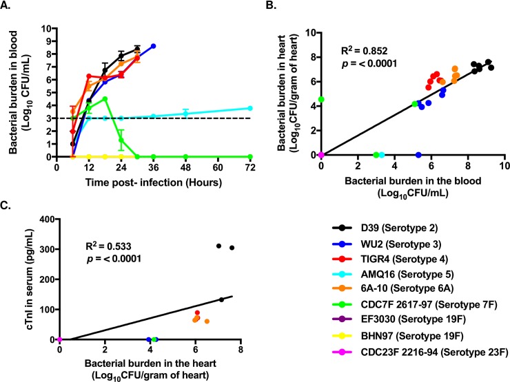
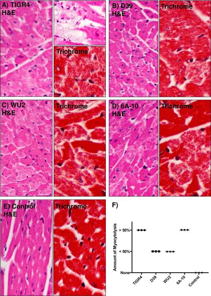
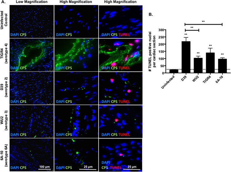
Streptococcus pneumoniae is an opportunistic Gram-positive pathogen that can cause invasive disease. Recent studies have shown that S. pneumoniae is able to invade the myocardium and kill cardiomyocytes, with one-in-five adults hospitalized for pneumococcal pneumonia having a pneumonia-associated adverse cardiac event. Furthermore, clinical reports have shown up to a 10-year increased risk of adverse cardiac events in patients formerly hospitalized for pneumococcal bacteremia. In this study, we investigated the ability of nine S. pneumoniae clinical isolates, representing eight unique serotypes, to cause cardiac damage in a mouse model of invasive disease. Following intraperitoneal challenge of C57BL/6 mice, four of these strains (D39, WU2, TIGR4, and 6A-10) caused high-grade bacteremia, while CDC7F:2617-97 and AMQ16 caused mid- and low-grade bacteremia, respectively. Three strains did not cause any discernible disease. Of note, only the strains capable of high-grade bacteremia caused cardiac damage, as inferred by serum levels of cardiac troponin-I. This link between bacteremia and heart damage was further corroborated by Hematoxylin & Eosin and Trichrome staining which showed cardiac cytotoxicity only in D39, WU2, TIGR4, and 6A-10 infected mice. Finally, hearts infected with these strains showed varying histopathological characteristics, such as differential lesion formation and myocytolysis, suggesting that the mechanism of heart damage varied between strains.


Conflict of interest statement

The authors have declared that no competing interests exist.

Figures

Fig 1
Fig 1. High-grade bacteremia is prerequisite to myocardial invasion and myocardial damage.
A) Bacterial burden of different strains of pneumococci circulating in the blood at designated time points following i.p. challenge of mice (n = 6 mice per strain). Points represent the average titer for the mice infected with each strain. Dashed line indicates limit of detection. B) Linear Regression analysis of pneumococcal burden recovered from the heart versus the pneumococcal burden in the blood of the same mice (n = 6 mice per strain). C) Linear Regression analysis of detectable troponin-I (cTn-I) in the blood of infected mice correlated versus pneumococcal burden recovered from the heart of the same mice (n = 3 mice per strain). Note, the limit of detection for the cTn-I ELISA is 0.156 ng/mL. Data are represented as mean ± SEM.
Fig 2
Fig 2. Pneumococcal invasion of the heart and microlesion formation is not required to incite cardiac damage.
Representative images of cardiac sections from mice infected with A) TIGR4, B) D39, C) WU2, and D) 6A-10, and E) an uninfected control. Tissue sections were either stained with H&E or Trichrome. Myocytolysis (i.e. degenerative cardiomyocyte damage visible as lightly stained areas in an otherwise homogenously stained myocardium) and hydropic degeneration of cardiomyocytes is clearly visible across all infected hearts. F) H&E stained heart sections from three mice per cohort were scored for extent of myocytolysis as < 50% of myocytes undergoing myocytolysis, > 50% of myocytes undergoing myocytolysis, or no myocytes undergoing myocytolysis.
Fig 3
Fig 3. TUNEL staining reveals cardiomyocyte death close to TIGR4, but dispersed damage in hearts from mice challenged with other strains.
A) Representative low and high magnification immunofluorescent microscopy images of cardiac sections form mice infected with TIGR4, D39, WU2, and 6A-10 pneumococci 30 hours post infection. The heart sections were probed for S. pneumoniae (green), nuclei (blue, DAPI) and fragmented nuclei (red, TUNEL probe). B) Enumeration of TUNEL positive nuclei per cardiac section of mice infected with TIGR4, D39, WU2, and 6A-10 pneumococci 30 hours post-infection. For each strain, 4–6 sections were examined. Statistical analyses were performed using a non-parametric one-way ANOVA. P value: ** ≤ 0.01, **** < 0.0001; data is represented as mean ± SEM.

References

    1. Shenoy AT, Orihuela CJ. Anatomical site-specific contributions of pneumococcal virulence determinants. Pneumonia. 2016; 8:7 10.1186/s41479-016-0007-9 - DOI - PMC - PubMed
    1. Butler JC, Schuchat A. Epidemiology of pneumococcal infections in the elderly. Drugs & Aging. 1999; 15: 11–19. 10.2165/00002512-199915001-00002 - DOI - PubMed
    1. Randle E, Ninis N, Inwald D. Invasive pneumococcal disease. Arch Dis Child Educ Pract Ed. 96: 183–190. 10.1136/adc.2010.191718 - DOI - PubMed
    1. Tan TQ. Pediatric invasive pneumococcal disease in the United States in the era of pneumococcal conjugate vaccines. Clin Microbiol Rev. 2012; 25(3): 409–419. 10.1128/CMR.00018-12 - DOI - PMC - PubMed
    1. Morrill HJ, Caffrey AR, Noh E, LaPlante KL. Epidemiology of pneumococcal disease in a national cohort of older adults. Infect Dis Ter. 2014; 3(1): 19–33. 10.1007/s40121-014-0025-y - DOI - PMC - PubMed

Publication types

MeSH terms