. 2019 Mar;90(3):295-305.
doi: 10.1002/JPER.18-0226. Epub 2018 Oct 7.

Periodontitis causes abnormalities in the liver of rats

Affiliations

Periodontitis causes abnormalities in the liver of rats

Any Carolina Cardoso Guimarães Vasconcelos et al. J Periodontol. 2019 Mar.

Abstract

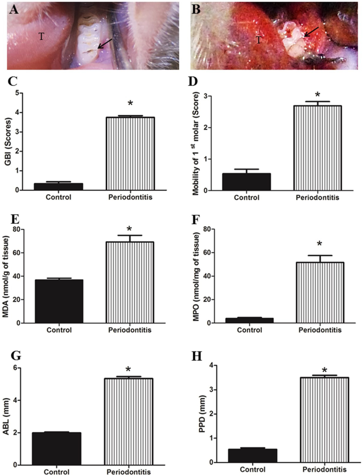
Background: Periodontitis not only causes injury to the periodontium, but also damages other tissues such as: articulate, renal, cardiac, and hepatic. The objective of this study was to investigate periodontitis induced alterations in liver function and structure using an experimental model.

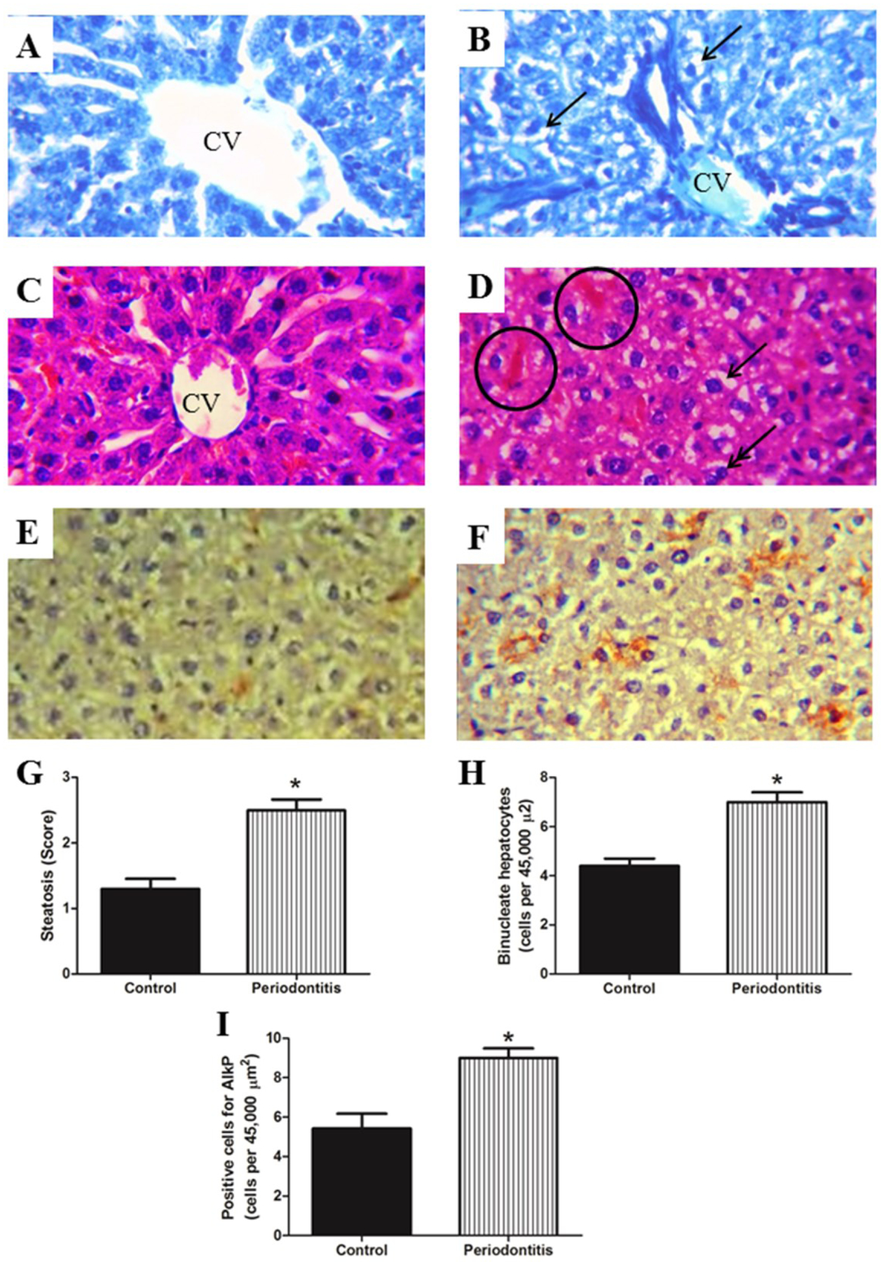
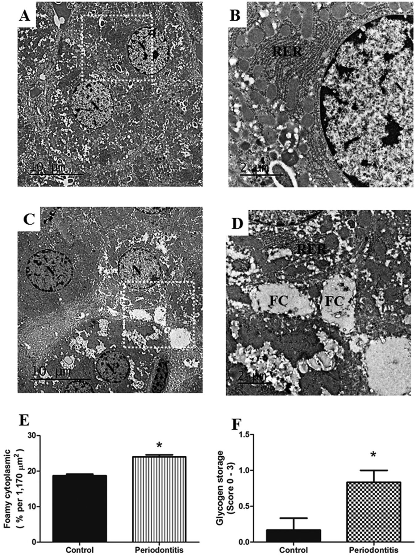
Methods: Twenty female rats (Rattus norvegicus) were allocated into two groups: control and periodontitis. Gingival bleeding index and oxidative stress parameters and specific circulating biomarkers were measured. Immunohistochemistry was carried out using alkaline phosphatase (AlkP) staining of the liver. Hepatic tissues, cytokines, and lipid contents were measured. Histopathologic evaluation of the liver was carried out using light and electron microscopy.

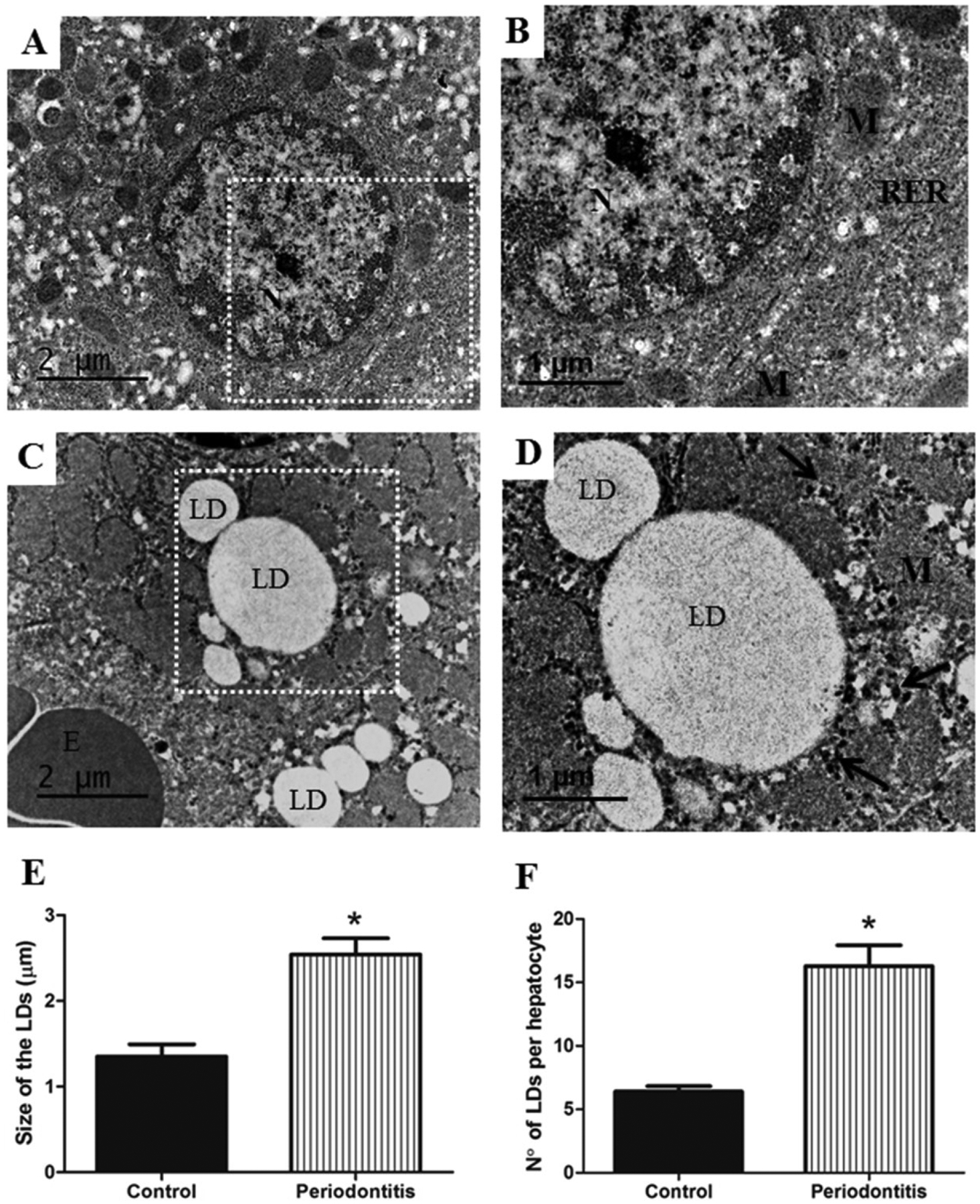
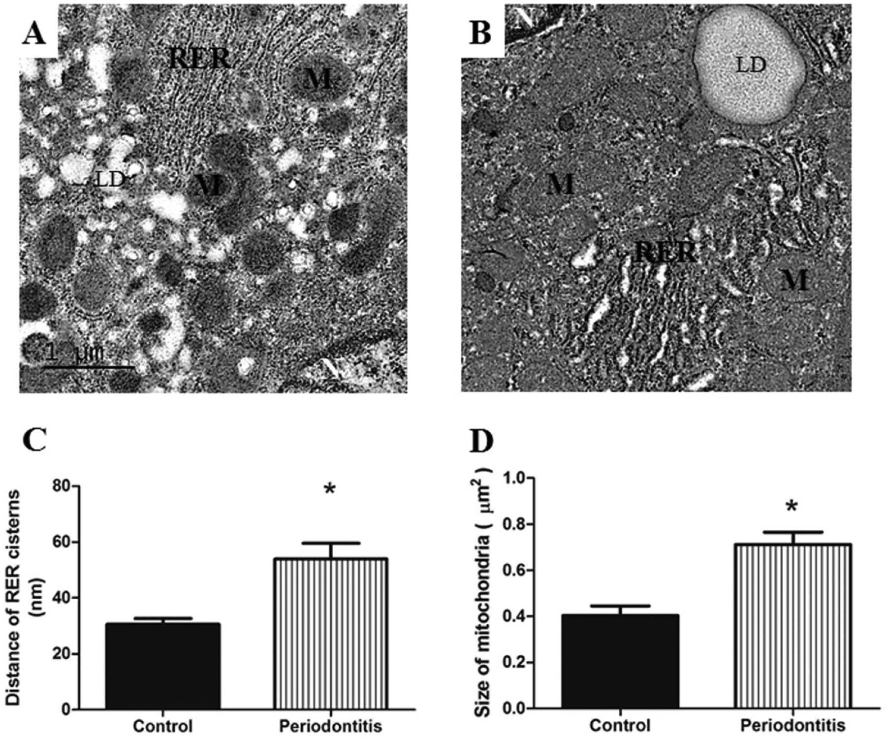
Results: Liver histopathologic and immunohistochemistry assessment showed increase in steatosis score, and presence of binucleate hepatocytes and positive cells for AlkP in periodontitis versus control group. Ultrastructural evaluation showed significant increase in size and number of lipid droplets (LD), distance between the cisterns of rough endoplasmic reticulum (RER), mitochondria size, foamy cytoplasm, and glycogen accumulation in the liver of the periodontitis group compared with the control group. In addition, plasma levels of AlkP, high-density lipoprotein (HDL), triglycerides, and total cholesterol were also changed.

Conclusion: Experimental periodontitis caused immunohistochemistry, histopathologic, ultrastructural, oxidative, and biochemical changes in the liver of rats.

Keywords: cytokines; inflammation; liver; oral medicine; periodontal diseases.


Conflict of interest statement

Conflict of interest

The authors declare there is no conflict of interest.

Figures

Figure 1.
Figure 1.
A) represents clinical aspect from control group, tongue is represented in T and the first molar by arrow. B) Clinical aspect of the periodontitis group, presenting gingival papilla with alteration in color, severe edema and bleeding after slight probe. C) Gingival Bleeding Index (GBI) scores. D) Mobility (M) of the 1st molar. E) MDA level. F) Myeloperoxidase (MPO) levels. G) Sum of alveolar bone loss (ABL). H) Probing Pocket Depth (PPD). GBI scores, M, MDA and GSH levels, ABL and PPD presented significantly higher values in periodontitis group than control group. * p < 0.05.
Figure 2.
Figure 2.
Liver in A) and C) demonstrating hepatocytes with normal organization surrounding the central vein (CV). Fatty liver (arrows) is illustrated in B) and D) for the periodontitis group, congestion in sinusoid blood vessels (circle) and binucleate hepatocytes (double arrow) are also observed. The periodontitis group presented higher values for G) steatosis score, H) number of binucleate hepatocytes and I) positive cell for AlkP compared with control group. E) illustrate positive cells for AlkP in the control group and F) in the periodontitis group. A and B) toluidine blue, C and D) hematoxylin and eosin, E and F) immunohistochemistry for AlkP, all photomicrographs are at 600x original magnification. * p < 0.05.
Figure 3.
Figure 3.
A) Hepatocyte with normal ultrastructures from control group, white rectangle shows B) area at higher magnification, demonstrating normal aspect of mitochondria (M), rough endoplasmic reticulum (RER) and nucleus (N). On the other hand, C) the periodontitis group shows hepatocyte with increase in size and number of lipid droplets (LD), D) area at higher magnification, representing detail from LD and glycogen accumulation (arrows). Higher values for E) size and F) number of LD were observed in the periodontitis group rather than in the control group. E, erythrocyte; M, mitochondria; N, nucleus. Images A) and C) of TEM are at ×10,000 original magnification, and B) and D), at ×15,000 original magnification. * p < 0.05.
Figure 4.
Figure 4.
A) Hepatocytes with normal mitochondria (M) and RER are observed in the control group. B) illustrates hepatocyte with dilatation of RER and mitochondria larger in the periodontitis group, when compared with control group; in addition, the mitochondria of periodontitis group were presented as less electron-dense with less distinct cristae. Periodontitis group presented C) dilation of cistern of the RER and D) larger mitochondria in comparison with the control group. LD, lipid droplets; N, nucleus; RER, rough endoplasmic reticulum. Images of TEM are at ×15,000 original magnification. * p < 0.05.
Figure 5.
Figure 5.
A) Two normal hepatocytes are observed in the center of the image of the control group, B) white rectangle shows the area at higher magnification, demonstrating normal RER, rough endoplasmic reticulum (RER), N, nucleus. C) shows hepatocytes with abundant foamy cytoplasm (FC) from the periodontitis group. D) presents the area from the white rectangle with higher magnification, demonstrating details of FC in the hepatocyte from rats with periodontitis. Periodontitis caused increase in the E) FC and F) in the glycogen storage into hepatocytes compared with hepatocytes from the control group. N, nucleus. Images A) and C) of TEM are at ×2,500 original magnification, and B) and D), at ×8,000 original magnification. * p < 0.05.

References

    1. Petersen PE, Bourgeois D, Ogawa H, Estupinan-Day S, Ndiaye C. The global burden of oral diseases and risks to oral health. Bull World Health Organ 2005; 83:661–9. - PMC - PubMed
    1. Eke PI, Dye BA, Wei L, et al. Update on prevalence of periodontitis in adults in the United States: NHANES 2009 to 2012. J Periodontol 2015; 86:611–22. - PMC - PubMed
    1. Chapple IL. Reactive oxygen species and antioxidants in inflammatory diseases. J Clin Periodontol 1997; 24:287–296. - PubMed
    1. White P, Sakellari D, Roberts H, et al. Peripheral blood neutrophil extracellular trap production and degradation in chronic periodontitis. J Clin Periodontol 2016; 12:1041–1049. - PubMed
    1. Oliveira RR, Fermiano D, Feres M, et al. Levels of candidate periodontal pathogens in subgingival biofilm. J Dent Res 2016; 95:711–8. - PMC - PubMed

Publication types