. 2018 Sep;32(5):1517-1529.
doi: 10.1111/jvim.15248. Epub 2018 Sep 14.

Atrioventricular accessory pathways in 89 dogs: Clinical features and outcome after radiofrequency catheter ablation

Affiliations

Atrioventricular accessory pathways in 89 dogs: Clinical features and outcome after radiofrequency catheter ablation

Kathy N Wright et al. J Vet Intern Med. 2018 Sep.

Abstract

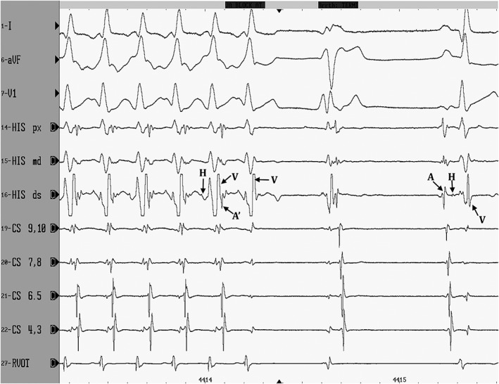
Background: Atrioventricular accessory pathways (APs) in dogs have been reported rarely. Data regarding clinical presentation and long-term outcome after radiofrequency catheter ablation (RFCA) are limited.

Hypothesis/objectives: To study clinical features, electrophysiologic characteristics, and outcome of RFCA in dogs with APs.

Animals: Eighty-nine dogs presented consecutively for RFCA of APs.

Methods: Case series.

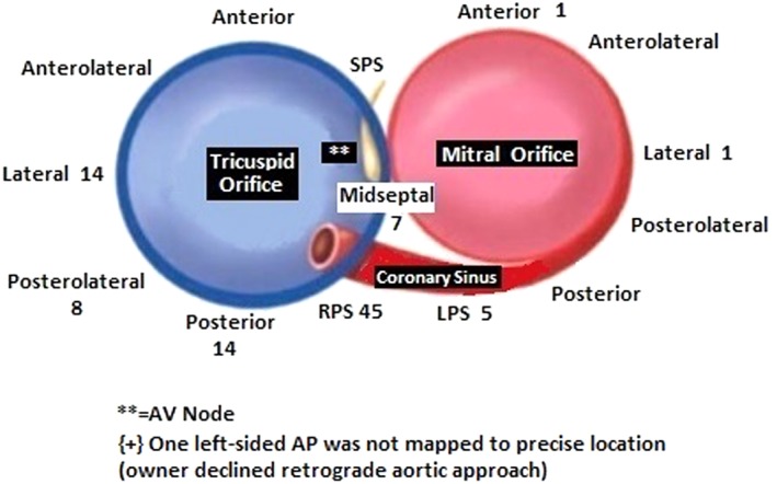
Results: Labrador retrievers (47.2% of dogs) and male dogs (67.4% of dogs) were most commonly affected. Labrador retrievers were more likely to be male than non-Labrador breeds (P = .043). Clinical signs were nonspecific and most commonly included lethargy and gastrointestinal signs. Concealed APs were more prevalent in Labrador retrievers than other breeds (P = .001). Right-sided APs (91.7%) predominated over left-sided (8.3%). Tachycardia-induced cardiomyopathy (TICM) occurred in 46.1% of dogs, with complete resolution or substantial improvement noted on one-month postablation echocardiograms. Radiofrequency catheter ablation successfully eliminated AP conduction long term in 98.8% of dogs in which it was performed. Complications occurred in 5/89 dogs. Recurrence in 3 dogs was eliminated long term with a second procedure.

Clinical importance/conclusions: Accessory pathways are challenging to recognize in dogs because of nonspecific clinical signs, frequency of concealed APs that show no evidence of their presence during sinus rhythm, and intermittent occurrence of tachyarrhythmias resulting from APs. Tachycardia-induced cardiomyopathy commonly occurs with AP-mediated tachycardias and should be considered in any dog presenting with a dilated cardiomyopathic phenotype because of its good long-term prognosis with rhythm control. Radiofrequency catheter ablation is a highly effective method for eliminating AP conduction and providing long-term resolution.

Keywords: accessory pathway; congestive heart failure; orthodromic atrioventricular reciprocating tachycardia; tachycardia; tachycardia-induced cardiomyopathy; ventricular preexcitation.


Figures

Figure 1
Figure 1
A 30° left anterior oblique fluoroscopic view of multielectrode catheter positioning for the initial EP study. HRA, high right atrium; HB, His bundle region; CS, coronary sinus; RVA, right ventricular apex. All multielectrode catheter poles are labeled by convention in numerical order starting at the distal (tip) electrode
Figure 2
Figure 2
A, Left anterior oblique fluoroscopic view of a right posteroseptal accessory pathway ablation. ABL: ablation catheter at the ninth electrode of the CS catheter, which was located at the coronary sinus os. All multielectrode catheter poles are labeled by convention in numerical order starting at the distal (tip) electrode. CS, coronary sinus decapolar catheter; His, decapolar catheter with the tip electrodes located at the His bundle region. RV, quadripolar catheter located in the right ventricle. B, Left anterior oblique fluoroscopic view of a left anterior accessory pathway ablation. ABL, ablation catheter that has been prolapsed retrograde across the aortic valve and is deflected under the septal mitral valve leaflet. CS, coronary sinus decapolar catheter. His, decapolar catheter with the tip electrodes originally located at the His bundle region; however, it had flipped away from the septum at the time of this image. RV, quadripolar catheter located in the right ventricle
Figure 3
Figure 3
A, Surface ECG leads and intracardiac electrograms demonstrating retrograde accessory pathway activation during orthodromic atrioventricular reciprocating tachycardia. The figure on the left displays 9 of the 12 ECG leads recorded in the awake state just prior to electrophysiologic study. A narrow complex tachycardia at a cycle length of 221 ms is present. Arrows point to representative retrograde P′ waves visible within the early ST segment following each QRS complex. Paper speed 50 mm/s, calibration 10 mm/mV. The figure on the right is at the time of electrophysiologic study. Three surface ECG leads and eight intracardiac dipole recordings are shown. Orthodromic atrioventricular tachycardia is present. Electrograms are labeled as follows: A′, retrograde atrial depolarization over the accessory pathway; V, ventricular depolarization; H, His bundle activation. The earliest retrograde atrial activation is seen in the proximal coronary sinus for this right posterolateral free wall accessory pathway. Sweep speed is 100 mm/s. B, Surface ECG leads and intracardiac electrograms demonstrating intermittent retrograde accessory pathway conduction during sinus rhythm. The figure on the left is a baseline ECG during sinus rhythm in the awaken state. Note that the second and fifth sinus complexes have a deflection within the ST segment (arrows) that is not present on the first, third, and fourth complexes. This deflection represents retrograde accessory pathway conduction of the sinus impulse once it reaches the ventricles. Atrial depolarization results and is seen as a retrograde P′ wave on the surface ECG. The RP′ interval is always the same. Paper speed 25 mm/s, calibration 10 mm/mV. The figure on the right is at the time of electrophysiologic study. Three surface ECG leads and nine intracardiac leads are shown (100 mm/s). The first sinus complex conducts retrograde over a concealed right posteroparaseptal accessory pathway, while the second sinus complex does not. A, sinus nodal depolarization of the atria; H, His bundle electrograms; V, ventricular depolarization; A′, retrograde depolarization of the atria over the accessory pathway. Note that no A′ is visible on the second sinus complex. C, Twelve‐lead ECG from a 3‐year‐old Boxer with a midseptal accessory pathway at the time of electrophysiologic study (50 mm/s, 10 mm/mV). The first, third, and fifth complexes of this sinus rhythm have a shorter PR interval, slurred upstroke to the QRS complex, and prolonged QRS duration compared to the second and fourth complexes. This represents intermittent ventricular preexcitation. Antegrade accessory pathway conduction manifests on the surface ECG in this dog for every other sinus complex
Figure 4
Figure 4
Diagram corresponding to a left anterior oblique fluoroscopic view showing the mitral and tricuspid valve annuli (11, 24). The number of accessory pathways found in this group of 89 dogs at each location is marked. RPS, right posteroparaseptal; LPS, left posteroparaseptal; SPS, superoparaseptal or anteroparaseptal; AV, atrioventricular. The location of the AV node is marked by the symbol **
Figure 5
Figure 5
Three surface ECG leads and eight intracardiac dipoles are displayed during radiofrequency catheter ablation (sweep speed 100 mm/s). The first 5 complexes represent orthodromic atrioventricular reciprocating tachycardia with retrograde atrial activation (labeled as A′ in the fifth complex) over a right posteroparaseptal accessory pathway. Earliest retrograde atrial activation in these standard leads occurs in the CS 9, 10 dipole. With the sixth complex, the accessory pathway is successfully ablated, as ventricular activation (V) is not followed by retrograde atrial activation. In this dog, OAVRT terminates 3.4 s after the onset of radiofrequency energy (RF onset is not shown in this figure), and sinus rhythm resumes after a ventricular complex. Small His bundle electrograms are marked with H. Sinus nodal activation of the atria is labeled A

References

    1. Chugh A, Morady F. Preexcitation, atrioventricular reentry, and variants In: Zipes DP, Jalife J, eds. Cardiac Electrophysiology: From Cell to Bedside. Philadelphia: Elsevier Saunders; 2014:755‐765.
    1. De la Fuente D, Sasyniuk B, Moe GK. Conduction through a narrow isthmus in canine atrial tissue. A model of the WPW syndrome. Circulation. 1971;44:803‐809. - PubMed
    1. Slama R, Coumel P, Bouvrain T. Type A Wolff‐Parkinson‐White syndromes, inapparent or latent in sinus rhythm. Arch Mol Coeur Vaiss. 1973;66:639‐653. - PubMed
    1. Josephson ME. Preexcitation syndromes In: Josephson's Clinical Cardiac Electrophysiology: Techniques and Interpretation. Baltimore: Wolters Kluwer; 2016:336‐440.
    1. Lishmanov A, Chockalingam P, Senthilkumar A, Chockalingam A. Tachycardia‐induced cardiomyopathy: evaluation and therapeutic options. Congest Heart Fail. 2010;16:122‐126. - PubMed