Review
. 2018 Sep 13;19(9):2756.
doi: 10.3390/ijms19092756.

Ambiguous Effects of Autophagy Activation Following Hypoperfusion/Ischemia

Affiliations
Review

Ambiguous Effects of Autophagy Activation Following Hypoperfusion/Ischemia

Michela Ferrucci et al. Int J Mol Sci. .

Abstract

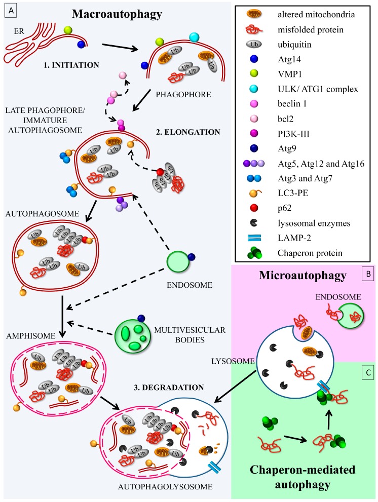
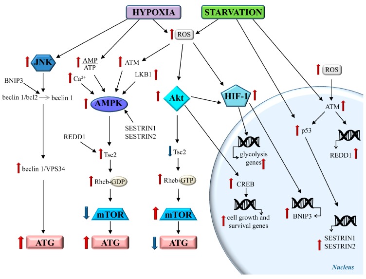
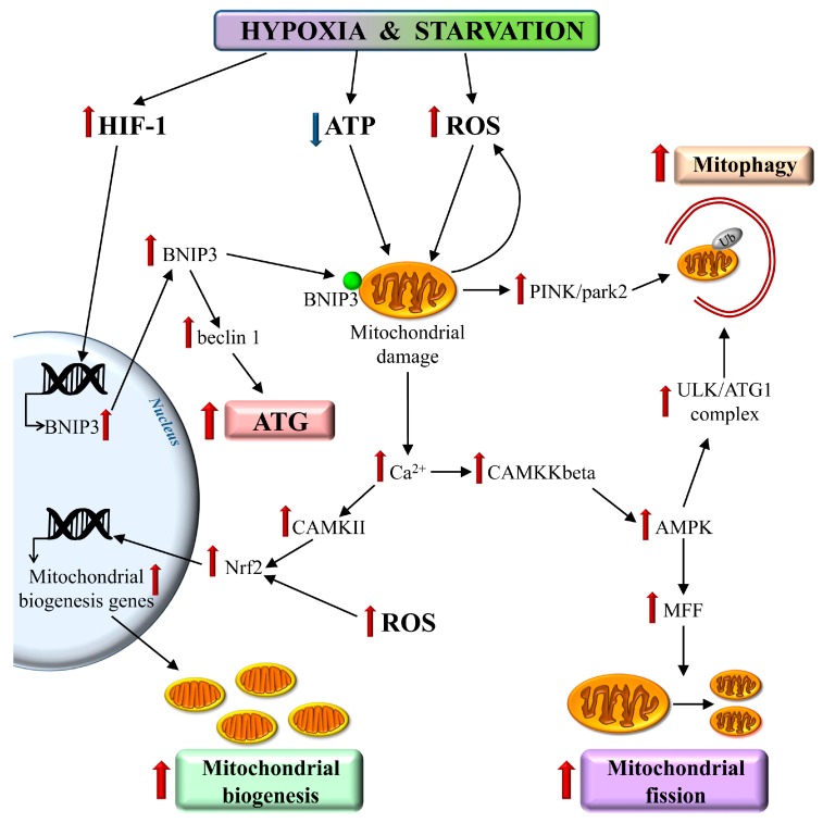
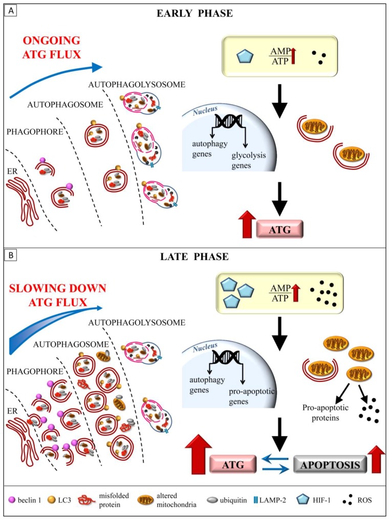
Autophagy primarily works to counteract nutrient deprivation that is strongly engaged during starvation and hypoxia, which happens in hypoperfusion. Nonetheless, autophagy is slightly active even in baseline conditions, when it is useful to remove aged proteins and organelles. This is critical when the mitochondria and/or proteins are damaged by toxic stimuli. In the present review, we discuss to that extent the recruitment of autophagy is beneficial in counteracting brain hypoperfusion or, vice-versa, its overactivity may per se be detrimental for cell survival. While analyzing these opposite effects, it turns out that the autophagy activity is likely not to be simply good or bad for cell survival, but its role varies depending on the timing and amount of autophagy activation. This calls for the need for an appropriate autophagy tuning to guarantee a beneficial effect on cell survival. Therefore, the present article draws a theoretical pattern of autophagy activation, which is hypothesized to define the appropriate timing and intensity, which should mirrors the duration and severity of brain hypoperfusion. The need for a fine tuning of the autophagy activation may explain why confounding outcomes occur when autophagy is studied using a rather simplistic approach.

Keywords: autophagy; brain ischemia; cerebral blood flow; hypoxia; mitophagy; neurodegeneration; starvation.


Conflict of interest statement

The authors declare no conflict of interest.

Figures

Figure 1
Figure 1
The autophagy (ATG) clearing pathway. (A) Macro-ATG follows the activation of the autophagy initiation complex, named the ATG1 or Unc-51 like autophagy activating kinase (ULK) (Unc-51 like autophagy activating kinase) complex. This leads to the phagophore, an incomplete double-membrane structure, which gems from the mitochondrial or endoplasmic reticulum carrying Atg14 and vacuolar membrane protein 1 (VMP1). The ULK/ATG1 complex phosphorylates Atg14, allowing the recruitment of beclin 1 from bcl2 on the phagophore membrane. Beclin 1 binds to phosphatidylinositol-3-kinase class III (PI3K-III)/VPS34, forming the active complex PI3K-III, which produces a focal increase of phosphatidylinositol-3-phosphate (PI3P). This leads to the followng: (i) the Atg9-mediated recruitment of endosomes and multivesicular bodies, and the (ii) induction of ubiquitin-like reactions. These consist of the formation of the Atg5/Atg12/Atg16 complex; or the assembly of the E1-like Atg7-mediated and the E2-like Atg3-mediated conjugation of LC3 to the phosphatidyletanolamine (PE), thus forming LC3-II (also called LC3 lipidation). Meanwhile, the late phagophore folds in order to enwrap the aged or damaged cell components, such as the ubiquitinated mitochondria and p62-bound poly-ubiquitinated proteins, which are sequestered within a mature autophagosome. The fusion between the autophagosome with the lysosome involves proteins belonging to the soluble N-ethylmaleimide sensitive factor attachment protein receptors (SNARE) complex. (B) Micro-ATG is traditionally described as a direct transport of several substrates into lysosomes for degradation; recently, it has been associated with the delivery of cargoes to late endosomes/multivesicular bodies. This latter process is dependent from ESCTR and Hsc70 proteins. (C) Chaperone-mediated ATG is a chaperone-mediated binding of specific substrates, which are delivered to the lysosomes through a lysosomal receptor, LAMP-2. Solid arrow: main step, Dotted arrow: molecule/vesicle trafficking.
Figure 2
Figure 2
The main metabolic pathways triggered by chronic brain hypoperfusion (CBH). Hypoxia and starvation occurring during CBH activate several cell pathways. The AMP activated protein kinase (AMPK) pathway is strongly activated during CBH. Increased AMP binds to AMPK, which is phosphorylated by several kinases, such as LKB1. Glucose deprivation increases p53, which stimulates the transcription of sestrin 1/sestrin 2 proteins, which, in turn, enhance the AMPK activation. Reactive oxygen species (ROS)-induced DNA damage increases the ataxia-telengiectasia-mutated (ATM) activity, thus leading to increased p53 and REDD1 transcription. Upon oxidation by ROS, cytosolic ATM forms homodimers and activates AMPK by phosphorylation. CBH-induced calcium overload activate AMPK through calcium calmodulin-dependent kinases. AMPK inhibits mammalian target of rapamycin (mTOR) by acting on the Tsc2 protein. Tsc2 is part of the Tsc1/Tsc2 complex, which converts the guanosine triphosphate (GTP) to guanosine diphosphate (GDP)-bound Rheb, which causes the inhibition of mTOR. In particular, AMPK phosphorylates Tsc2 at two sites, thus enhancing the ability of the Tsc1/Tsc2 complex to block Rheb-dependent mTOR activation. Finally, mTOR inhibition leads to ATG activation. The hypoxia-induced Akt pathway stimulates mTOR by inhibiting Tsc2. This occurs through Akt-dependent phosphorylation of Tsc2, thus inducing its dissociation from the complex Tsc1/Tsc2. This stabilizes Rheb to activate mTOR, thereby inhibiting ATG. The activation of Akt also triggers a phosphorylation cascade, producing various effects. A key protein targeted by Akt is the transcription factor cyclic AMP-responsive element binding protein (CREB), which induces the expression of genes related to cell growth and survival, such as brain derived neurotrophic factor (BDNF) and the anti-apoptotic protein bcl2. Moreover, Akt increases the expression of the alpha subunit of the transcription factor HIF-1, which is essential for hypoxia-inducible factor-1 (HIF-1) transcriptional activity. In turn, HIF-1 regulates the transcription of the genes related to anaerobic metabolism, thus promoting the shift towards the glycolytic metabolism. CBH-induced hypoxia and starvation also activate ATG throughout mechanisms that do not involve mTOR. The hypoxia-induced HIF-1 increases the expression BNIP3, which, via beclin 1, promotes ATG. An increased amount of beclin1 occurs through the c-Jun N-terminal kinase (JNK) pathway under starvation and oxidative stress. Red arrow: increased activity/levels, Blue arrow: decreased activity/levels, Grey arrow: transition.
Figure 3
Figure 3
CBH induces mitophagy. Hypoxia heavily affects the mitochondria by interfering with the mitochondrial respiratory chain, and by dramatically decreasing the mitochondrial ATP production. In these conditions, dysfunctional mitochondria massively produce ROS to be released into the cytosol. Moreover, a loss of the mitochondrial membrane potential occurs. Thus, a mitochondrial depolarized membrane causes an inversion of calcium pumps and cytosolic calcium overload. The intracellular calcium activates the calcium-sensitive kinases, which activate AMPK. These calcium dependent kinases contribute to the activation of the Nrf2/SKN1 protein, which promotes the genes involved in mitochondrial biogenesis. The depolarized mitochondria are rapidly removed by ATG through the PINK1/parkin-dependent mitophagy. This is also induced by hypoxia-induced HIF-1. In fact, HIF-1 increases the BNIP3 protein, which acts on the mitochondrial membrane as a mitophagy receptor through its binding to LC3-II. Finally, the removal of dysfunctional mitochondria by mitophagy is also induced by AMPK-dependent ULK activation. Interestingly, the AMPK activation also promotes mitochondrial fission by enhancing the mitochondrial fission factor (MFF)activity. CAMKKbeta—calcium/calmodulin dependent protein kinase kinase beta; CAMKII—calcium/calmodulin dependent protein kinase type II. Red arrow: increased activity/levels, Blue arrow: decreased activity/levels.
Figure 4
Figure 4
A dynamic model for the protective role of ATG in CBH. (A) The early phase of CBH is characterized by a sudden increase of the ATG activation. In this phase, the O2 and nutrients deprivation rapidly induce ATG, which is effective in the removal of altered cell components. Oxidative stress damages mitochondria, which are suddenly cleared by mitophagy. In this early phase of CBH, the ATG flux is normally ongoing and the ATG activation is key in delaying the cell death. (B) In the late phase of CBH, the protracted hypoxia, the severe energy deprivation, along with a further increase in the ROS levels, seriously worsen the cell functions. In these conditions, ATG further increased, but the broad cargoes overwhelm the ATG capacity, leading to a defective cell clearance. In particular, damaged mitochondria accumulate within the cytosol and become by themselves a source of additional ROS. Moreover, altered mitochondria release cytochrome c and other pro-apoptotic proteins. On the other hand, in this late phase of CBH, apoptosis is induced by a variety of stimuli. In particular, the apoptosis is triggered by the severe hypoxia, associated with mitochondrial damage and oxidative DNA injury. In this condition, a concomitant strong increase of both the ATG and apoptosis occurs. Moreover, the apoptosis and ATG are strictly interconnected and reciprocally regulated by several mechanisms. Therefore, the late CBH features as accumulation of ATG vacuoles, because of the relented ATG flux and increased apoptotic cell death. Red arrow: increased activity/levels, Blue straight arrow: modulatory effect, Blue curved arrow: intensity of ATG flux.

Similar articles

Cited by

References

    1. Rubinsztein D.C. The roles of intracellular protein-degradation pathways in neurodegeneration. Nature. 2006;443:780–786. doi: 10.1038/nature05291. - DOI - PubMed
    1. Balch W.E., Morimoto R.I., Dillin A., Kelly J.W. Adapting proteostasis for disease intervention. Science. 2008;319:916–919. doi: 10.1126/science.1141448. - DOI - PubMed
    1. Douglas P.M., Dillin A. Protein homeostasis and aging in neurodegeneration. J. Cell Biol. 2010;190:719–729. doi: 10.1083/jcb.201005144. - DOI - PMC - PubMed
    1. Koga H., Kaushik S., Cuervo A.M. Protein homeostasis and aging: The importance of exquisite quality control. Ageing Res. Rev. 2011;10:205–215. doi: 10.1016/j.arr.2010.02.001. - DOI - PMC - PubMed
    1. Lopez-Otin C., Blasco M.A., Partridge L., Serrano M., Kroemer G. The hallmarks of aging. Cell. 2013;153:1194–1217. doi: 10.1016/j.cell.2013.05.039. - DOI - PMC - PubMed