

Treponema pallidum promotes macrophage polarization and activates the NLRP3 inflammasome pathway to induce interleukin-1β production
- PMID: 30217146
- PMCID: PMC6137923
- DOI: 10.1186/s12865-018-0265-9
Treponema pallidum promotes macrophage polarization and activates the NLRP3 inflammasome pathway to induce interleukin-1β production
Abstract
Background: The involvement of inflammasome activation and macrophage polarization during the process of syphilis infection remains unknown. In this study, A series of experiments were performed using human macrophages to research the role of NLRP3 inflammasome regulation in interleukin (IL)-1β production and its influence on macrophage polarization triggered by T. pallidum.
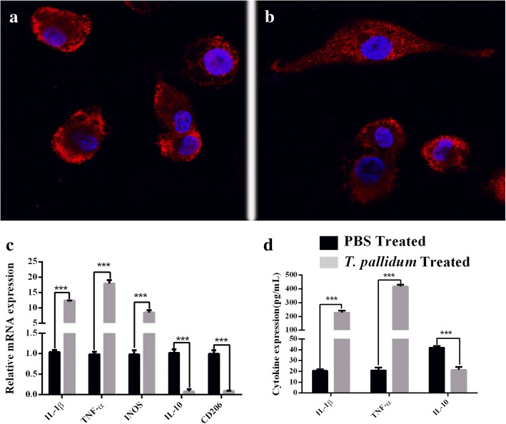
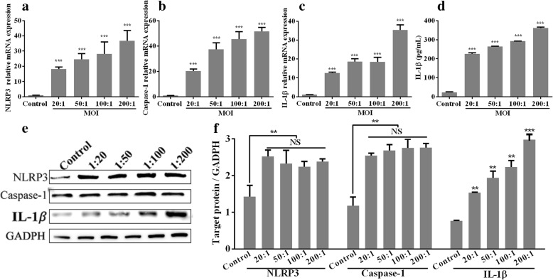
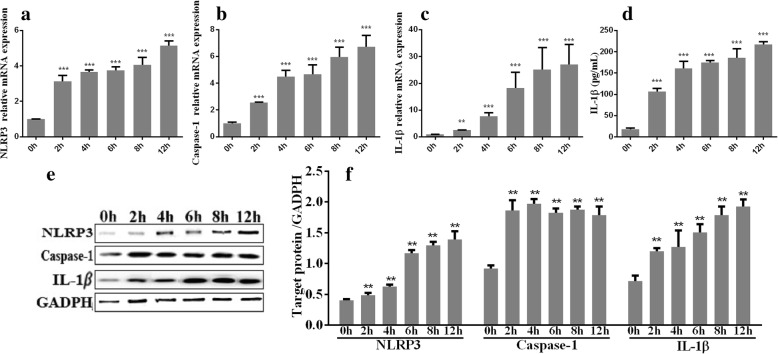
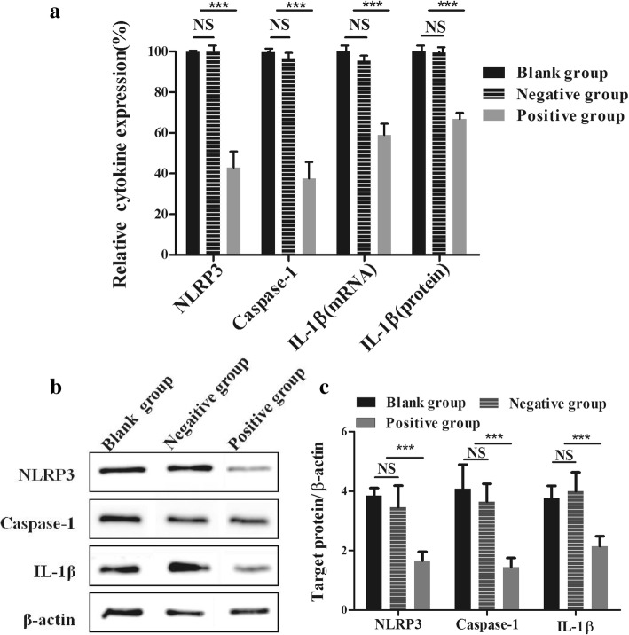
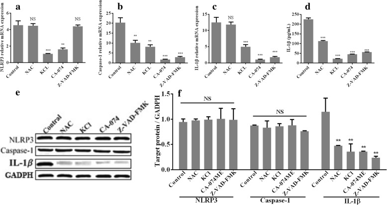
Results: The results showed that in M0 macrophages treated with T. pallidum, the M1-associated markers inducible nitric oxide synthase (iNOS), IL-1β and TNF-α were upregulated, and the M2-associated markers CD206 and IL-10 were downregulated. In addition, we observed NLRP3 inflammasome activation and IL-1β secretion in T. pallidum-treated macrophages, and the observed production of IL-1β occurred in a dose- and time-dependent manner. Moreover, the secretion of IL-1β by macrophages after T. pallidum treatment was notably reduced by anti-NLRP3 siRNA and caspase-1 inhibitor treatment. NAC, KCl, and CA074-ME treatment also suppressed IL-1β release from T. pallidum-treated macrophages.
Conclusions: These findings showed that T. pallidum induces M0 macrophages to undergo M1 macrophage polarization and elevate IL-1β secretion through NLRP3. Moreover, the process of NLRP3 inflammasome activation and IL-1β production in macrophages in response to T. pallidum infection involves K+ efflux, mitochondrial ROS production and cathepsin release. This study provides a new insight into the innate immune response to T. pallidum infection.
Keywords: IL-1β; Macrophage; NLRP3; Polarization; Treponema pallidum.
Conflict of interest statement
Ethics approval
This study was approved by the animal experimental ethics committee of the Medical College of Xiamen University.
Consent for publication
Not applicable.
Competing interests
The authors declare that they have no competing interests.
Publisher’s Note
Springer Nature remains neutral with regard to jurisdictional claims in published maps and institutional affiliations.
Figures
Similar articles
-
Development of tissue inflammation accompanied by NLRP3 inflammasome activation in rabbits infected with Treponema pallidum strain Nichols.BMC Infect Dis. 2018 Mar 1;18(1):101. doi: 10.1186/s12879-018-2993-0. BMC Infect Dis. 2018. PMID: 29490620 Free PMC article.
-
Leptospira interrogans infection leads to IL-1β and IL-18 secretion from a human macrophage cell line through reactive oxygen species and cathepsin B mediated-NLRP3 inflammasome activation.Microbes Infect. 2018 Apr;20(4):254-260. doi: 10.1016/j.micinf.2018.01.010. Epub 2018 Feb 9. Microbes Infect. 2018. PMID: 29432801
-
Treponema pallidum (Syphilis) Antigen TpF1 Induces Activation of Macrophages and Accelerates P2X7R-Induced NLRP3-Dependent Release of IL-1β.Endocr Metab Immune Disord Drug Targets. 2022;22(4):425-432. doi: 10.2174/1871530321666211015091109. Endocr Metab Immune Disord Drug Targets. 2022. PMID: 34649493
-
The role of lysosomal cysteine cathepsins in NLRP3 inflammasome activation.Arch Biochem Biophys. 2019 Jul 30;670:32-42. doi: 10.1016/j.abb.2019.02.015. Epub 2019 Feb 23. Arch Biochem Biophys. 2019. PMID: 30807742 Review.
-
Research Progress of Mitochondrial Mechanism in NLRP3 Inflammasome Activation and Exercise Regulation of NLRP3 Inflammasome.Int J Mol Sci. 2021 Oct 8;22(19):10866. doi: 10.3390/ijms221910866. Int J Mol Sci. 2021. PMID: 34639204 Free PMC article. Review.
Cited by
-
Interleukin-1β and cathepsin D modulate formation of the terminal complement complex in cultured human disc tissue.Eur Spine J. 2021 Aug;30(8):2247-2256. doi: 10.1007/s00586-021-06901-5. Epub 2021 Jun 24. Eur Spine J. 2021. PMID: 34169354
-
Syphilis vaccine: challenges, controversies and opportunities.Front Immunol. 2023 Apr 6;14:1126170. doi: 10.3389/fimmu.2023.1126170. eCollection 2023. Front Immunol. 2023. PMID: 37090699 Free PMC article. Review.
-
Aldehyde dehydrogenase 2 and NOD-like receptor thermal protein domain associated protein 3 inflammasome in atherosclerotic cardiovascular diseases: A systematic review of the current evidence.Front Cardiovasc Med. 2023 Feb 23;10:1062502. doi: 10.3389/fcvm.2023.1062502. eCollection 2023. Front Cardiovasc Med. 2023. PMID: 36910525 Free PMC article. Review.
-
2-Bromopalmitate decreases spinal inflammation and attenuates oxaliplatin-induced neuropathic pain via reducing Drp1-mediated mitochondrial dysfunction.PLoS One. 2022 Oct 31;17(10):e0275428. doi: 10.1371/journal.pone.0275428. eCollection 2022. PLoS One. 2022. PMID: 36315519 Free PMC article.
-
Absent in melanoma 2-mediating M1 macrophages facilitate tumor rejection in renal carcinoma.Transl Oncol. 2021 Apr;14(4):101018. doi: 10.1016/j.tranon.2021.101018. Epub 2021 Jan 22. Transl Oncol. 2021. PMID: 33493800 Free PMC article.
References
-
- Lin LR, Xiao Y, Liu W, Chen YY, Zhu XZ, Gao ZX, Gao K, Tong ML, Zhang HL, Li SL. Development of tissue inflammation accompanied by NLRP3 inflammasome activation in rabbits infected with Treponema pallidum strain Nichols. BMC Infect Dis. 2018;18(1):101. doi: 10.1186/s12879-018-2993-0. - DOI - PMC - PubMed
Publication types
MeSH terms
Substances
LinkOut - more resources
Full Text Sources
Other Literature Sources
Medical
