. 2018 Sep 14;17(1):146.
doi: 10.1186/s12934-018-0994-8.

Evaluation of cell-surface displayed synthetic consensus dengue EDIII cells as a potent oral vaccine candidate

Affiliations

Evaluation of cell-surface displayed synthetic consensus dengue EDIII cells as a potent oral vaccine candidate

Jyotiranjan Bal et al. Microb Cell Fact. .

Abstract

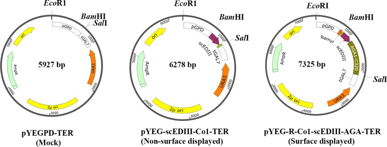
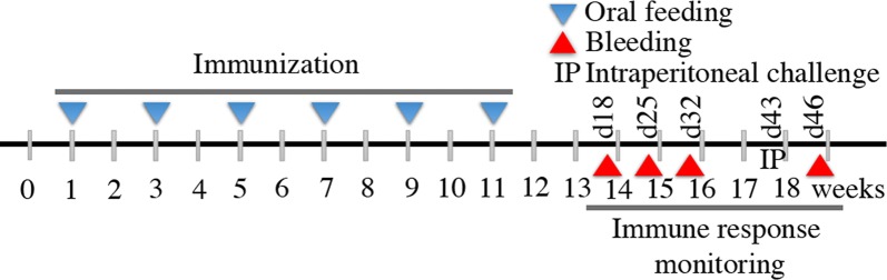
Background: Dengue is a rapidly spreading mosquito borne tropical viral disease affecting hundreds of millions of people across the globe annually. The dengue virus (DENV) includes four genetically distinct serotypes that cause serious life-threatening infections, including dengue hemorrhagic fever/dengue shock syndrome. Dengue vaccine development is complicated by the possibility of vaccine-enhanced severe dengue disease due to antibody-dependent enhancement by pre-existing cross-reactivity, as well as homotypic antibodies. Thus, the development of an efficacious dengue vaccine conferring simultaneous and durable immunity to each of the four DENV serotypes has not yet been developed despite years of research. For mass immunization in deeply affected resource-limited countries, oral vaccination is considered more beneficial than conventional approaches. Therefore, in a continuing effort towards designing economical and potent vaccine candidates, the current study applied yeast surface display technology to develop an oral dengue vaccine candidate using whole recombinant yeast cells displaying the recombinant fusion protein of M cell targeting ligand Co1 fused to the synthetic consensus dengue envelope domain III (scEDIII). Female Balb/c mice were orally fed with recombinant yeast cells and immunogenicity in terms of systemic and mucosal immune responses was monitored.

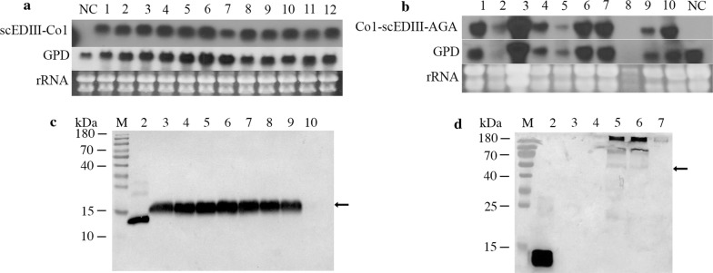
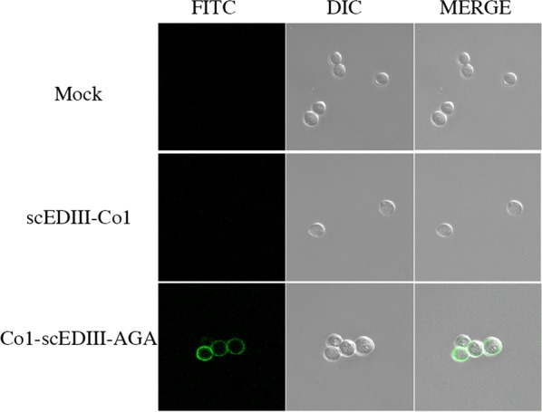
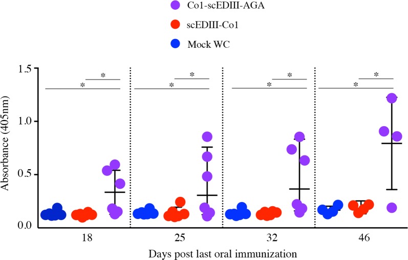
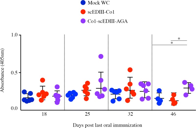
Results: Immunofluorescence microscopy with dengue specific antibody and fluorescein isothiocyanate-conjugated anti-mouse IgG antibody clearly showed that recombinant protein Co1-scEDIII-AGA was localized on the cell surface of the respective clones in comparison with scEDIII-Co1 and Mock cells with no fluorescence. Oral dosage applications of surface displayed Co1-scEDIII-AGA stimulated a systemic humoral immune response in the form of dengue-specific serum IgG, as well as a mucosal immune response in the form of secretory immunoglobulin A (sIgA). Antigen-specific B cell responses in isolated lymphoid cells from the spleen and Peyer's patches further supported an elevated mucosal immune response. In addition, surface displayed Co1-scEDIII-AGA feeding elicited strong immune responses in comparison with scEDIII-Co1 and Mock following intraperitoneal booster with purified scEDIII antigen.

Conclusions: Surface displayed preparations of Co1-scEDIII-AGA induced strong immunogenicity compared with non-displayed scEDIII-Co1. Prior studies have supported the neutralization potential of scEDIII constructs against all four serotypes. Thus, the oral administration of genetically engineered yeast whole cells displaying biologically active Co1-scEDIII fusion protein without any further processing shows prospective as a potent oral vaccine candidate against dengue viral infection.

Keywords: Dengue; Mucosal immunity; Oral vaccine; Saccharomyces cerevisiae; Surface display; scEDIII.


Figures

Fig. 1
Fig. 1
Expression vector design. Schematic illustration of the expression plasmids pYEGPD-TER (Mock), pYEG-scEDIII-Co1-TER (non-surface displayed), and pYEG-R-Co1-scEDIII-AGA-TER (surface displayed)
Fig. 2
Fig. 2
Schematic representation of the oral immunization protocol. Each mouse in a group of six was orally gavaged with 1.6 g fresh weight of cells resuspended in a final volume of 2.4 mL of phosphate-buffered saline. A total of six doses were orally administered every alternate week with each dose consisting of cell suspensions that were split equally and administered four times per day. Subsequent immune response monitoring was performed through retro-orbital bleeding and fecal matter collection at 4 days after the final feeding and continued every week up to 32 days, followed by intraperitoneal booster injection with purified antigen on day 43 and subsequent bleeding on day 46. d# represents days post final vaccination
Fig. 3
Fig. 3
Expression analysis of recombinant proteins. a Northern blot analysis showing levels of Co1-scEDIII transcripts from selected transformants. Lane NC contains RNA from mock-transformed cells. Lanes 1–12 consist of RNA from the corresponding pYEG-Co1-scEDIII-TER transformants. Glyceraldehyde-3-phosphate dehydrogenase (GPD) transcription is shown as an internal control. Ribosomal RNA (rRNA) was used as a loading control. b Northern blot analysis showing levels of Co1-scEDIII-AGA transcripts from selected transformants. Lanes 1–10 consist of RNA from the corresponding pYEG-Co1-scEDIII-AGA-TER transformants. Lane NC contains RNA from mock-transformed cells. GPD transcription is shown as an internal control and rRNA was used as a loading control. c Western blot analysis of recombinant protein expression using anti-dengue IgG antibodies (lanes 2–10). Lane marked as ‘M’ represents pre-stained protein size marker. Their sizes (in kDa) are indicated to the left of the first lane. The arrow indicates the position of the recombinant scEDIII-Co1 protein. Lane 2 contains 1 µg of cell-free extract (CFE) from Escherichia coli-expressed scEDIII [14]. Lanes 3–9 contain 40 µg of CFE from recombinant Co1-scEDIII strains. Lane 10 contains 40 µg of CFE from Mock as negative controls. d Western blot analysis to confirm the expression of surface displayed recombinant Co1-scEDIII-AGA protein using anti-dengue IgG antibodies (lanes 2–7). Lane marked as ‘M’ represents pre-stained protein size marker. Their sizes (in kDa) are indicated to the left of the first lane. The arrow indicates the position of the recombinant Co1-scEDIII-AGA protein. Lane 2 contains 1 µg of CFE from E. coli-expressed scEDIII. Lane 3 contain 40 µg of CFE from wild type Saccharomyces cerevisiae 2805 strain. Lane 4 contain 40 µg of CFE from the Mock control. Lanes 5 and 6 contain CFEs and cell wall fractions from recombinant Co1-scEDIII-AGA strains, respectively. Lane 7 contains 10 mL of culture filtrate extracted from Co1-scEDIII-AGA strains
Fig. 4
Fig. 4
Immunofluorescence labeling of recombinant yeast cells. Immunofluorescence microscopic observation of displayed Co1-scEDIII-AGA on S. cerevisiae. Fluorescence microscopic images after staining recombinant yeast cells using the anti-dengue mAb followed by fluorescein isothiocyanate-conjugated anti-mouse IgG. The surface displayed protein is visualized as a ring on the cell wall of yeast cells
Fig. 5
Fig. 5
Humoral IgG responses in orally immunized mice. Dengue-specific serum IgG was induced in mice that received oral administration of scEDIII-Co1 and Co1-scEDIII-AGA, and this induction was observed at 4 days after the last immunization until day 32, as measured at weekly intervals. On day 46, following booster immunization with purified E. coli expressed scEDIII protein, dengue-specific serum IgG levels were also observed. An unpaired Student’s t-test was used to calculate p-values, and *p < 0.05 indicates significant differences between the groups
Fig. 6
Fig. 6
Mucosal sIgA responses upon oral immunization. scEDIII-specific fecal sIgA was induced in mice immunized orally with recombinant scEDIII-Co1 and Co1-scEDIII-AGA at 4 days after the last immunization until day 32, observed at weekly intervals. Dengue-specific fecal sIgA levels were also observed on day 46, 3 days after booster immunization with purified E. coli expressed scEDIII protein. An unpaired Student’s t-test was used to calculate p-values, and *p < 0.05 indicates significant differences between the groups
Fig. 7
Fig. 7
Frequency of antigen-specific immune cells in orally immunized mice. Enzyme-linked immunosorbent spot assay showing (a) scEDIII-specific IgG and (b) scEDIII-specific IgA antibody spot forming cells in lymphocytes isolated from spleens and Peyer’s patches of immunized mice, respectively. Each group represents two mice analyzed individually in triplicate. This is a representative result of two independent experiments showing similar results

Similar articles

Cited by

References

    1. Achee NL, Gould F, Perkins TA, Reiner RC, Jr, Morrison AC, Ritchie SA, Gubler DJ, Teyssou R, Scott TW. A critical assessment of vector control for dengue prevention. PLoS Negl Trop Dis. 2015;9:e0003655. doi: 10.1371/journal.pntd.0003655. - DOI - PMC - PubMed
    1. WHO. Dengue: guidelines for diagnosis, treatment, prevention, and control. Spec Program Res Train Trop Dis. 2009;409:147. WHO reference number: WHO/HTM/NTD/DEN/2009.1.
    1. Bhatt S, Gething PW, Brady OJ, Messina JP, Farlow AW, Moyes CL, Drake JM, Brownstein JS, Hoen AG, Sankoh O, et al. The global distribution and burden of dengue. Nature. 2013;496:504–507. doi: 10.1038/nature12060. - DOI - PMC - PubMed
    1. McArthur MA, Sztein MB, Edelman R. Dengue vaccines: recent developments, ongoing challenges and current candidates. Expert Rev Vaccines. 2013;12:933–953. doi: 10.1586/14760584.2013.815412. - DOI - PMC - PubMed
    1. Yauch LE, Shresta S. Dengue virus vaccine development. Adv Virus Res. 2014;88:315–372. doi: 10.1016/B978-0-12-800098-4.00007-6. - DOI - PubMed

LinkOut - more resources