Case Reports
. 2018 Sep 14:2018:bcr2018224893.
doi: 10.1136/bcr-2018-224893.

Management of oesophageal intramucosal carcinoma

Affiliations
Case Reports

Management of oesophageal intramucosal carcinoma

Darius Ashrafi et al. BMJ Case Rep. .

Abstract

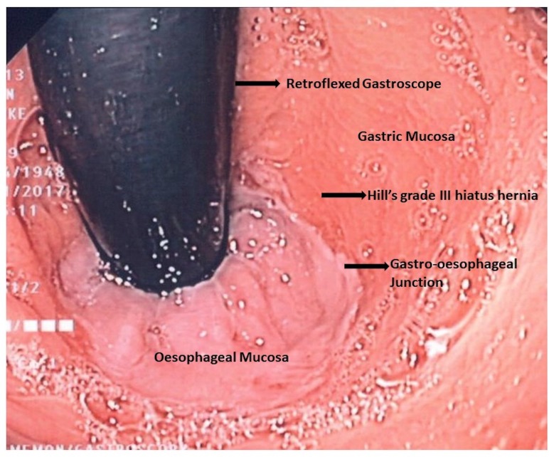
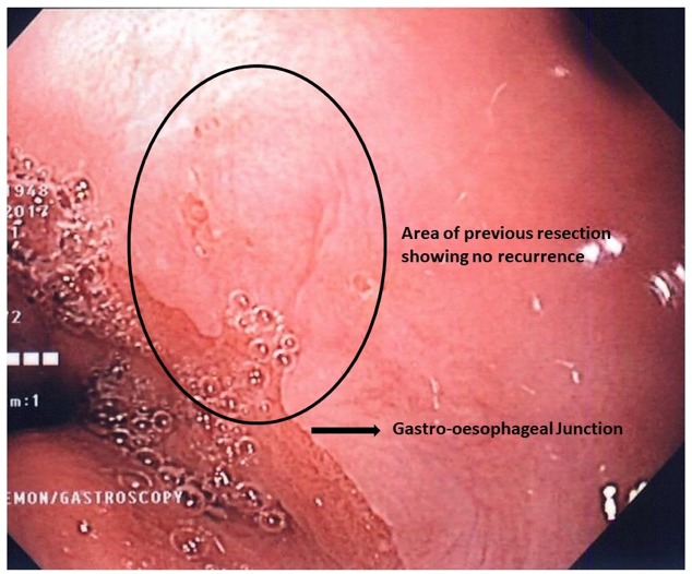
We present an interesting case of an intramucosal carcinoma (IMC) in the setting of Barrett's oesophagus in a 66-year-old woman. Her clinical course highlights the shifting paradigm in the approach to management of Barrett's oesophagus and IMC. With innovation in imaging and endoscopic treatment modalities, patients are detected earlier and managed prior to development of malignancy. The patient was treated with endoscopic modalities, and after 3 years' follow-up, she remains recurrence free.

Keywords: gastric cancer; gastrointestinal surgery; general surgery.


Conflict of interest statement

Competing interests: None declared.

Figures

Figure 1
Figure 1
Diaphragmatic representation of depth of invasion of intramucosal carcinoma.
Figure 2
Figure 2
Hill’s classification of hiatal hernias. (4)
Figure 3
Figure 3
Oesophagogastroduodenoscopy showing intramucosal carcinoma.
Figure 4
Figure 4
Histological specimens at ×100 and ×400 zoom, respectively. Evidence of intramucosal adenocarcinoma with grade 2 moderately differentiated cells. There is invasion into the outer muscularis mucosa (M3). There is no evidence of lymphovascular invasion.
Figure 5
Figure 5
Postresection oesophagogastroduodenoscopy showing Hill’s grade IV hiatus hernia with no recurrence.
Figure 6
Figure 6
Magnified view of previous resection site showing no recurrence.

Similar articles

Cited by

References

    1. Pennathur A, Gibson MK, Jobe BA, et al. . Oesophageal carcinoma. Lancet 2013;381:400–12. 10.1016/S0140-6736(12)60643-6 - DOI - PubMed
    1. Rajendra S, Sharma P. Management of Barrett’s oesophagus and intramucosal oesophageal cancer: a review of recent development. Therap Adv Gastroenterol 2012;5:285–99. 10.1177/1756283X12446668 - DOI - PMC - PubMed
    1. Cameron AJ, Lomboy CT, Pera M, et al. . Adenocarcinoma of the esophagogastric junction and Barrett’s esophagus. Gastroenterology 1995;109:1541–6. 10.1016/0016-5085(95)90642-8 - DOI - PubMed
    1. Shimada H, Nabeya Y, Matsubara H, et al. . Prediction of lymph node status in patients with superficial esophageal carcinoma: analysis of 160 surgically resected cancers. Am J Surg 2006;191:250–4. 10.1016/j.amjsurg.2005.07.035 - DOI - PubMed
    1. Leers JM, DeMeester SR, Oezcelik A, et al. . The prevalence of lymph node metastases in patients with T1 esophageal adenocarcinoma a retrospective review of esophagectomy specimens. Ann Surg 2011;253:271–8. 10.1097/SLA.0b013e3181fbad42 - DOI - PubMed

Publication types

MeSH terms