. 2018 Nov 9;293(45):17349-17361.
doi: 10.1074/jbc.RA118.005369. Epub 2018 Sep 14.

Structural and spectroscopic analyses of the sporulation killing factor biosynthetic enzyme SkfB, a bacterial AdoMet radical sactisynthase

Affiliations

Structural and spectroscopic analyses of the sporulation killing factor biosynthetic enzyme SkfB, a bacterial AdoMet radical sactisynthase

Tsehai A J Grell et al. J Biol Chem. .

Abstract

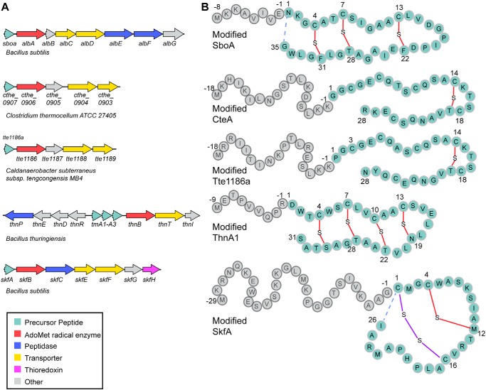
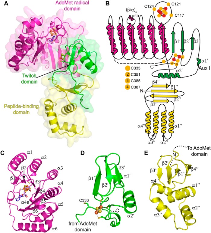
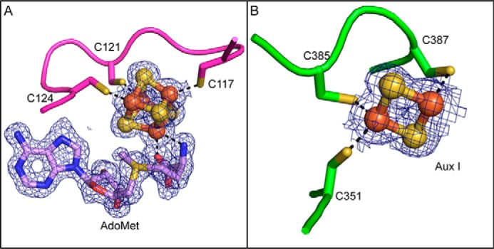
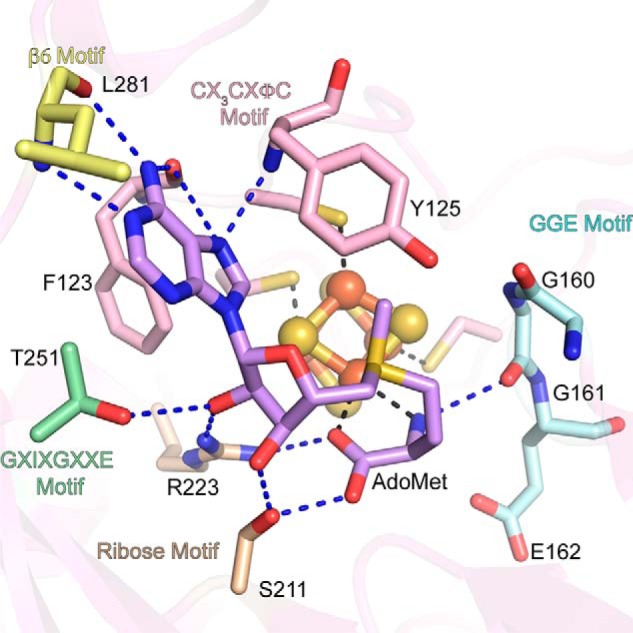
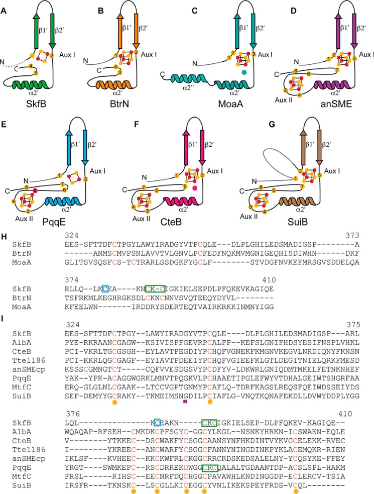
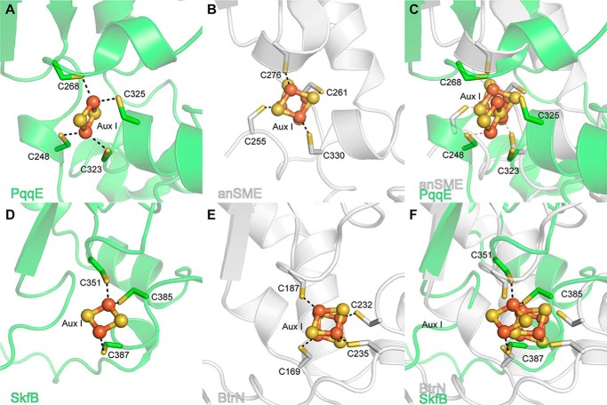
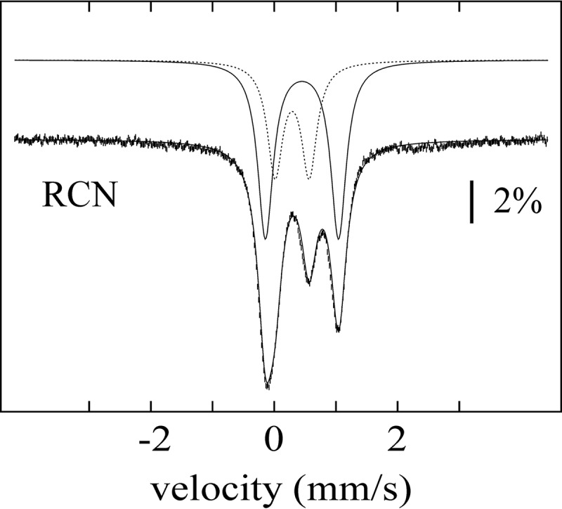
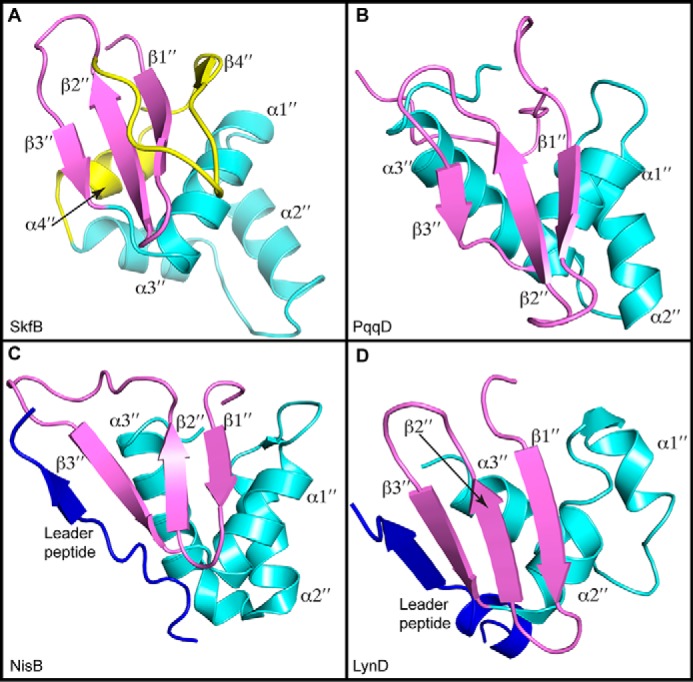
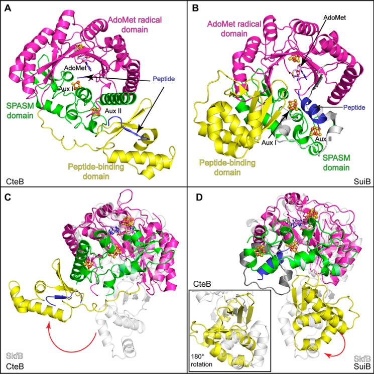
Sactipeptides are a subclass of ribosomally synthesized and post-translationally modified peptides (RiPPs). They contain a unique thioether bond, referred to as a sactionine linkage, between the sulfur atom of a cysteine residue and the α-carbon of an acceptor residue. These linkages are formed via radical chemistry and are essential for the spermicidal, antifungal, and antibacterial properties of sactipeptides. Enzymes that form these linkages, called sactisynthases, are AdoMet radical enzymes in the SPASM/Twitch subgroup whose structures are incompletely characterized. Here, we present the X-ray crystal structure to 1.29-Å resolution and Mössbauer analysis of SkfB, a sactisynthase from Bacillus subtilis involved in making sporulation killing factor (SKF). We found that SkfB is a modular enzyme with an N-terminal peptide-binding domain comprising a RiPP recognition element (RRE), a middle domain that forms a classic AdoMet radical partial (β/α)6 barrel structure and displays AdoMet bound to the [4Fe-4S] cluster, and a C-terminal region characteristic of the so-called Twitch domain housing an auxiliary iron-sulfur cluster. Notably, both crystallography and Mössbauer analyses suggest that SkfB can bind a [2Fe-2S] cluster at the auxiliary cluster site, which has been observed only once before in a SPASM/Twitch auxiliary cluster site in the structure of another AdoMet radical enzyme, the pyrroloquinoline quinone biosynthesis enzyme PqqE. Taken together, our findings indicate that SkfB from B. subtilis represents a unique enzyme containing several structural features observed in other AdoMet radical enzymes.

Keywords: AdoMet radical enzymes; catalysis; crystal structure; enzyme catalysis; enzyme structure; iron sulfur clusters; metalloenzyme; radical SAM; sactionine linkage; sporulation killing factor.


Conflict of interest statement

The authors declare that they have no conflicts of interest with the contents of this article

Figures

Figure 1.
Figure 1.
Sactipeptide biosynthesis. A, gene scheme of several RiPP biosynthetic gene clusters involved in the biosynthesis of sactipeptides. These biosynthetic clusters encode precursor peptides (cyan); AdoMet radical enzymes (red), which are responsible for installing the unique sactionine linkages characteristic of the sactipeptide natural products; peptidases (blue), which are responsible for cleaving the leader peptide; and ABC transporters, which are involved in transporting the sactipeptide out of the producing organism. B, the precursor peptides for each biosynthetic gene cluster in A are shown with the leader peptide sequence, which binds to the biosynthetic enzymes, in gray and the mature peptide, which undergoes post-translational modifications, in cyan. The sactionine linkages formed between the sulfur atom of a cysteine residue and the α-carbon of an acceptor residue are shown in red. Final maturation steps of SboA and SkfA involve an N- to C-terminal macrocyclization (dashed blue lines) step concurrent with a leader peptide cleavage step. Maturation of SkfA also includes disulfide bond formation (purple).
Figure 2.
Figure 2.
Overall architecture of SkfB. A, the partial (β/α)6 TIM barrel of the AdoMet radical domain (magenta) houses the [4Fe-4S] AdoMet radical cluster and an AdoMet molecule (lilac). The C-terminal domain adopts a Twitch domain architecture (green), providing three visible ligands to a [2Fe-2S] auxiliary cluster, Aux I. The N-terminal peptide-binding domain (yellow) displays the architecture of the RRE motif identified in peptide-binding domains of RiPP biosynthetic enzymes. B, topology diagram of SkfB colored by domain with the AdoMet radical domain in magenta, the Twitch domain in green, and the RRE domain in yellow. Yellow circles indicate cysteine residues, and orange and yellow circles represent the iron and sulfur atoms, respectively, of the clusters shown as ball and stick representations. C–E show a closer look at the individual domains of SkfB. C, the active site of SkfB is located within the inner cavity of the AdoMet radical domain and comprises six parallel β-strands. The binding sites of the AdoMet radical cluster and AdoMet (lilac) are located at the top of the partial barrel. D, the C-terminal Twitch domain (green) contains the canonical elements of a SPASM/Twitch fold, a β-hairpin, β1′ and β2′, followed by α2′. In SkfB, a short β-strand, β3′, and α-helix α1′ are found in between β2′ and α2′. E, the N-terminal peptide-binding domain of SkfB (yellow) folds into a three-stranded antiparallel β-sheet (β1″–β3″) and a consecutive helical bundle (α1″–α4″), reminiscent of the winged helix turn helix motif that comprises the RRE. The N-terminal domain ends with a parallel β-strand, β4″ adjacent to β1″. The clusters are shown as ball and stick representations with iron atoms colored orange and sulfur atoms colored yellow.
Figure 3.
Figure 3.
Iron-sulfur clusters and AdoMet density. A, the characteristic CX3CXφC motif, Cys117, Cys121, and Cys124, coordinates the AdoMet radical cluster in SkfB. An AdoMet molecule ligates the site-differentiated iron through a bidentate interaction with the nitrogen of the α-amino moiety and oxygen from the α-carboxyl moiety. B, a [2Fe-2S] cluster is observed in the Aux I site of the Twitch domain, bound by a CX33CXC sequence, Cys351, Cys385, and Cys387. 2FoFc composite omit density is shown in blue and contoured at 1σ.
Figure 4.
Figure 4.
AdoMet-binding motifs are conserved in SkfB. The AdoMet-binding pocket is located within the partial TIM barrel (translucent pink) and includes interactions from the GGE motif, Gly160, Gly161, and Glu162 (cyan); the ribose motif, Ser211 and Arg223 (tan); GXIXGXXE or β5 motif, Thr251 (green); and β6 motif (yellow), Leu281. Arg223 plays a dual role, positioning both the carboxyl group and the ribose ring. Hydrophobic interactions from the CX3CXφC motif Phe123, and the adjacent residue, Tyr125 (pink), orient the adenine ring of AdoMet (lilac) in the active site. The AdoMet radical cluster is shown as a ball and stick representation with iron atoms colored orange and sulfur atoms colored yellow.
Figure 5.
Figure 5.
Comparison of auxiliary cluster binding by SPASM/Twitch domains. The canonical structural elements of the Twitch (A–C) and SPASM (D–F) domain are shown for each enzyme. A, SkfB (green) binds a [2Fe-2S] cluster using one cysteine following the β-hairpin and two cysteines following α2′, similar to BtrN. SkfB appears to have an open coordination site: although the cysteine corresponding to the cluster ligand before the β-hairpin motif is present, this cysteine is ∼12 Å away from the cluster. B, the Twitch domain of BtrN (orange) provides four cysteine residues to fully ligate a [4Fe-4S] auxiliary cluster. C, MoaA (teal) uses a Twitch domain to bind a [4Fe-4S] cluster with an open coordination site. The unique iron is ligated by substrate, GTP, (teal circle). D, anSME (purple) utilizes a SPASM motif to bind two fully ligated [4Fe-4S] clusters, Aux I and Aux II. E, PqqE (blue) binds two fully ligated clusters, a [2Fe-2S] Aux I cluster and a [4Fe-4S] Aux II cluster, using a unique aspartic acid (red circle) ligand. F, CteB (pink) also binds two [4Fe-4S] auxiliary clusters with an open coordination site on Aux I to which a cysteine residue from the substrate, CteA (pink circle), is bound in the structure. G, the SPASM domain of SuiB ligates two auxiliary clusters. The first cysteine ligand to Aux I is provided by the linker region connecting the SPASM domain to the AdoMet radical domain. The cluster-binding cysteines are shown as yellow circles, and the cluster iron and sulfur atoms are represented as orange and yellow circles, respectively. The linker regions are denoted in gray. H, Twitch domain sequence alignment. Residue numbers are for SkfB. Cysteine ligands to Aux I are indicated in red, yellow spheres represent the first four cysteines of the seven-cysteine motif (CX9–15GX4CgapCX2CX5CX3CgapC), and a purple rectangle denotes the conserved glycine residue. The CXC of SkfB is indicated by a green box, and a blue box designates the cysteine previously proposed to bind the Aux I cluster of SkfB (CX4CXC) (7). I, sequence alignments of SPASM domains with the sequence of SkfB (sequence numbers indicated for SkfB). Cluster-coordinating cysteines are denoted in red, cysteines corresponding to the seven-cysteine motif (CX9–15GX4CgapCX2CX5CX3CgapC) are indicated by yellow circles, and the conserved glycine residue of this motif is denoted by a purple rectangle. Green boxes denote the CXC of SkfB and PqqE, and the blue box indicates the cysteine previously proposed to bind the Aux I cluster of SkfB (CX4CXC) (7).
Figure 6.
Figure 6.
Comparison of Aux I iron-sulfur clusters in SPASM/Twitch AdoMet radical enzymes. A, the Aux I site of the PqqE SPASM domain (green) has been shown biochemically to bind either a [4Fe-4S] cluster or a [2Fe-2S] cluster. In the recent structure of PqqE (PDB code 6C8V), the Aux I cluster-binding site is populated by a [2Fe-2S] cluster with two of the ligands provided by a CXC motif, Cys323 and Cys325. B, anSME (PDB code 4K37), shown in white, contains a [4Fe-4S] cluster at the Aux I cluster-binding site ligated by Cys255, Cys261, Cys276, and Cys330. C, when overlaid with anSME (white), the cysteine positions of the PqqE (green) active site appear to be amenable for binding a [4Fe-4S] cluster. D, the Twitch Aux I site of SkfB (green) can bind a [2Fe-2S] cluster with cysteines from the CXC motif, Cys385 and Cys387, binding different irons of the cluster. E, the Twitch Aux I site of BtrN (PDB code 4M7T), shown in white, fully ligates a [4Fe-4S] auxiliary cluster. F, the Aux I site of SkfB (green) overlays well with the Aux I site BtrN (white). To allow for [4Fe-4S] cluster binding, the loop containing residues of the CXC motif of SkfB would have to move slightly. This rearrangement appears possible as this loop corresponds to the C-terminal end of the structure and is highly flexible.
Figure 7.
Figure 7.
Mössbauer spectrum of SkfB. The spectrum of a sample of SkfB reconstituted with 57Fe (vertical bars) was recorded with the sample kept at 4.2 K in a 53-mT magnetic field applied parallel to the γ-beam. The solid line overlaid with the data is a simulation with two quadrupole doublets using parameters quoted in the text. The individual contributions of the quadrupole doublets associated with the [2Fe-2S]2+ and [4Fe-4S]2+ clusters are shown as dashed and solid lines above the data, respectively. RCN, reconstituted.
Figure 8.
Figure 8.
Structural comparisons of RRE domains. A, SkfB exhibits a canonical RRE comprising a three-strand antiparallel β-sheet (β1″–β3″ in purple) and three consecutive α-helices (α1″–α3″ in cyan). Following the RRE motif, SkfB folds into an additional α-helix, α4″, and β-strand, β4″ (yellow). B, the small peptide-binding protein PqqD (PDB code 5SXY) is a standalone RRE domain. C, the RRE domain of NisB (PDB code 4WD9) binds the leader peptide sequence of the peptide substrate NisA (blue) by extending the antiparallel β-sheet or the wing. D, the leader peptide of PatE (blue) also binds to the RRE domain of LynD (PDB code 4V1T) through interactions with the wing.
Figure 9.
Figure 9.
Structural comparisons of SkfB with other AdoMet radical enzymes involved in RIPP biosynthesis. A, CteB (PDB code 5WGG) exhibits a trimodular fold composed of a peptide-binding or RRE domain (yellow) followed by an AdoMet-binding domain (magenta), which binds the AdoMet radical cluster and AdoMet (lilac). The C-terminal end of CteB binds two clusters, Aux I and Aux II, using the SPASM domain architecture (green). The leader sequence of CteA (blue) binds to the RRE domain by extending the antiparallel β-sheet. B, SuiB (PDB code 5V1T) demonstrates a similar modular fold to CteB (A). Interestingly, the substrate for SuiB, SuiA (blue), is observed making contacts with the insertion (gray) between the AdoMet radical domain (magenta) and the SPASM domain (green) and not with the peptide-binding domain (yellow). C, the AdoMet radical and Twitch domains of SkfB (white) overlay well with CteB, but the peptide-binding domains are located on opposite sides of the AdoMet radical domain. D, SkfB (white) overlays well with SuiB, and the positions of the N-terminal domains show modest differences. A view of the overlaid N-terminal domains SkfB and SuiB, rotated 180°, is shown in the inset.
Figure 10.
Figure 10.
SPASM/Twitch subclass sequence similarity network. The protein similarity network (51) for the SPASM/Twitch subclass is visualized in Cytoscape (52) at a blast probability of 10−20. Sequences were obtained from the Structure Function Linkage Database (53) (http://sfld.rbvi.ucsf.edu/django; Please note that the JBC is not responsible for the long-term archiving and maintenance of this site or any other third party-hosted site.), and each node represents sequences that share 50% identity. Nodes corresponding to the previously solved members of the SPASM/Twitch subclass are represented as blue diamonds (11, 19, 20, 24–26), and green indicates family members that have been biochemically characterized but not structurally characterized (8). SkfB, which is structurally characterized here, is shown as a purple diamond.

Similar articles

Cited by

References

    1. Flühe L., and Marahiel M. A. (2013) Radical S-adenosylmethionine enzyme catalyzed thioether bond formation in sactipeptide biosynthesis. Curr. Opin. Chem. Biol. 17, 605–612 10.1016/j.cbpa.2013.06.031 - DOI - PubMed
    1. Rea M. C., Sit C. S., Clayton E., O'Connor P. M., Whittal R. M., Zheng J., Vederas J. C., Ross R. P., and Hill C. (2010) Thuricin CD, a posttranslationally modified bacteriocin with a narrow spectrum of activity against Clostridium difficile. Proc. Natl. Acad. Sci. U.S.A. 107, 9352–9357 10.1073/pnas.0913554107 - DOI - PMC - PubMed
    1. Sit C. S., van Belkum M. J., McKay R. T., Worobo R. W., and Vederas J. C. (2011) The 3D solution structure of thurincin H, a bacteriocin with four sulfur to α-carbon crosslinks. Angew. Chem. Int. Ed. Engl. 50, 8718–8721 10.1002/anie.201102527 - DOI - PubMed
    1. Liu W.-T., Yang Y.-L., Xu Y., Lamsa A., Haste N. M., Yang J. Y., Ng J., Gonzalez D., Ellermeier C. D., Straight P. D., Pevzner P. A., Pogliano J., Nizet V., Pogliano K., and Dorrestein P. C. (2010) Imaging mass spectrometry of intraspecies metabolic exchange revealed the cannibalistic factors of Bacillus subtilis. Proc. Natl. Acad. Sci. U.S.A. 107, 16286–16290 10.1073/pnas.1008368107 - DOI - PMC - PubMed
    1. Datta S., Mori Y., Takagi K., Kawaguchi K., Chen Z.-W., Okajima T., Kuroda S., Ikeda T., Kano K., Tanizawa K., and Mathews F. S. (2001) Structure of a quinohemoprotein amine dehydrogenase with an uncommon redox cofactor and highly unusual crosslinking. Proc. Natl. Acad. Sci. U.S.A. 98, 14268–14273 10.1073/pnas.241429098 - DOI - PMC - PubMed

Publication types

MeSH terms

LinkOut - more resources