. 2018 Oct 2;115(40):10118-10123.
doi: 10.1073/pnas.1811866115. Epub 2018 Sep 14.

Interleukin-22 promotes phagolysosomal fusion to induce protection against Salmonella enterica Typhimurium in human epithelial cells

Affiliations

Interleukin-22 promotes phagolysosomal fusion to induce protection against Salmonella enterica Typhimurium in human epithelial cells

Jessica L Forbester et al. Proc Natl Acad Sci U S A. .

Abstract

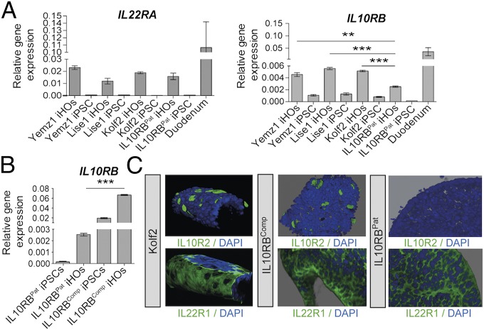
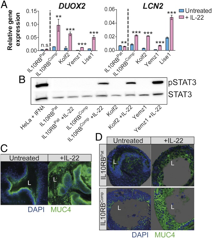
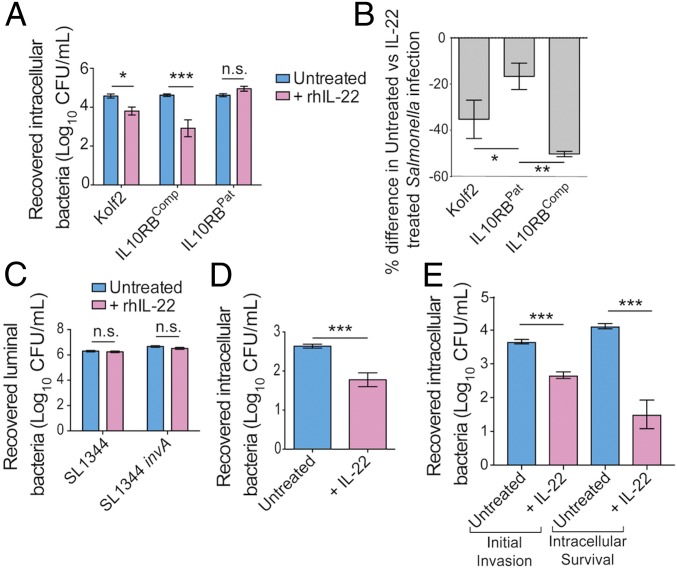
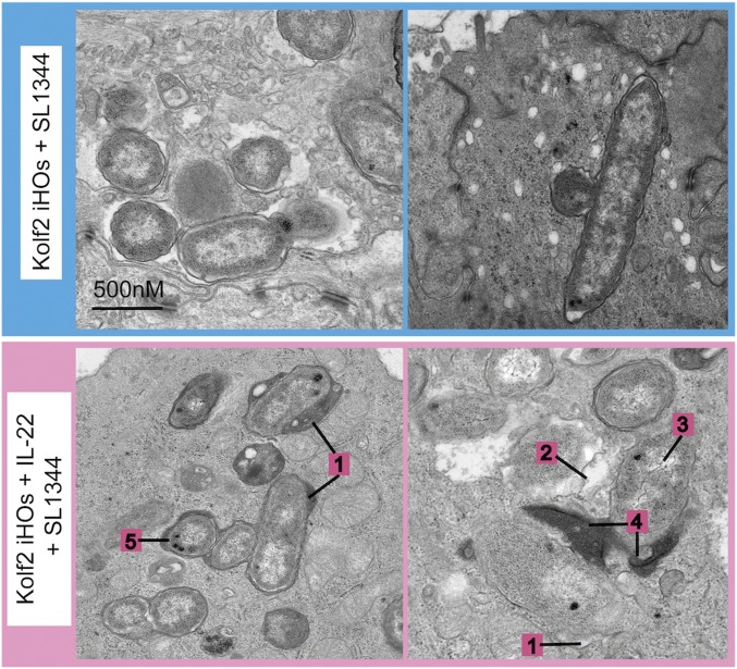
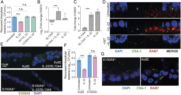
Intestinal epithelial cells (IECs) play a key role in regulating immune responses and controlling infection. However, the direct role of IECs in restricting pathogens remains incompletely understood. Here, we provide evidence that IL-22 primed intestinal organoids derived from healthy human induced pluripotent stem cells (hIPSCs) to restrict Salmonella enterica serovar Typhimurium SL1344 infection. A combination of transcriptomics, bacterial invasion assays, and imaging suggests that IL-22-induced antimicrobial activity is driven by increased phagolysosomal fusion in IL-22-pretreated cells. The antimicrobial phenotype was absent in hIPSCs derived from a patient harboring a homozygous mutation in the IL10RB gene that inactivates the IL-22 receptor but was restored by genetically complementing the IL10RB deficiency. This study highlights a mechanism through which the IL-22 pathway facilitates the human intestinal epithelium to control microbial infection.

Keywords: Salmonella; interleukin-22; intestinal organoids.


Conflict of interest statement

The authors declare no conflict of interest.

Figures

Fig. 1.
Fig. 1.
Analysis of expression of IL-22R subunits IL22R1 and IL10R2 demonstrated that iHOs generated from healthy control hIPSCs express the receptors for IL-22, IL22R1, and IL10R2, in contrast to iHOs generated from a patient with infantile IBD, which lack the IL10R2 subunit. To generate IL10RBComp, an isogenic control line for patient iPSCs (IL10RBPat), TALEN-mediated gene integration was used to integrate a copy of the IL10RB gene into the AAVS1 site in IL10RBPat iPSCs. (A) mRNA levels determined by RT-qPCR for IL22RA and IL10RB in three healthy control iHO lines (Yemz1, Lise1, and Kolf2), IL10RBPat iHOs, and control duodenal tissue. IL10RB is significantly up-regulated in control iHOs in comparison with IL10RBPat (P = 0.0005, P < 0.0001, and P < 0.0001, respectively; Student’s t tests). (B) RT-qPCR analysis shows significant up-regulation of IL10RB expression in IL10RBComp iHOs in comparison with IL10RBPat iHOs (P < 0.0001; unpaired, two-tailed Student’s t test). RT-qPCR was performed with TaqMan gene-expression assays and analyzed via the comparative cycle threshold (CT) method with GAPDH as an endogenous control. Data are presented from four technical replicates, with assays repeated at least three times from independent iHO batches. **P < 0.001; ***P < 0.0001. (C) Z-stacked immunostaining for IL-22 receptors IL10R2 or IL22R1 (green) and DAPI (blue) on healthy control Kolf2, IL10RBComp, and IL10RBPat iHOs showing localization of IL22R1 and IL10R2 on the basal IEC surface, with IL10R2 not detected in IL10RBPat iHOs. (Original magnification: 20×.)
Fig. 2.
Fig. 2.
Expression of IL10RB was required for iHO responses to IL-22. iHOs were treated with 100 ng/mL recombinant human IL-22 (rhIL-22) added to the iHO medium. (A) In iHO lines with a functional copy of IL10RB, transcripts for IL-22–regulated genes lipocalin 2 (LCN2) and dual oxidase 2 (DUOX2) are significantly up-regulated after the addition of 100 ng/mL rhIL-22 for 18 h, in comparison with unstimulated iHOs (**P < 0.001; ***P < 0.0001; unpaired, two-tailed, Student’s t tests; n.s., not significant). Data are presented from four technical replicates; assays were repeated with at least three biological replicates. RT-qPCR was performed with TaqMan gene-expression assays and analyzed via the comparative CT method with GAPDH as an endogenous control. (B) Phospho-STAT3 level was detected by Western blot after stimulation of healthy control (Yemz1 and Kolf2), IL10RBPat, and IL10RBComp iHOs for 30 min with rhIL-22 and preparation of whole-cell extracts. To verify equal protein loading, the blot was stripped and reprobed with STAT3 antibody. Lysate from HeLa cells stimulated with IFNλ were used as a positive control. (C) Kolf2 iHOs challenged with IL-22 for 18 h or left untreated were examined for Mucin 4 (MUC4; green) and DAPI (blue) by immunofluorescence. (D) Z-stacked immunostaining for Mucin 4 in IL10RBPat and IL10RBComp iHOs challenged with IL-22 for 18 h or were left untreated. (Original magnification: 20×; L = iHO lumen.)
Fig. 3.
Fig. 3.
Pretreatment with rhIL-22 of iHOs that express both IL22R1 and IL10R2 restricts S. enterica Typhimurium SL1344 invasion into IECs. For protection assays iHOs were treated with 100 ng/mL IL-22 18 h before infection or were left untreated. After injection of S. enterica Typhimurium SL1344 into the luminal cavity, iHOs were incubated for 90 min unless otherwise stated. Bacteria were recovered either from the luminal cavity or from IECs. The data presented show the mean from three technical replicates from the combined total of 25 iHOs per replicate, ± SEM, unless otherwise stated. For significance testing unpaired, two-tailed, Mann–Whitney U tests were used for all assays. *P < 0.05; **P < 0.001; ***P < 0.0001; n.s., not significant. (A) Gentamicin protection assays in iHOs show that pretreatment with IL-22 results in significantly less invasion after microinjection of S. enterica Typhimurium SL1344 in healthy control lines with functional IL10R2 (Kolf2: P = 0.0012; IL10R2Comp: P < 0.0001), but this phenotype is not observed in IL10RBPat iHOs (P = 0.2). (B) Percentage difference in TIMERbac Salmonella-infected cells assayed by flow cytometry recovered from untreated or IL-22–treated iHOs (40 iHOs per condition), with gating on live cells. There were significant differences between Kolf2 samples (P = 0.0214) or IL10RBComp samples and IL10RBPat samples (P = 0.0002; Kruskal–Wallis test with Dunn’s multiple comparison test). (C) Log numbers of cfu/mL recovered from lumens of iHOs after microinjection of untreated or IL-22–pretreated Kolf2 iHOs with SL1344 or an invasion-deficient strain, S. enterica Typhimurium SL1344 invA. There was no significant difference in numbers recovered. (D) Protection against S. enterica Typhimurium infection after IL-22 pretreatment is observed in human primary duodenal organoids. Data presented show the mean from three biological replicates with 10 organoids injected per replicate, ± SEM (P < 0.0001). (E) Modified gentamicin protection assays in Kolf2 iHOs show that pretreatment with IL-22 results in significantly less initial invasion (30-min incubation) after microinjection of S. enterica Typhimurium SL1344 (P < 0.0001) and also significantly less intracellular survival, when infected iHO IECs were incubated for an additional 90 min after gentamicin treatment and before IEC lysis (P < 0.0001).
Fig. 4.
Fig. 4.
IL-22 protection was mediated through enhanced lysosomal fusion with SCVs in IL-22–treated iHOs. TEM images of S. enterica Typhimurium SL1344 internalized into IECs 90 min after injection into the lumen of Kolf2 iHOs pretreated for 18 h with 100 ng/mL IL-22 (Lower) or left untreated (Upper), showing healthy bacteria in untreated organoids and degraded bacteria, often associated with lysosomes, in IL-22–pretreated iHOs. Representative images for each treatment condition were selected from 30 iHOs injected and processed per condition. Numbers indicate characterizations of bacterial cell damage/stress used for scoring: widening of periplasmic space (1); membrane damage and ragged appearance (2); decrease in cytosol density (3); direct contact with lysosomes (4); and presence of volutin granules (5).
Fig. 5.
Fig. 5.
IL-22–induced calgranulin B enhanced iHO colonization resistance to S. enterica Typhimurium infection. (A) Direct blocking of phagolysosomal fusion with phagolysosomal inhibitors restricts IL-22–mediated protection in iHOs. Kolf2 iHOs were pretreated with 100 ng/mL IL-22 for 18 h and then were treated with the phagolysosomal inhibitors W7 at 50 μM for 6 h or concanamycin A (CCMA) at 100 nM for 4 h or were left untreated, after which gentamicin protection assays were performed. There was no significant difference in intracellular recovered bacteria from W7- or concanamycin A-treated iHOs and untreated iHOs (W7, P = 0.7015; CCMA P = 0.0631, IL-22, ***P < 0.0001; unpaired, two-tailed Mann–Whitney U tests). Data presented show the mean from four biological replicates with three technical replicates per assay, ± SEM. (B and C) RT-qPCR was performed with a TaqMan gene-expression assay specific for RAB7A (B) or S100A9 (C) and was analyzed via the comparative CT method with GAPDH as an endogenous control. Data presented show the mean fold change between untreated samples and infected samples from three biological replicates, ± SEM. There was a significant difference in RAB7A and S100A9 expression in iHOs pretreated with IL-22 in comparison with iHOs left untreated before infection (RAB7A: P = 0.0001; S100A9: P < 0.0001; unpaired, two-tailed Student’s t tests). (D) Kolf2 iHOs challenged with IL-22 for 18 h or left untreated were microinjected with S. enterica Typhimurium SL1344 and examined for RAB7 (red), CSA-1 (green), and DAPI (blue) by immunofluorescence with colocalization between RAB7 and CSA-1 (yellow) visible in IL-22–pretreated samples. (Original magnification: 63×.) (E) Expression of S100A9 (green) by IL-22/S. enterica Typhimurium-treated Kolf2 and S100A9−/− iHOs was examined using immunofluorescence. (F) Protection assays in S100A9−/− iHOs show that pretreatment with IL-22 results in no significant difference in recovered intracellular bacterial after microinjection of S. enterica Typhimurium SL1344, in contrast to Kolf2 iHOs. (G) S100A9−/− or Kolf2 iHOs treated with IL-22 for 18 h and then microinjected with S. enterica Typhimurium SL1344 were examined for RAB7 (red), CSA-1 (green), and DAPI (blue) by immunofluorescence; RAB7 was not visible in S100A9−/− samples. (Original magnification: 40×.) n.s., not significant; ***P < 0.0001.

References

    1. Villa-Diaz LG, Ross AM, Lahann J, Krebsbach PH. Concise review: The evolution of human pluripotent stem cell culture: From feeder cells to synthetic coatings. Stem Cells. 2013;31:1–7. - PMC - PubMed
    1. McKernan R, Watt FM. What is the point of large-scale collections of human induced pluripotent stem cells? Nat Biotechnol. 2013;31:875–877. - PMC - PubMed
    1. An MC, et al. Genetic correction of Huntington’s disease phenotypes in induced pluripotent stem cells. Cell Stem Cell. 2012;11:253–263. - PMC - PubMed
    1. Bellin M, et al. Isogenic human pluripotent stem cell pairs reveal the role of a KCNH2 mutation in long-QT syndrome. EMBO J. 2013;32:3161–3175. - PMC - PubMed
    1. Glocker EO, et al. Inflammatory bowel disease and mutations affecting the interleukin-10 receptor. N Engl J Med. 2009;361:2033–2045. - PMC - PubMed

Publication types

MeSH terms

Substances

LinkOut - more resources