An MRI measure of degenerative and cerebrovascular pathology in Alzheimer disease
- PMID: 30217936
- PMCID: PMC6177275
- DOI: 10.1212/WNL.0000000000006310
An MRI measure of degenerative and cerebrovascular pathology in Alzheimer disease
Abstract
Objective: To develop, replicate, and validate an MRI-based quantitative measure of both cerebrovascular and neurodegeneration in Alzheimer disease (AD) for clinical and potentially research purposes.
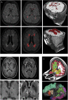
Methods: We used data from a cross-sectional and longitudinal community-based study of Medicare-eligible residents in northern Manhattan followed every 18-24 months (n = 1,175, mean age 78 years). White matter hyperintensities, infarcts, hippocampal volumes, and cortical thicknesses were quantified from MRI and combined to generate an MRI measure associated with episodic memory. The combined MRI measure was replicated and validated using autopsy data, clinical diagnoses, and CSF biomarkers and amyloid PET from the Alzheimer's Disease Neuroimaging Initiative.
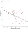
Results: The quantitative MRI measure was developed in a group of community participants (n = 690) and replicated in a similar second group (n = 485). Compared with healthy controls, the quantitative MRI measure was lower in patients with mild cognitive impairment and lower still in those with clinically diagnosed AD. The quantitative MRI measure correlated with neurofibrillary tangles, neuronal loss, atrophy, and infarcts at postmortem in an autopsy subset and was also associated with PET amyloid imaging and CSF levels of total tau, phosphorylated tau, and β-amyloid 42. The MRI measure predicted conversion to MCI and clinical AD among healthy controls.
Conclusion: We developed, replicated, and validated an MRI measure of cerebrovascular and neurodegenerative pathologies that are associated with clinical and neuropathologic diagnosis of AD and related to established biomarkers.
© 2018 American Academy of Neurology.
Figures


Comment in
-
An MRI biomarker of mixed pathology.Neurology. 2018 Oct 9;91(15):682-683. doi: 10.1212/WNL.0000000000006305. Epub 2018 Sep 14. Neurology. 2018. PMID: 30217934 No abstract available.
References
-
- Diaz JF, Merskey H, Hachinski VC, et al. . Improved recognition of leukoaraiosis and cognitive impairment in Alzheimer's disease. Arch Neurol 1991;48:1022–1025. - PubMed
-
- Hachinski V. Stroke and Alzheimer disease: fellow travelers or partners in crime? Arch Neurol 2011;68:797–798. - PubMed
-
- Thal DR, Ghebremedhin E, Orantes M, Wiestler OD. Vascular pathology in Alzheimer disease: correlation of cerebral amyloid angiopathy and arteriosclerosis/lipohyalinosis with cognitive decline. J Neuropathol Exp Neurol 2003;62:1287–1301. - PubMed
Publication types
MeSH terms
Substances
Grants and funding
LinkOut - more resources
Full Text Sources
Other Literature Sources
Medical
