. 2018 Sep 15:24:6480-6488.
doi: 10.12659/MSM.910050.

N-Methyl Pyrrolidone (NMP) Alleviates Lipopolysaccharide (LPS)-Induced Inflammatory Injury in Articular Chondrocytes

Affiliations

N-Methyl Pyrrolidone (NMP) Alleviates Lipopolysaccharide (LPS)-Induced Inflammatory Injury in Articular Chondrocytes

Xianping Zhu et al. Med Sci Monit. .

Abstract

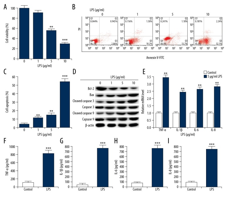
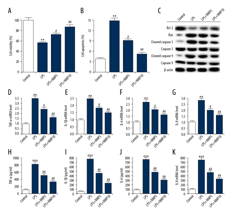
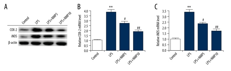
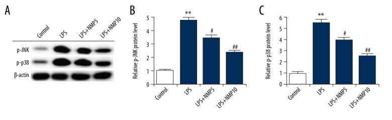
BACKGROUND Studies on the chondrocyte inflammatory injury are very important for understanding the pathogenesis and clinical treatment of osteoarthritis (OA). Evidence suggests that N-methyl pyrrolidone (NMP) may be used as an adjuvant therapy alongside established methods of OA treatment. This study investigated the effect of NMP on chondrocyte inflammatory injury and explored the underlying molecular mechanism. MATERIAL AND METHODS To mimic the inflammatory injury in vitro, the articular chondrocyte line ATDC5 was simulated with lipopolysaccharide (LPS). ATDC5 cells were treated with various concentrations of NMP (0, 5, and 10 nM). Cell viability was measured using CCK-8 assay; cell apoptosis was detected using FCM; related protein and mRNA expressions were determined using Western blot assay and qRT-PCR assay; and inflammatory factors (tumor necrosis factor (TNF)-α, interleukin (IL)-1β, IL-6, and IL-8) productions were measured by performing ELISA assay. RESULTS The results showed that LPS simulation repressed ATDC5 cell viability, prompted cell apoptosis, and enhanced the secretion of inflammatory factors. NMP treatment reduced inflammatory injury induced by LPS in a dose-dependent manner. Furthermore, NMP inhibited the activation of JNK and p38 pathways. In addition, inhibition of NF-κB activation was observed following NMP treatment. CONCLUSIONS NMP prevents inflammatory reaction of articular chondrocytes via repressing the MAPK/NF-kB pathway. Our findings provide a promising therapeutic agent for OA treatment.


Conflict of interest statement

Conflict of interests

None.

Figures

Figure 1
Figure 1
LPS induced chondrocyte inflammatory injury in ATDC5 cells. ATDC5 cells were treated with various doses of LPS (1, 5, and 10 μg/ml) to simulate inflammatory lesions. Cells without LPS administration (0 μg/ml) were used as control. (A) CCK-8 was performed to measure cell viability; (B, C) Relative apoptotic cells were measured by flow cytometry (cell apoptosis=early apoptosis late apoptosis); (D) The protein levels of apoptosis-related factors were detected by Western blotting. (E) The mRNA expression levels of pro-inflammatory cytokines (TNF-α, IL-1β, IL-6, and IL-8) were detected by qRT-PCR. (F–I) ELISA was used to measure the productions of TNF-α, IL-1β, IL-6 and IL-8. *, **, *** p<0.05, 0.01, 0.001 compared with control.
Figure 2
Figure 2
NMP reduced LPS induced inflammatory injury. ATDC5 cells were pre-treated with various concentrations of NMP (0, 5, and 10 nM) and then treated with 5 μg/ml LPS. (A) CCK-8 was performed to measure cell viability; (B) Relative apoptotic cells were measured by flow cytometry (cell apoptosis=early apoptosis late apoptosis); (C) The protein levels of apoptosis-related factors were detected by Western blotting. (D–G) The mRNA expression levels of pro-inflammatory cytokines (TNF-α, IL-1β, IL-6, and IL-8) were detected by qRT-PCR; (H–K) ELISA was used to measure the productions of TNF-α, IL-1β, IL-6, and IL-8. Control: cells without any treatment; LPS: cells were treated with 5 μg/ml LPS for 6 h at 37°C; LPS+NMP5: cells were treated with 5 mM NMP for 48 h at 37°C and then 5 μg/ml LPS for 6 h at 37°C; LPS+NMP10: cells were treated with 10 mM NMP for 48 h at 37°C and then 5 μg/ml LPS for 6 h at 37°C. ** p<0.01 vs. control; #, ## p<0.05, 0.01 vs. LPS group.
Figure 3
Figure 3
NMP significantly inhibits the LPS-induced increase of COX-2 and iNOS expression. ATDC5 cells were pre-treated with various concentrations of NMP (0, 5, and 10 nM) and then treated with 5 μg/ml LPS. The protein (A) and mRNA (B, C) levels of COX-2 and iNOS was determined using Western blot and qRT-PCR, respectively. Control: cells without any treatment; LPS: cells were treated with 5 μg/ml LPS for 6 h at 37°C; LPS+NMP5: cells were treated with 5 mM NMP for 48 h at 37°C and then 5 μg/ml LPS for 6 h at 37°C; LPS+NMP10: cells were treated with 10 mM NMP for 48 h at 37°C and then 5 μg/ml LPS for 6 h at 37°C. ** p<0.01 vs. control; #, ## p<0.05, 0.01 vs. LPS group.
Figure 4
Figure 4
NMP significantly inhibits the LPS-induced over-activation of JNK and p38. ATDC5 cells were pre-treated with various concentrations of NMP (0, 5, and 10 nM) and then treated with 5 μg/ml LPS. The protein level of p-JNK and p-p38 was determined using Western blot assay. Control: cells without any treatment; LPS: cells were treated with 5 μg/ml LPS for 6 h at 37°C; LPS+NMP5: cells were treated with 5 mM NMP for 48 h at 37°C and then 5 μg/ml LPS for 6 h at 37°C; LPS+NMP10: cells were treated with 10 mM NMP for 48 h at 37°C and then 5 μg/ml LPS for 6 h at 37°C. ** p<0.01 vs. control; #, ## p<0.05, 0.01 vs. LPS group.
Figure 5
Figure 5
NMP significantly inhibits the LPS-induced over-activation of NF-κB pathway. ATDC5 cells were pre-treated with various concentrations of NMP (0, 5, and 10 nM) and then treated with 5 μg/ml LPS. The protein level of p-p65 was determined using Western blot assay. Control: cells without any treatment; LPS: cells were treated with 5 μg/ml LPS for 6 h at 37°C; LPS+NMP5: cells were treated with 5 mM NMP for 48 h at 37°C and then 5 μg/ml LPS for 6 h at 37°C; LPS+NMP10: cells were treated with 10 mM NMP for 48 h at 37°C and then 5 μg/ml LPS for 6 h at 37°C. ** p<0.01 vs. control; #, ## p<0.05, 0.01 vs. LPS group.

References

    1. Pelletier JP, Mantel-Pelletier JF, Malemud CJ. Immunological analysis of proteoglycan changes in the early stage of osteoarthritic canine cartilage lesions. J Orthop Res. 1992;10:511–23. - PubMed
    1. Colen S, Bekerom MPJVD, Mulier M. Intra-articular viscosupplementation in patients with hip osteoarthritis. Eur Musculoskelet Rev. 2011;6:79–82.
    1. Arden N, Blanco F, Cooper C, et al. Atlas of osteosteoarthritis. Springer Healthcare Ltd; 2014. pp. 69–82.
    1. Goldring MB, Goldring SR. Osteoarthritis. J Cell Physiol. 2007;213:626–34. - PubMed
    1. Fitzgerald GK, Piva SR, Irrgang JJ. Reports of joint instability in knee osteoarthritis: Its prevalence and relationship to physical function. Arthritis Rheum. 2004;51(6):941–46. - PubMed