Review
. 2018 Dec;36(8):2051-2059.
doi: 10.1016/j.biotechadv.2018.09.001. Epub 2018 Sep 12.

Towards rationally designed biomanufacturing of therapeutic extracellular vesicles: impact of the bioproduction microenvironment

Affiliations
Review

Towards rationally designed biomanufacturing of therapeutic extracellular vesicles: impact of the bioproduction microenvironment

Divya B Patel et al. Biotechnol Adv. 2018 Dec.

Abstract

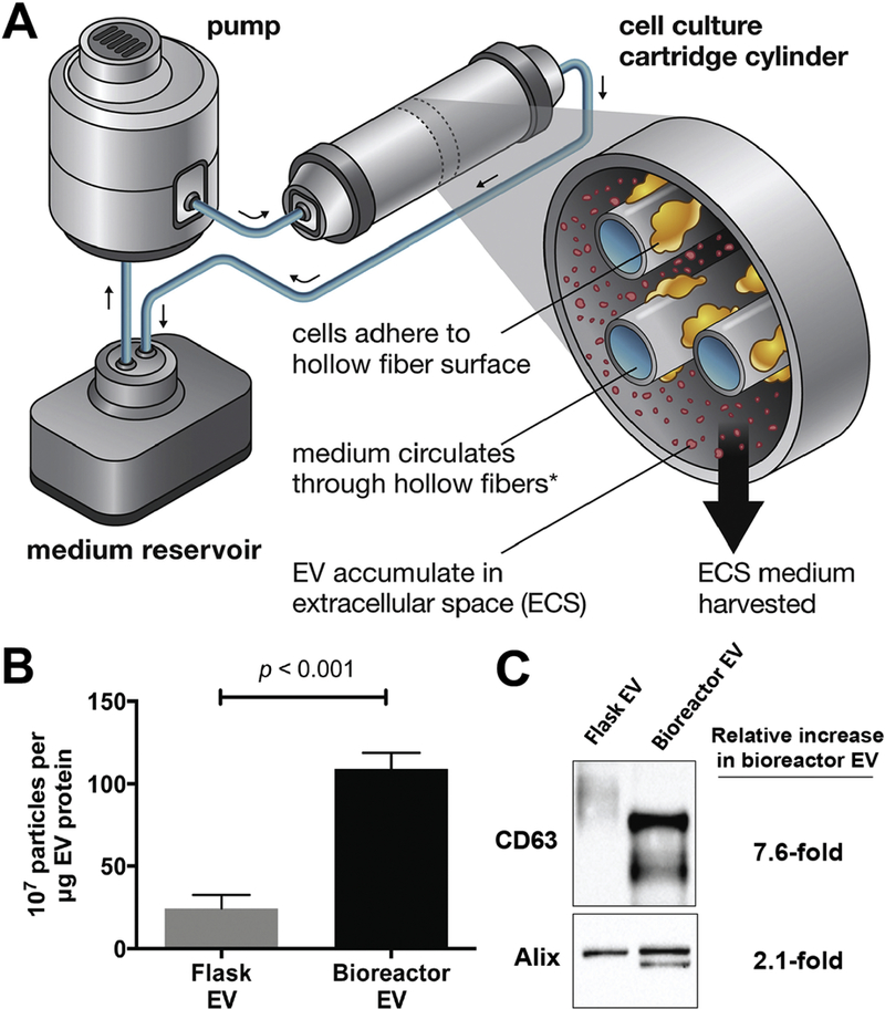
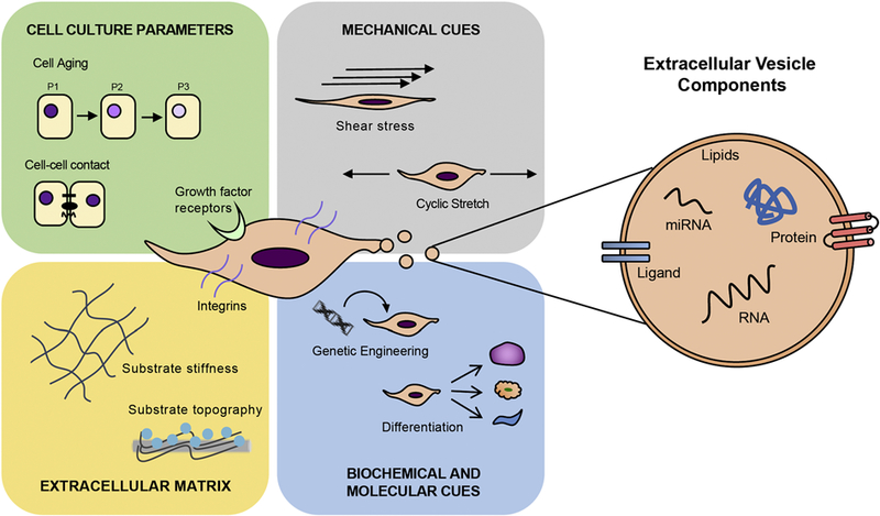
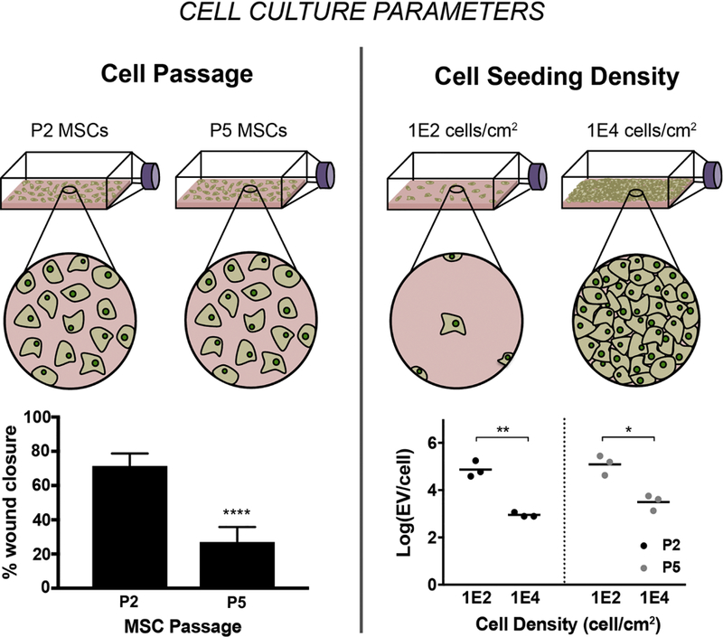
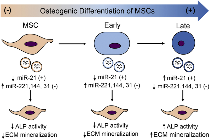
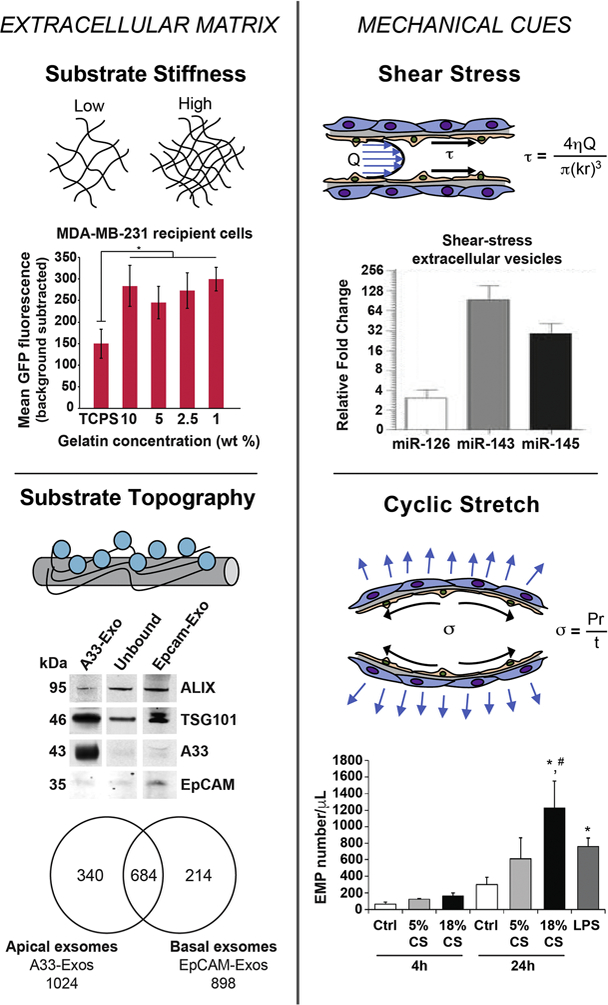
Extracellular vesicles (EVs), including exosomes, microvesicles, and others, have emerged as potential therapeutics for a variety of applications. Pre-clinical reports of EV efficacy in treatment of non-healing wounds, myocardial infarction, osteoarthritis, traumatic brain injury, spinal cord injury, and many other injuries and diseases demonstrate the versatility of this nascent therapeutic modality. EVs have also been demonstrated to be effective in humans, and clinical trials are underway to further explore their potential. However, for EVs to become a new class of clinical therapeutics, issues related to translation must be addressed. For example, approaches originally developed for cell biomanufacturing, such as hollow fiber bioreactor culture, have been adapted for EV production, but limited knowledge of how the cell culture microenvironment specifically impacts EVs restricts the possibility for rational design and optimization of EV production and potency. In this review, we discuss current knowledge of this issue and delineate potential focus areas for future research towards enabling translation and widespread application of EV-based therapeutics.

Keywords: Biomanufacturing; Exosome; Extracellular vesicle; Mesenchymal stem cell; Microenvironment.


Figures

Figure 1.
Figure 1.. Hollow fiber bioreactor and 3D culture impact on EV production and bioactivity.
(A) Schematic of the hollow fiber bioreactor system. (B) HEK293 cells cultured in a hollow fiber bioreactor produced ~4-fold more EVs than cells cultured in conventional tissue culture flasks. Data presented as mean ± SEM and statistical significance was compared by ANOVA. (B) Western blot analysis showed 7.6-fold and 2.1-fold increase in EV-associated markers of EVs (20µg) from hollow-fiber bioreactor compared to tissue culture flask. Additional bands observed for CD63 and Alix in Bioreactor EVs is hypothesized to be due to differential glycosylation pattern and phosphorylation, respectively. Adapted via open access from Watson, D.C. et al. (2016).
Figure 2.
Figure 2.
Schematic representations of factors influencing MSC-derived EV production and cargo composition.
Figure 3.
Figure 3.. Impact of MSC passage and seeding density on EV production and vascular bioactivity.
(A) EVs were isolated from MSCs at P2, P3, P4, and P5 were seeded at 1E2, 5E2, 1E3, 1E4 cells/cm2. For MSCs at all passages, increase in EV production is observed per cell with decreasing seeding densities. Statistical significance was calculated using two-way ANOVA with Tukey’s multiple comparison test (* p<0.05, ** p<0.01). Vascular bioactivity of EVs from different densities (B) and different passages (C) was analyzed using a gap closure assay with endothelial cells. (B) No significant difference in gap closure was observed for EVs from MSCs seeded at 1E2 and 1E4 cells/cm2. (C) Bioactivity of EVs significantly diminishes with increasing passage. Data are representative of three independent experiments with three replicates each (n = 3). Statistical significance was calculated using two-way ANOVA with Tukey’s multiple comparison test (* p<0.05, ** p<0.01, **** p<0.0001). Figure reprinted from Patel et al., 2017 via open access.
Figure 4.
Figure 4.. Schematic describing that EVs from varying stages of differentiating MSCs induce osteogenic differentiation of MSCs differently.
Osteogenic differentiation was induced in MSCs using osteogenic differentiation media. EVs were isolated from conditioned media collected at different stages after media change, namely, early and late stages. Exosomes from MSCs in growth media were used as control. Using an array-based method, expression of miRNA associated with osteogenic differentiation of MSCs was measured. ALP activity, ECM calcium, and phosphate was measured to evaluate osteogenic differentiation of MSCs after EV treatment. Adapted from data presented in Wang et al., (2018) via open access.
Figure 5.
Figure 5.. ECM characteristics and mechanical stimulation of cells can dictate EV production, composition, and uptake profiles.
In the top left figure, breast cancer cell lines MCF-7 and MDA-MB-231 were seeded on gelatin substrate with varying stiffness. MDA-MB-231 cells seeded on gelatin matrix with different stiffness were treated with CD63-GFP-labled EVs. EV uptake was monitored following 2 hours of treatment through live-cell imaging. Seeding cell on softer gelatin matrices compared to tissue culture polystyrene (TCPS), resulted in significantly increased EV uptake for both cell lines. Adapted from Tauro et al., (2013) via open access. Bottom left figure shows western blot analysis of exosomes (10 µg) derived from LIM1863 cells from apical (EpCAM-Exos) or basolateral (A33-Exos) surfaces, which were enriched in EpCAM and A33, respectively. Both sources were also positive for exosomal markers TSG101 and Alix. Venn diagram shows differences in protein expression between EpCAM-Exos and A33-Exos. In total, 340 and 214 proteins were specifically found in A33-and EpCAM-Exos, respectively. In comparison, 684 proteins were present in both exosome sources. Adapted from Stranford et al., (2017) with permission. For the top right figure, EVs were isolated from HUVECs with or without exposure to shear-stress of 20 dynes/cm2 for 3 days. RNA analysis revealed that EVs derived from HUVECs under shear stress were highly enriched in miR-143/145, which are known to modulate SMC phenotype. Adapted from Hergenreider et al., (2012) with permission. For the bottom right figure, confluent lung-ECs were subjected to 5% CS or 18% CS for 4 and 24 hours or to LPS (1 µg/mL) for 24 hours. Lung-EC-derived microparticles (EMPs) (0.1–1 µm) were isolated from conditioned media and quantified by flow cytometry. Significant increase in EMP production is observed after 24 h exposure to 18% CS and LPS compared to the static control. Reprinted from Letsiou et al., (2015) with permission.

References

    1. Abagnale G, Steger M, Nguyen VH, Hersch N, Sechi A, Joussen S, Denecke B, Merkel R, Hoffmann B, Dreser A, Schnakenberg U, Gillner A, Wagner W, 2015. Surface topography enhances differentiation of mesenchymal stem cells towards osteogenic and adipogenic lineages. Biomaterials 61, 316–326. https://doi.org/http://doi.org/10.1016/j.biomaterials.2015.05.030 - DOI - PubMed
    1. Ader M, Tanaka EM, 2014. Modeling human development in 3D culture. Curr. Opin. Cell Biol 31, 23–28. https://doi.org/https://doi.org/10.1016/j.ceb.2014.06.013 - DOI - PubMed
    1. Aharon A, Tamari T, Brenner B, 2008. Monocyte-derived microparticles and exosomes induce procoagulant and apoptotic effects on endothelial cells. Thromb. Haemost 100, 878–885. https://doi.org/10.1160/TH07-11-0691 - DOI - PubMed
    1. Alvarez-Erviti L, Seow Y, Yin H, Betts C, Lakhal S, Wood MJA, 2011. Delivery of siRNA to the mouse brain by systemic injection of targeted exosomes. Nat Biotech 29, 341–345. - PubMed
    1. Aswad H, Jalabert A, Rome S, 2016. Depleting extracellular vesicles from fetal bovine serum alters proliferation and differentiation of skeletal muscle cells in vitro. BMC Biotechnol 16, 32 https://doi.org/10.1186/s12896-016-0262-0 - DOI - PMC - PubMed

Publication types