. 2018 Oct;20(10):1045-1058.
doi: 10.1016/j.neo.2018.08.008. Epub 2018 Sep 13.

PDZ-RhoGEF Is a Signaling Effector for TROY-Induced Glioblastoma Cell Invasion and Survival

Affiliations

PDZ-RhoGEF Is a Signaling Effector for TROY-Induced Glioblastoma Cell Invasion and Survival

Zonghui Ding et al. Neoplasia. 2018 Oct.

Abstract

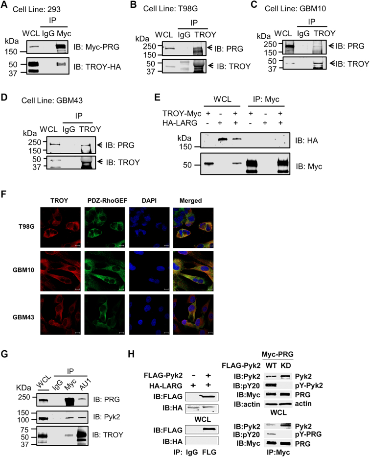
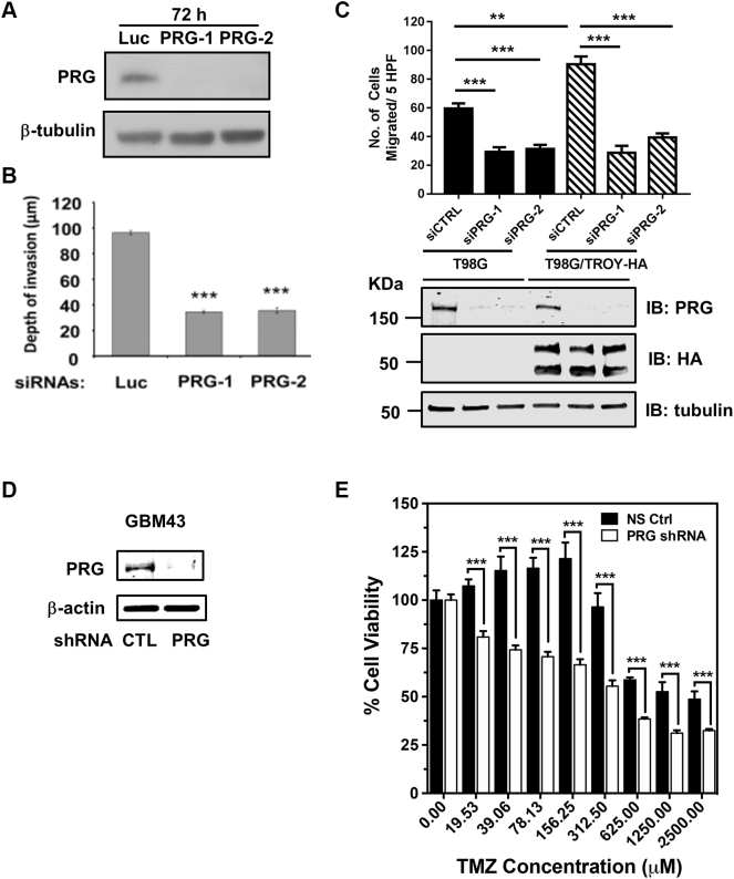
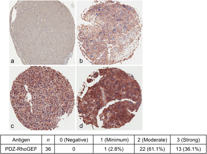
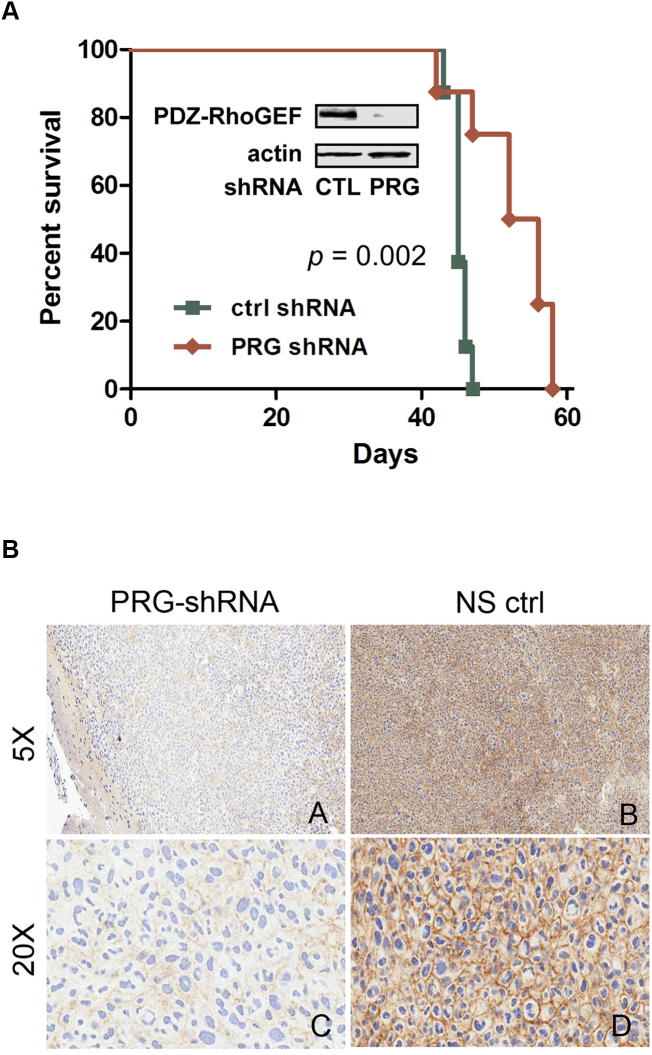
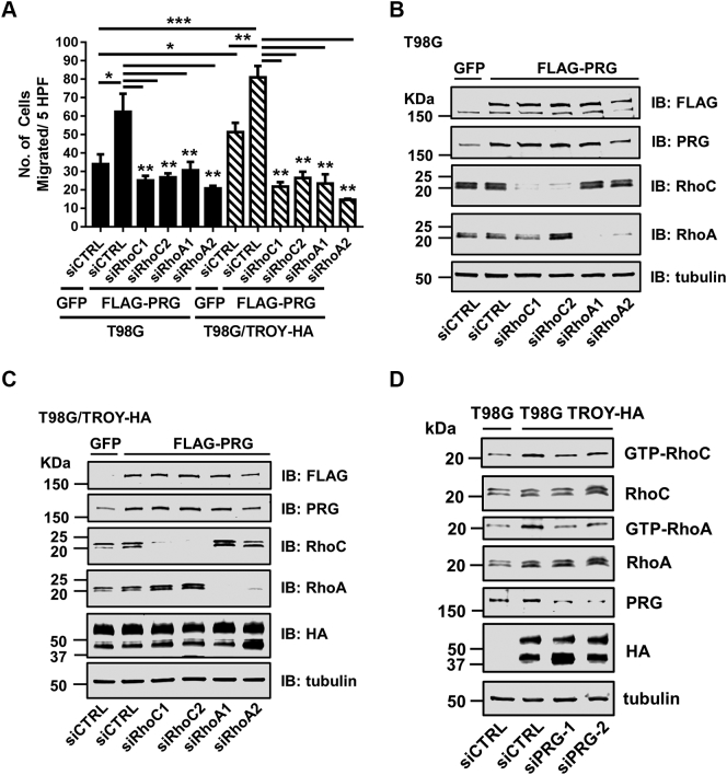
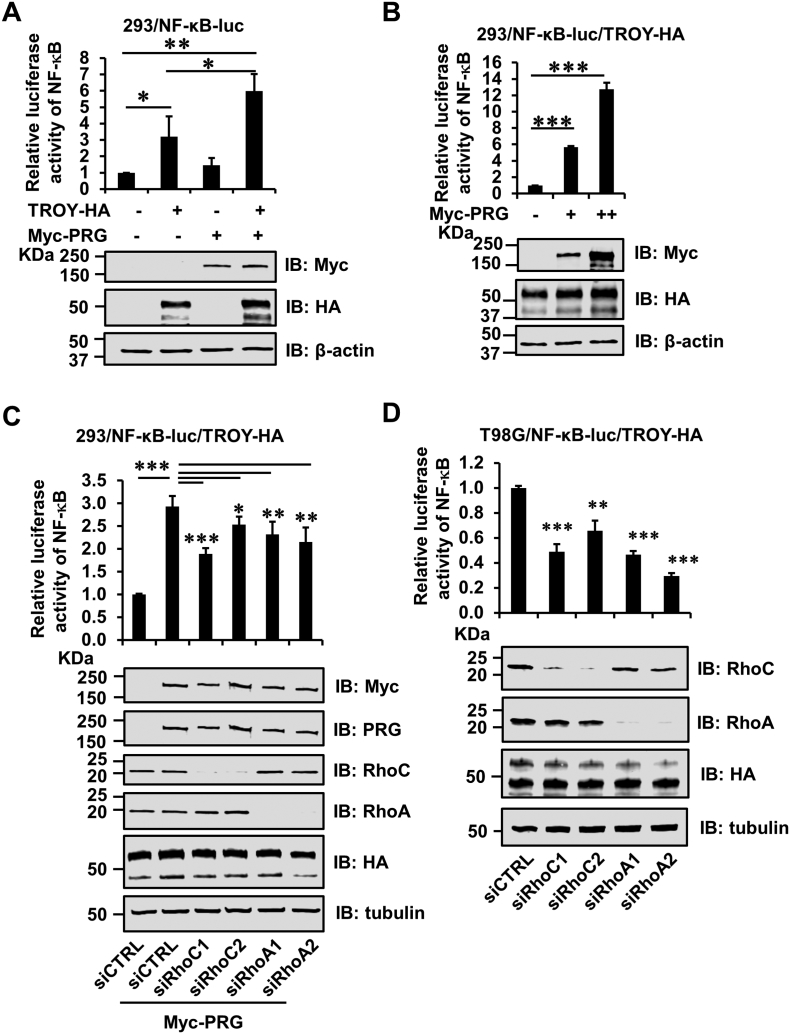
Glioblastoma multiforme (GBM) is the most common type of malignant brain tumors in adults and has a dismal prognosis. The highly aggressive invasion of malignant cells into the normal brain parenchyma renders complete surgical resection of GBM tumors impossible, increases resistance to therapeutic treatment, and leads to near-universal tumor recurrence. We have previously demonstrated that TROY (TNFRSF19) plays an important role in glioblastoma cell invasion and therapeutic resistance. However, the potential downstream effectors of TROY signaling have not been fully characterized. Here, we identified PDZ-RhoGEF as a binding partner for TROY that potentiated TROY-induced nuclear factor kappa B activation which is necessary for both cell invasion and survival. In addition, PDZ-RhoGEF also interacts with Pyk2, indicating that PDZ-RhoGEF is a component of a signalsome that includes TROY and Pyk2. PDZ-RhoGEF is overexpressed in glioblastoma tumors and stimulates glioma cell invasion via Rho activation. Increased PDZ-RhoGEF expression enhanced TROY-induced glioma cell migration. Conversely, silencing PDZ-RhoGEF expression inhibited TROY-induced glioma cell migration, increased sensitivity to temozolomide treatment, and extended survival of orthotopic xenograft mice. Furthermore, depletion of RhoC or RhoA inhibited TROY- and PDZ-RhoGEF-induced cell migration. Mechanistically, increased TROY expression stimulated Rho activation, and depletion of PDZ-RhoGEF expression reduced this activation. Taken together, these data suggest that PDZ-RhoGEF plays an important role in TROY signaling and provides insights into a potential node of vulnerability to limit GBM cell invasion and decrease therapeutic resistance.


Figures

Figure 1
Figure 1
TROY associates with PDZ-RhoGEF (PRG). (A) 293 cells were co-transfected with HA-tagged TROY plasmid and Myc-tagged PDZ-RhoGEF plasmid, and the lysates were immunoprecipitated with an anti-Myc antibody or mouse IgG. The immunoprecipitates were analyzed for the presence of PDZ-RhoGEF by immunoblot analysis. The expression level of Myc-tagged PDZ-RhoGEF and HA-tagged TROY in the cells is shown by immunoblotting of whole cell lysates (WCL). (B-D) The lysates of T98G, GBM10, and GBM43 cells were immunoprecipitated with an anti-TROY antibody or rabbit IgG. The immunoprecipitates were analyzed for the presence of PDZ-RhoGEF by immunoblot analysis. The expression level of PDZ-RhoGEF and TROY in the cells is shown by immunoblotting of WCL. (E) TROY does not co-immunoprecipitate with LARG in 293 cells. 293 cells were transfected with HA-tagged LARG or Myc-tagged TROY, or co-transfected with HA-tagged LARG and Myc-tagged TROY. Twenty-four hours after transfection, cells were lysed and immunoprecipitated with anti-Myc antibody. Immunoprecipitates or WCL were immunoblotted with the indicated antibodies. (F) Colocalization of TROY and PDZ-RhoGEF in T98G, GBM10, and GBM43 cells. Cells were stained and examined by confocal microscopy. TROY was visualized with Alexa Fluor 546–conjugated antibody, and PDZ-RhoGEF was visualized with Alexa Fluor 488–conjugated antibody. The colocalization of TROY (red) with PDZ-RhoGEF (green) appears as a yellow color in the merged image. Cells were counterstained with DAPI (blue) (bars = 10 μm). (G) 293 cells were co-transfected with AU1-tagged TROY, Myc-tagged PRG, and HA-tagged Pyk2, and 24 hours later, cells were lysed and immunoprecipitated with goat anti-Myc or goat anti-AU1 antibodies or goat IgG. Immunoprecipitates or WCL were immunoblotted (IB) with the indicated antibodies. (H) Left panel, 293 cells were transfected with HA-tagged LARG and/or FLAG-tagged Pyk2. Twenty-four hours later, the cells were lysed and immunoprecipitated with anti-FLAG antibody or mouse IgG. Immunoprecipitates or WCL were IB with anti-FLAG or anti-HA antibodies. Right panel, 293 cells were co-transfected with Myc-tagged PRG and FLAG-tagged WT Pyk2 or kinase-deficient (KD) Pyk2 for 24 hours. Cells were lysed and immunoprecipitated with anti-Myc antibody. Immunoprecipitates or WCL were IB with the indicated antibodies.
Figure 2
Figure 2
Depletion of PDZ-RhoGEF expression inhibits glioma cell migration and invasion and TROY-stimulated glioma cell migration. (A) U87MG cells were transfected with two independent siRNAs targeting PDZ-RhoGEF (PRG-1, PRG-2) or with a control siRNA targeting firefly luciferase (Luc). Cell lysates were analyzed for PDZ-RhoGEF by immunoblotting. Immunoblotting of β-tubulin protein was used as a loading control. (B) Invasion of U87MG cells transfected with siRNAs targeting PDZ-RhoGEF (PRG-1, PRG-2) or luciferase (Luc). Forty-eight hours after cell seeding onto the brain slice, glioma cell invasion into the brain slices was quantified using confocal microscopy. The data are depicted as the mean values (+/− SEM) from three separate experiments. ***P < .001. (C) Migration of T98G cells and T98G cells overexpressing TROY (T98G/TROY-HA) transfected with siRNAs targeting PDZ-RhoGEF (siPRG-1, siPRG-2) or a nonsilencing control (siCTRL). Data represent the mean values (+/− SEM) (n = 3, **, P < .01; ***, P < .001). Cell lysates were analyzed by immunoblotting with indicated antibodies. Immunoblotting of tubulin was used as a loading control. (D) Knockdown of PDZ-RhoGEF expression in patient xenograft GBM43 cells transduced with the shRNA-targeting PDZ-RhoGEF (PRG) or the control (CTL) nontargeting shRNA. Cell lysates were analyzed by immunoblotting with indicated antibodies. Immunoblotting of β-actin protein was used as a loading control. (E) Primary xenograft GBM43 cells transduced with a control nonsilencing shRNA (NS Ctrl) or shRNA targeting PDZ-RhoGEF (PRG) were treated with the indicated concentration of TMZ. Cell viability was assessed after 72 hours by CellTiter-Glo assay. Data are depicted as the mean values (+/− SD) of six replicates. ***P < .001.
Figure 3
Figure 3
PDZ-RhoGEF expression is increased in GBM. (A) IHC analysis of PDZ-RhoGEF expression in non-neoplastic brain and GBM biopsy samples (upper panel). Samples are representative of 36 matched biopsy samples on a tissue microarray. a, negative, score 0; b, minimum, score 1; c, moderate, score 2; d, strong, score 3. Neuropathology scoring of the percentage distribution of staining intensity of PDZ-RhoGEF in GBM patient specimens (lower panel).
Figure 4
Figure 4
Knockdown of PDZ-RhoGEF expression increases xenograft survival. (A) Kaplan-Meier survival curves of athymic nude mice with intracranial xenografts of primary GBM10 cells transduced with a control nontargeting shRNA (CTL) or a shRNA-targeting PDZ-RhoGEF (PRG). Curves show a significant survival benefit for mice with xenografts with silencing PDZ-RhoGEF expression (P = .002). Insert: Western blot of whole cell lysates of transduced GBM10 cells used for the intracranial xenografts immunoblotted with the indicated antibodies. (B) IHC of GBM10 tumors transduced with shRNA targeting PDZ-RhoGEF (PRG-shRNA) or a nonsilencing control shRNA (NS ctrl).
Figure 5
Figure 5
Depletion of Rho suppresses TROY and PDZ-RhoGEF-induced glioma cell migration. (A) T98G and T98G/TROY-HA cells were transfected with a nontargeting control siRNA (siCTRL) or two independent siRNAs targeting PDZ-RhoGEF (siPRG-1, siPRG-2) for 24 hours and infected with FLAG-tagged PRG lentivirus or GFP lentivirus for additional 24 hours. The cells were serum starved overnight and then seeded on top of a Transwell chamber. The number of migrated cells post 24 hours was quantified using DAPI staining. Data represent the mean values (+/− SEM) (n = 3, *P < .05; **P < .01; ***P < .001). (B) The lysates of T98G used for migration assay in A were immunoblotted with indicated antibodies. (C) The lysates of T98G/TROY-HA used for migration assay in A were immunoblotted with indicated antibodies. (D) T98G and T98G/TROY-HA cells were transfected with a nontargeting control siRNA (siCTRL) or two independent siRNAs targeting PDZ-RhoGEF (siPRG-1, siPRG-2) for 2 days and then serum starved overnight. Cells were lysed, and the Rho activities in lysates were measured using GST-Rhotekin-RBD pull-down assay. Immunoblot is a representation of two independent experiments.
Figure 6
Figure 6
PDZ-RhoGEF (PRG) enhances TROY-induced NF-κB activation. (A) 293 cells expressing a NF-κB-luciferase reporter (293/NF-κB-luc) were transfected with the indicated plasmids. Twenty-four hours after transfection, cells were serum starved (0.1% BSA) for 16 hours and lysed, and NF-κB-luc reporter expression was measured using a luciferase reporter assay kit. Luciferase activity was normalized to the vector-transfected cells. The data are depicted as the mean values (+/− SD) (n = 3, *P < .05; **P < .01) (upper panel). The expression of HA-tagged TROY and Myc-tagged PDZ-RhoGEF in lysates was detected by immunoblotting (bottom panel). (B) 293/NF-κB-luc reporter cells overexpressing TROY (293/NF-κB-luc/TROY-HA) were transfected with either vector or 0.8 or 1.6 μg Myc-tagged PDZ-RhoGEF. Twenty-four hours after transfection, cells were serum starved (0.1% BSA) for 16 hours and lysed, and luciferase activity was measured using luciferase reporter assay kit. Luciferase activity was normalized to the vector-transfected cells. The data are depicted as the mean values (+/− SD) (n = 3, ***P < .001) (upper panel). Expression levels of PDZ-RhoGEF and TROY were determined by immunoblot analysis (bottom panel). (C) 293/NF-κB-luc/TROY-HA cells were transfected with either two independent siRNAs targeting RhoC or RhoA or a control siRNA (siCTRL). Twenty-four hours after transfection, cells were transfected with Myc-tagged PRG plasmid for an additional 24 hours. Cells were serum starved (0.1% BSA) for 16 hours and lysed, and luciferase activity was measured using a luciferase reporter assay kit. Luciferase activity was normalized to the control cells. The data are depicted as the mean values (+/− SD) (n = 3, *P < .05; **P < .01; ***P < .001) (upper panel). Knockdown of RhoC and RhoA expression and overexpression of PDZ-RhoGEF was verified by immunoblotting (bottom panel). (D) T98G/NF-κB-luc/TROY-HA cells were transfected with either two independent siRNAs targeting RhoC or RhoA or a control siRNA (siCTRL). Twenty-four hours after transfection, cells were serum starved (0.1% BSA) for additional 16 hours and lysed, and luciferase activity was measured using a luciferase reporter assay kit. Luciferase activity was normalized to the control cells. The data are depicted as the mean values (+/− SD) (n = 3, **P < .01; ***P < .001) (upper panel). Knockdown of RhoC and RhoA expression was verified by immunoblotting (bottom panel).

Similar articles

Cited by

References

    1. Stupp R, Mason WP, van den Bent MJ, Weller M, Fisher B, Taphoorn MJ, Belanger K, Brandes AA, Marosi C, Bogdahn U. Radiotherapy plus concomitant and adjuvant temozolomide for glioblastoma. N Engl J Med. 2005;352:987–996. - PubMed
    1. Van Meir EG, Hadjipanayis CG, Norden AD, Shu HK, Wen PY, Olson JJ. Exciting new advances in neuro-oncology: the avenue to a cure for malignant glioma. CA Cancer J Clin. 2010;60:166–193. - PMC - PubMed
    1. Giese A, Bjerkvig R, Berens ME, Westphal M. Cost of migration: invasion of malignant gliomas and implications for treatment. J Clin Oncol. 2003;21:1624–1636. - PubMed
    1. Lefranc F, Brotchi J, Kiss R. Possible future issues in the treatment of glioblastomas: special emphasis on cell migration and the resistance of migrating glioblastoma cells to apoptosis. J Clin Oncol. 2005;23:2411–2422. - PubMed
    1. Hu S, Tamada K, Ni J, Vincenz C, Chen L. Characterization of TNFRSF19, a novel member of the tumor necrosis factor receptor superfamily. Genomics. 1999;62:103–107. - PubMed

Publication types

MeSH terms