

Primary intracranial leiomyosarcoma among patients with AIDS in the era of new chemotherapeutic and biological agents
- PMID: 30219779
- PMCID: PMC6144167
- DOI: 10.1136/bcr-2018-225714
Primary intracranial leiomyosarcoma among patients with AIDS in the era of new chemotherapeutic and biological agents
Abstract
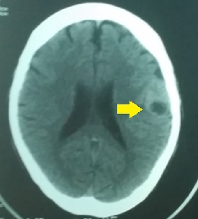
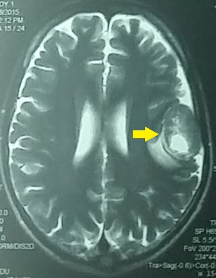
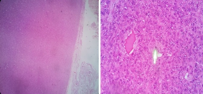
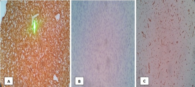
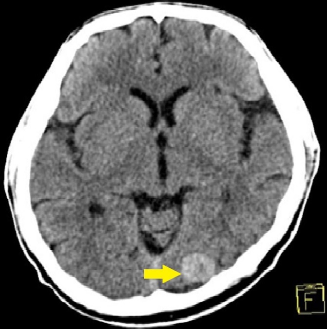
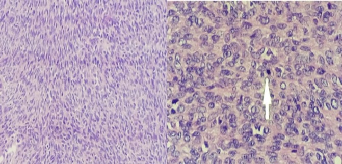
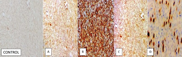
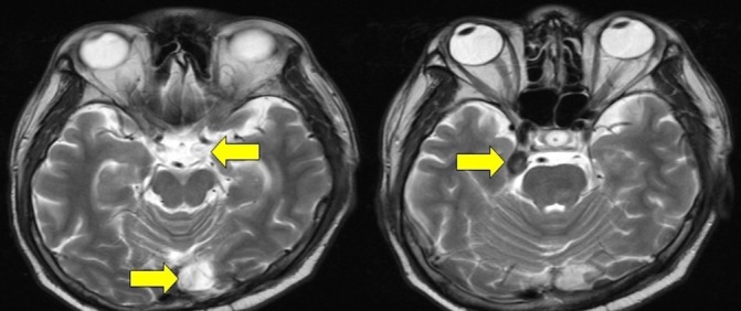
Primary intracranial leiomyosarcoma (PIL) is a rare non-infectious aetiology of focal mass lesions among HIV-infected individuals. With only 16 published cases worldwide, information on its pathophysiology, risk factors, clinical course and management options is limited. We report two cases of PIL in HIV-infected Filipino men who presented with 1-3 months history of persistent headache, progressing in severity. Both had cranial MRI revealing intracranial mass diagnosed as leiomyosarcoma by excision biopsy and immunohistochemical staining. Both patients underwent adjuvant cranial radiotherapy and chemotherapy. Biologics were initiated in one patient. Both patients were alive with evidence of the disease.
Keywords: Hiv / Aids; neurooncology.
© BMJ Publishing Group Limited 2018. No commercial re-use. See rights and permissions. Published by BMJ.
Conflict of interest statement
Competing interests: None declared.
Figures
Similar articles
-
Primary intracranial leiomyosarcoma in an immunocompetent patient: Case report and review of the literature.Clin Neurol Neurosurg. 2018 Feb;165:76-80. doi: 10.1016/j.clineuro.2017.12.014. Epub 2018 Jan 6. Clin Neurol Neurosurg. 2018. PMID: 29324399 Review.
-
Primary cerebral leiomyosarcoma.Clin Neurol Neurosurg. 1997 Aug;99(3):210-2. doi: 10.1016/s0303-8467(97)00018-8. Clin Neurol Neurosurg. 1997. PMID: 9350403
-
Intracranial leiomyosarcoma in a patient with AIDS.Neuroradiology. 1999 Jan;41(1):35-9. doi: 10.1007/s002340050701. Neuroradiology. 1999. PMID: 9987766
-
A rare case of primitive epithelioid leiomyosarcoma of the conjunctiva.Orbit. 2011 Jun;30(3):169-71. doi: 10.3109/01676830.2011.574771. Orbit. 2011. PMID: 21574810
-
A late systemic and brain metastasis from subcutaneous leiomyosarcoma of the right forearm: a case report and review of the literature.J Med Case Rep. 2021 Jan 19;15(1):14. doi: 10.1186/s13256-020-02625-0. J Med Case Rep. 2021. PMID: 33461603 Free PMC article. Review.
Cited by
-
Primary Intracranial Leiomyosarcoma Secondary to Glioblastoma: Case Report and Literature Review.Front Oncol. 2021 May 20;11:642683. doi: 10.3389/fonc.2021.642683. eCollection 2021. Front Oncol. 2021. PMID: 34094927 Free PMC article.
-
Primary Leiomyosarcoma of the Calvarium with Intracranial Extension: a Case Report.Indian J Surg Oncol. 2020 Sep;11(Suppl 2):165-169. doi: 10.1007/s13193-020-01129-z. Epub 2020 Jul 1. Indian J Surg Oncol. 2020. PMID: 33364689 Free PMC article. No abstract available.
-
Giant primary intracranial multi-fossa leiomyosarcoma involving the frontal sinus, ethmoid air cells, anterior fossa, middle fossa, and intraventricular space: A case report and literature review.Surg Neurol Int. 2023 Oct 27;14:384. doi: 10.25259/SNI_647_2023. eCollection 2023. Surg Neurol Int. 2023. PMID: 37941634 Free PMC article.
References
-
- Bhigjee AI, Naidoo K, Patel VB, et al. . Intracranial mass lesions in HIV-positive patients--the KwaZulu/natal experience. Neuroscience AIDS research group. S Afr Med J 1999;89:1284–8. - PubMed
-
- McGuire D, 2013. Neurologic complications of HIV. HIV In site http://hivinsite.ucsf.edu/InSite?page=kb-04-01-02.
Publication types
MeSH terms
LinkOut - more resources
Full Text Sources
Other Literature Sources
Medical