. 2018 Oct;188(10):2392-2405.
doi: 10.1016/j.ajpath.2018.06.020. Epub 2018 Sep 13.

Inhibition of Atypical Protein Kinase C Reduces Inflammation-Induced Retinal Vascular Permeability

Affiliations

Inhibition of Atypical Protein Kinase C Reduces Inflammation-Induced Retinal Vascular Permeability

Cheng-Mao Lin et al. Am J Pathol. 2018 Oct.

Abstract

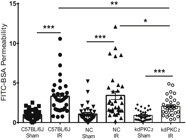
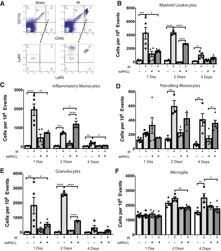
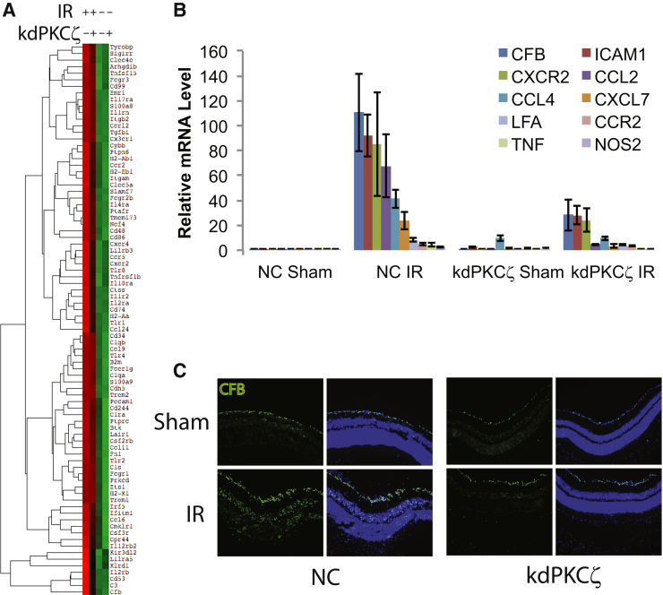
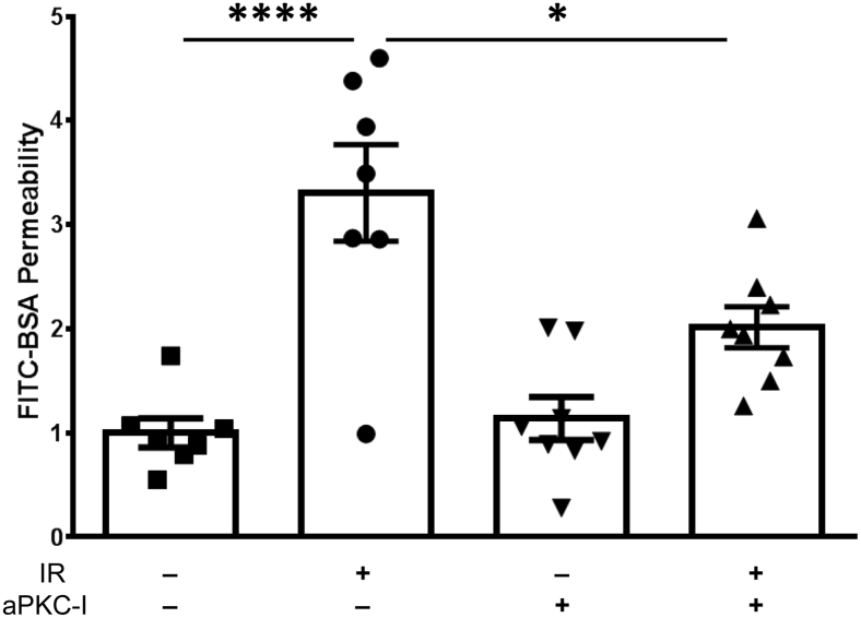
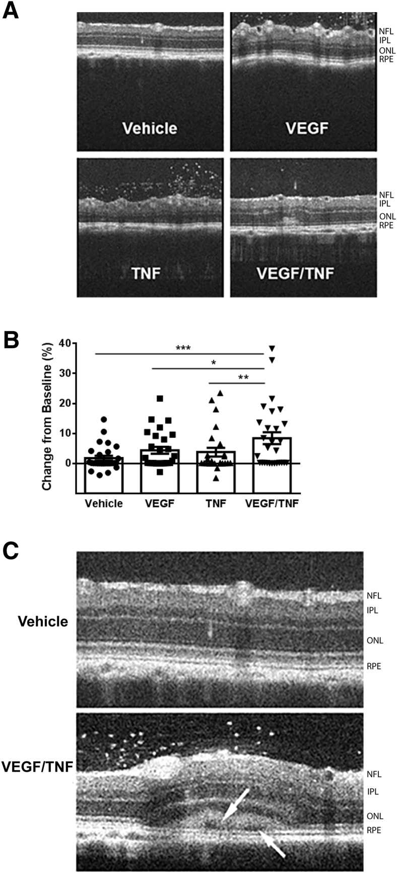
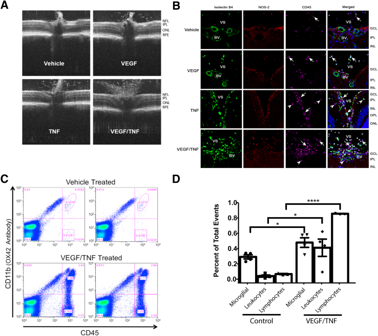
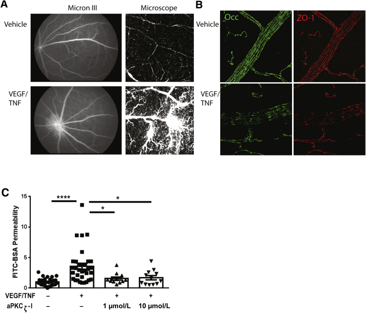
Changes in permeability of retinal blood vessels contribute to macular edema and the pathophysiology of numerous ocular diseases, including diabetic retinopathy, retinal vein occlusions, and macular degeneration. Vascular endothelial growth factor (VEGF) induces retinal permeability and macular thickening in these diseases. However, inflammatory agents, such as tumor necrosis factor-α (TNF-α), also may drive vascular permeability, specifically in patients unresponsive to anti-VEGF therapy. Recent evidence suggests VEGF and TNF-α induce permeability through distinct mechanisms; however, both require the activation of atypical protein kinase C (aPKC). We provide evidence, using genetic mouse models and therapeutic intervention with small molecules, that inhibition of aPKC prevented or reduced vascular permeability in animal models of retinal inflammation. Expression of a kinase-dead aPKC transgene, driven by a vascular and hematopoietic restricted promoter, reduced retinal vascular permeability in an ischemia-reperfusion model of retinal injury. This effect was recapitulated with a small-molecule inhibitor of aPKC. Expression of the kinase-dead aPKC transgene dramatically reduced the expression of inflammatory factors and blocked the attraction of inflammatory monocytes and granulocytes after ischemic injury. Coinjection of VEGF with TNF-α was sufficient to induce permeability, edema, and retinal inflammation, and treatment with an aPKC inhibitor prevented VEGF/TNF-α-induced permeability. These data suggest that aPKC contributes to inflammation-driven retinal vascular pathology and may be an attractive target for therapeutic intervention.


Figures

Figure 1
Figure 1
Expression of kdPKCζ in mouse tissues. A: Plasmid map of Tek-driven kdPKCζ DNA construct with enhancer. B: Expression of kdPKCζ in mouse tissues by PCR; noncarrier (NC) versus kdPKCζ water or primer alone. C: Analysis of kdPKCζ expression in the retina by immunofluorescence. Six-week–old kdPKCζ carrier and NC littermate mice were analyzed for the expression of hemagglutinin (HA) tag (red) along with endothelial marker CD31 (green) and zonula occludens protein 1 (ZO-1; far red) in retinal cross sections. Yellow arrows indicate expression of kdPKCζ in retinal blood vessel. D: Expression of kdPKCζ in the circulating leukocytes of kdPKCζ mice but not NC controls. The product was observed only after cDNA synthesis and not from RNA alone, indicating no genomic contamination. Original magnification, ×630 (C). GCL, ganglion cell layer; INL, inner nuclear layer; IPL, inner plexiform layer; M, marker.
Figure 2
Figure 2
Expression of kdPKCζ blocks retinal permeability induced by ischemia-reperfusion (IR). C57BL/6J, kdPKCζ, and noncarrier (NC) mice were subjected to retinal ischemia for 90 minutes in the experimental eye, followed by natural reperfusion or sham needle punctured in the contralateral eye. At 48 hours after IR, fluorescein isothiocyanate–conjugated bovine serum albumin (FITC-BSA) assay was used to assess retinal vascular permeability. Results are expressed relative to the C57BL/6J-sham. Statistical analysis was performed using analysis of variance with Tukey's post hoc test. Data are expressed as means ± SEM. P < 0.05, ∗∗P < 0.01, and ∗∗∗P < 0.001.
Figure 3
Figure 3
Expression of kdPKCζ reduces ischemia-reperfusion (IR)–induced inflammation. Noncarrier and kdPKCζ mice were subjected to IR injury. At 1, 2, and 4 days after IR injury, retina samples were harvested for flow cytometry to quantify leukocytes: sample scatter gram (A), myeloid leukocytes (B), Ly6C high inflammatory monocytes (C), Ly6C-negative patrolling monocytes (D), granulocytes (E), and microglia (F). Treatment groups were compared within each day by analysis of variance, followed by Sidak's post hoc test. P < 0.05, ∗∗P < 0.01, ∗∗∗P < 0.001, and ∗∗∗∗P < 0.0001.
Figure 4
Figure 4
Expression of kdPKCζ attenuates ischemia-reperfusion (IR)–induced inflammation. Noncarrier (NC) and kdPKCζ mice were subjected to IR injury. At 48 hours after IR, retina samples were harvested for RNA isolation, followed by mRNA quantification by nCounter Analysis System using Immunology v2 kit. A: Heat map represents expression level from low (green) to high (red). B: Quantitative RT-PCR analysis of mRNA profile of various proinflammatory cytokines from a separate IR experiment with sample collection at 48 hours after injury. Every gene shown represents an increase after IR in the NC animals, which was significantly attenuated by expression of kdPKCζ, as determined by analysis of variance and Tukey's post hoc test. Data table and statistics are shown in Supplemental Table S1. C: Immunofluorescence staining of retinal cross section for CFB (green) and nuclei (blue) 48 hours after IR. Original magnification, ×200 (C). CCL, chemokine (C-C motif) ligand; CFB, complement factor B; CXCR, C-X-C chemokine receptor; ICAM, intercellular adhesion molecule; LFA, lymphocyte function-associated antigen; NOS, nitric oxide synthase; TNF, tumor necrosis factor.
Figure 5
Figure 5
aPKC inhibitor blocks ischemia-reperfusion (IR)–induced permeability. Rats were injected intravitreally with vehicle [0.1% bovine serum albumin (BSA) in phosphate-buffered saline] or 1 μmol/L of aPKC inhibitor (aPKC-I) at 30 minutes before IR injury. At 24 hours after IR, animals received another intravitreal injection of vehicle or aPKC-I. Fluorescein isothiocyanate–conjugated (FITC) BSA (100 mg/kg body weight) was injected 30 minutes later and allowed to circulate for 2 hours, and the retinal dye accumulation was determined as a measure of vascular permeability. Results are expressed relative to the vehicle-sham. Statistical analysis was performed using analysis of variance with Tukey's post hoc test. Data are expressed as means ± SEM. P < 0.05, ∗∗∗∗P < 0.0001.
Figure 6
Figure 6
Intravitreal injection of vascular endothelial growth factor (VEGF) and tumor necrosis factor (TNF) increases retinal thickness. Rats were intravitreally injected with vehicle (0.1% bovine serum albumin in phosphate-buffered saline), VEGF (50 ng), TNF (10 ng), or a combination of VEGF/TNF (50/10 ng, respectively). Retinal thickness was assessed with spectral domain–optical coherence tomography after 24 hours. A: Representative annular scan images at 500 μm from the optic nerve head. B: Retinal thickness quantification from the annular scan as the change in retinal thickness 24 hours after injection relative to baseline scan, analyzed by analysis of variance with Tukey's post hoc test. C: Example of VEGF/TNF injection leading to subretinal fluid accumulation and retinal detachment. Arrows indicate cystoid space formation observed near the retinal pigment epithelium (RPE). Data are expressed as means ± SEM (B). P < 0.05, ∗∗P < 0.01, and ∗∗∗P < 0.001. IPL, inner plexiform layer; NFL, nerve fiber layer; ONL, outer nuclear layer.
Figure 7
Figure 7
Vascular endothelial growth factor (VEGF)/tumor necrosis factor (TNF)–induced retinal inflammation. Rats were intravitreally injected with vehicle (0.1% bovine serum albumin in phosphate-buffered saline) or VEGF/TNF (50/ng). A: Optical coherence tomography scan above the optic nerve head reveals increased hyperreflective foci in the vitreous 24 hours after VEGF/TNF coinjection. B: Immunostaining for isolectin B4 (green), NOS2 (red), and CD45 (far red) reveals leukocyte infiltration 24 hours after VEGF/TNF injection. Arrows indicate leukocytes in the vitreous space (VS). Arrowheads indicate infiltrated leukocytes in the retina and the blood vessels (BVs). C: Example of duplicate scatter grams at 24 hours after intravitreal injection of VEGF/TNF; retinal leukocytes were quantified by flow analysis and compared with controls. D: Quantification of flow cytometry of microglia, myeloid leukocytes, and lymphocytes, and analyzed by t-test. P < 0.05, ∗∗∗∗P < 0.0001. Original magnification, ×630 (B). GCL, ganglion cell layer; INL, inner nuclear layer; IPL, inner plexiform layer; NFL, nerve fiber layer; ONL, outer nuclear layer; OPL, outer plexiform layer; RPE, retinal pigment epithelium.
Figure 8
Figure 8
Vascular endothelial growth factor (VEGF)/tumor necrosis factor (TNF) induces retinal permeability and tight junction loss at the cell border. Rats were injected intravitreally with vehicle [0.1% bovine serum albumin (BSA) in phosphate-buffered saline (PBS)] or VEGF/TNF (50/10 ng). A: Fluorescent angiography using Micron III was performed at 5 hours after intravitreal cytokine injection, followed by intravenous injection of fluorescein isothiocyanate–conjugated (FITC) BSA (100 mg/kg body weight) and 10-minute circulation. Generally increased retinal fluorescence, with clear changes at the optic nerve head, is readily observed (Micron III). After angiography, retinas were dissected and flat mounted for microscopic visualization, where vascular leak from capillaries was readily observed (microscope). B: Flat-mount retinas were analyzed for immunoreactivity of occludin (Occ) and zonula occludens protein 1 (ZO-1). Loss of occludin and ZO-1 immunostaining at endothelial cell borders was observed in the VEGF/TNF-treated retinas. C: aPKCζ inhibitor (aPKCζ-I) blocks VEGF/TNF-induced permeability. Rats were intravitreally injected with vehicle (0.1% BSA in PBS), VEGF/TNF (50/10 ng), or indicated dose of aPKCζ-I in combination with VEGF/TNF. At 3 hours after intravitreal injection, animals received an i.v. injection of FITC-BSA (100 mg/kg body weight). Two hours later, animals were perfused with warm saline and retinas were removed for quantification of FITC-BSA accumulation. Results are expressed relative to vehicle control. Differences between groups were analyzed by analysis of variance with Tukey's post hoc test. Data are expressed as means ± SEM (C). P < 0.05, ∗∗∗∗P < 0.0001. Original magnification, ×630 (A, right column, and B).

Comment in

References

    1. Campochiaro P.A. Anti-vascular endothelial growth factor treatment for retinal vein occlusions. Ophthalmologica. 2012;227 Suppl 1:30–35. - PubMed
    1. Emerson M.V., Lauer A.K. Emerging therapies for the treatment of neovascular age-related macular degeneration and diabetic macular edema. BioDrugs. 2007;21:245–257. - PubMed
    1. Markomichelakis N.N., Theodossiadis P.G., Pantelia E., Papaefthimiou S., Theodossiadis G.P., Sfikakis P.P. Infliximab for chronic cystoid macular edema associated with uveitis. Am J Ophthalmol. 2004;138:648–650. - PubMed
    1. Antonetti D.A., Klein R., Gardner T.W. Diabetic retinopathy. N Engl J Med. 2012;366:1227–1239. - PubMed
    1. Johnson M.W. Etiology and treatment of macular edema. Am J Ophthalmol. 2009;147:11–21.e1. - PubMed

Publication types

MeSH terms

Substances

LinkOut - more resources