

Phosphotyrosine profiling of human cerebrospinal fluid
- PMID: 30220890
- PMCID: PMC6136184
- DOI: 10.1186/s12014-018-9205-1
Phosphotyrosine profiling of human cerebrospinal fluid
Abstract
Background: Cerebrospinal fluid (CSF) is an important source of potential biomarkers that affect the brain. Biomarkers for neurodegenerative disorders are needed to assist in diagnosis, monitoring disease progression and evaluating efficacy of therapies. Recent studies have demonstrated the involvement of tyrosine kinases in neuronal cell death. Thus, neurodegeneration in the brain is related to altered tyrosine phosphorylation of proteins in the brain and identification of abnormally phosphorylated tyrosine peptides in CSF has the potential to ascertain candidate biomarkers for neurodegenerative disorders.
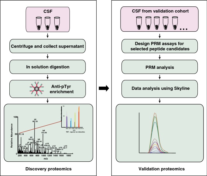
Methods: In this study, we used an antibody-based tyrosine phosphopeptide enrichment method coupled with high resolution Orbitrap Fusion Tribrid Lumos Fourier transform mass spectrometer to catalog tyrosine phosphorylated peptides from cerebrospinal fluid. The subset of identified tyrosine phosphorylated peptides was also validated using parallel reaction monitoring (PRM)-based targeted approach.
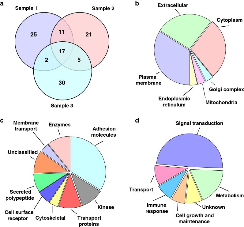
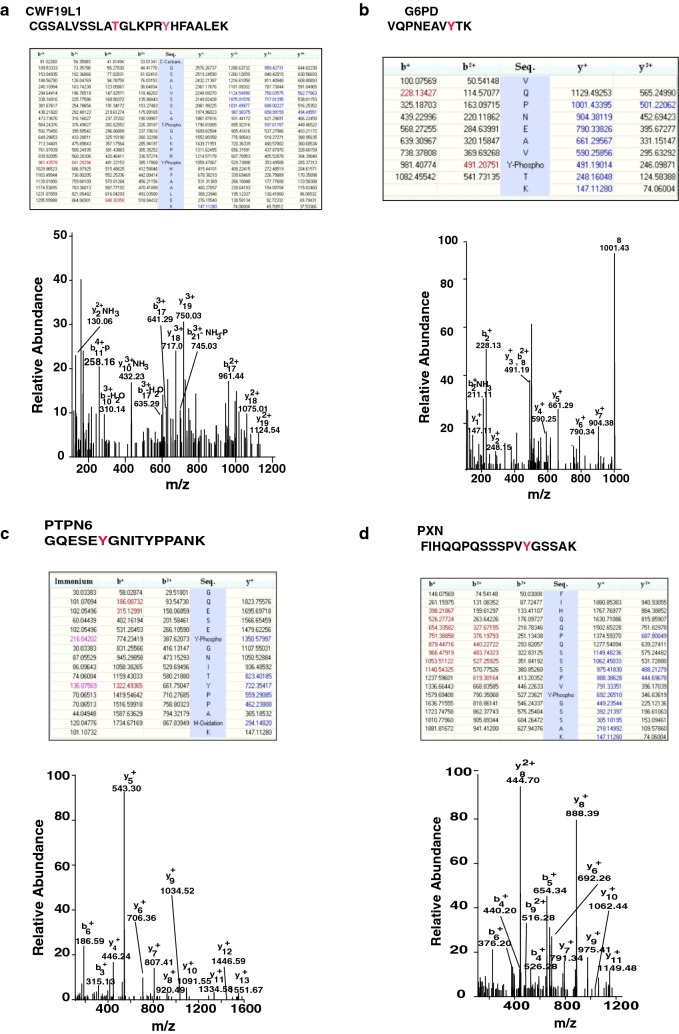
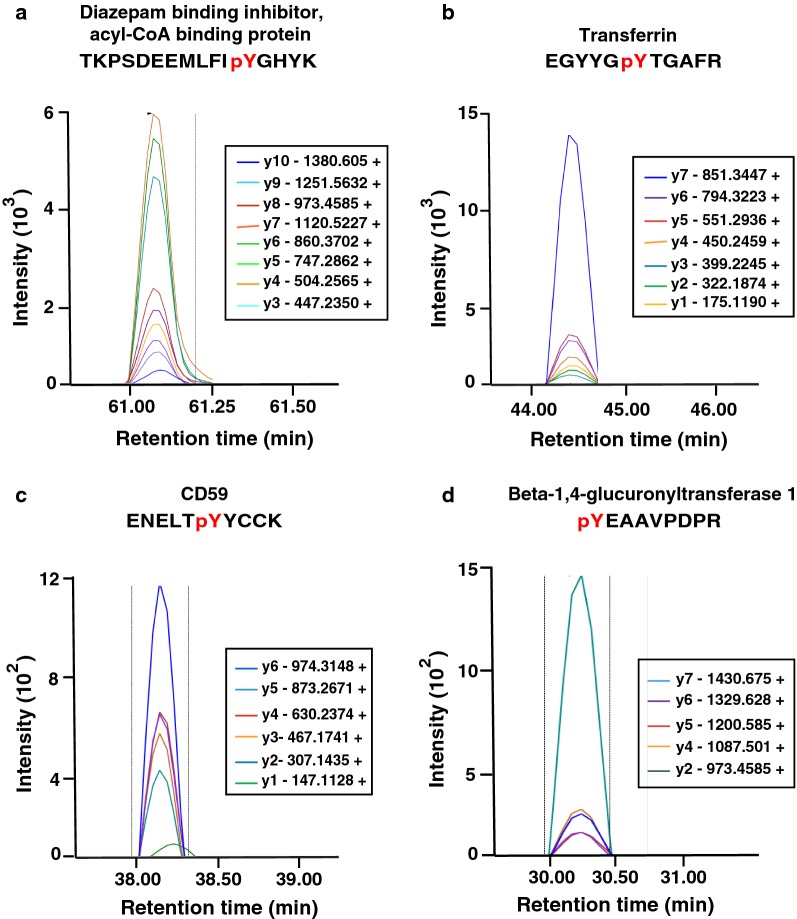
Results: To date, there are no published studies on global profiling of phosphotyrosine modifications of CSF proteins. We carried out phosphotyrosine profiling of CSF using an anti-phosphotyrosine antibody-based enrichment and analysis using high resolution Orbitrap Fusion Lumos mass spectrometer. We identified 111 phosphotyrosine peptides mapping to 66 proteins, which included 24 proteins which have not been identified in CSF previously. We then validated a set of 5 tyrosine phosphorylated peptides in an independent set of CSF samples from cognitively normal subjects, using a PRM-based targeted approach.
Conclusions: The findings from this deep phosphotyrosine profiling of CSF samples have the potential to identify novel disease-related phosphotyrosine-containing peptides in CSF.
Keywords: CSF; Phosphotyrosine; Proteome.
Figures
Similar articles
-
Development of a novel method for the quantification of tyrosine 39 phosphorylated α- and β-synuclein in human cerebrospinal fluid.Clin Proteomics. 2020 May 4;17:13. doi: 10.1186/s12014-020-09277-8. eCollection 2020. Clin Proteomics. 2020. PMID: 32390785 Free PMC article.
-
Distinguishing Sulfotyrosine Containing Peptides from their Phosphotyrosine Counterparts Using Mass Spectrometry.J Am Soc Mass Spectrom. 2018 Mar;29(3):455-462. doi: 10.1007/s13361-017-1854-1. Epub 2018 Jan 8. J Am Soc Mass Spectrom. 2018. PMID: 29313205
-
Proteomic analysis of cerebrospinal fluid extracellular vesicles: a comprehensive dataset.J Proteomics. 2014 Jun 25;106:191-204. doi: 10.1016/j.jprot.2014.04.028. Epub 2014 Apr 24. J Proteomics. 2014. PMID: 24769233
-
Biological confounders for the values of cerebrospinal fluid proteins in Parkinson's disease and related disorders.J Neurochem. 2016 Oct;139 Suppl 1:290-317. doi: 10.1111/jnc.13390. Epub 2016 Feb 10. J Neurochem. 2016. PMID: 26452984 Review.
-
Profiling the global tyrosine phosphorylation state.Mol Cell Proteomics. 2003 Apr;2(4):215-33. doi: 10.1074/mcp.R300002-MCP200. Epub 2003 May 17. Mol Cell Proteomics. 2003. PMID: 12754303 Review.
Cited by
-
Quantitative proteomic analysis of the frontal cortex in Alzheimer's disease.J Neurochem. 2021 Mar;156(6):988-1002. doi: 10.1111/jnc.15116. Epub 2020 Jul 22. J Neurochem. 2021. PMID: 32614981 Free PMC article.
-
Metagenomic next-generation sequencing and proteomics analysis in pediatric viral encephalitis and meningitis.Front Cell Infect Microbiol. 2023 Apr 21;13:1104858. doi: 10.3389/fcimb.2023.1104858. eCollection 2023. Front Cell Infect Microbiol. 2023. PMID: 37153144 Free PMC article.
-
FAM20C Overview: Classic and Novel Targets, Pathogenic Variants and Raine Syndrome Phenotypes.Int J Mol Sci. 2021 Jul 27;22(15):8039. doi: 10.3390/ijms22158039. Int J Mol Sci. 2021. PMID: 34360805 Free PMC article. Review.
-
Quantitative Analysis of Tyrosine Phosphorylation from FFPE Tissues Reveals Patient-Specific Signaling Networks.Cancer Res. 2021 Jul 15;81(14):3930-3941. doi: 10.1158/0008-5472.CAN-21-0214. Epub 2021 May 20. Cancer Res. 2021. PMID: 34016623 Free PMC article.
-
Revealing Dynamics of Protein Phosphorylation: A Study on the Cashmere Fineness Disparities in Liaoning Cashmere Goats.Mol Biotechnol. 2025 Jul;67(7):2832-2845. doi: 10.1007/s12033-024-01244-0. Epub 2024 Aug 8. Mol Biotechnol. 2025. PMID: 39117978
References
Grants and funding
LinkOut - more resources
Full Text Sources
Other Literature Sources
Medical