

The First Report of Small Cell Cancer of the Uvula Presenting With Ectopic Adrenocorticotropic Hormone Syndrome
- PMID: 30220949
- PMCID: PMC6134991
- DOI: 10.14740/wjon1130w
The First Report of Small Cell Cancer of the Uvula Presenting With Ectopic Adrenocorticotropic Hormone Syndrome
Abstract
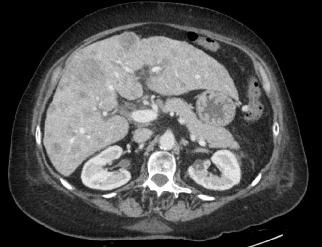
Small cell carcinoma (SmCC) of the head and neck is a rare occurrence. We herein present a case of a 62-year-old female who was diagnosed with small cell cancer of the uvula. The patient developed increased body swelling, elevated blood pressure, persistent hypokalemia and new onset diabetes mellitus. Further workup confirmed a diagnosis of Cushing's syndrome secondary to ectopic adrenocorticotropic hormone (ACTH) production. To our knowledge, this is only the second case of SmCC of the uvula described in literature, and the first associated with any paraneoplastic syndrome. By reporting this case, we aim to characterize the tumor clinical course and highlight the aggressive nature of its growth.
Keywords: ACTH; Ectopic; Small cell cancer; Uvula.
Figures


Similar articles
-
Ectopic Cushing's Syndrome as the First Presenting Sign of Small Cell Lung Carcinoma.J Brown Hosp Med. 2023 Jul 2;2(3):77572. doi: 10.56305/001c.77572. eCollection 2023. J Brown Hosp Med. 2023. PMID: 40026469 Free PMC article.
-
Ectopic production of ACTH and corticotropin-releasing hormone (CRH).J Steroid Biochem Mol Biol. 1992 Oct;43(5):403-8. doi: 10.1016/0960-0760(92)90076-u. J Steroid Biochem Mol Biol. 1992. PMID: 1327073 Review.
-
An Unlikely Cause of Hypokalemia.J Emerg Med. 2017 May;52(5):e187-e191. doi: 10.1016/j.jemermed.2016.12.011. Epub 2017 Jan 28. J Emerg Med. 2017. PMID: 28139270
-
Association of hypertension and hypokalemia with Cushing's syndrome caused by ectopic ACTH secretion: a series of 58 cases.Ann N Y Acad Sci. 2002 Sep;970:134-44. doi: 10.1111/j.1749-6632.2002.tb04419.x. Ann N Y Acad Sci. 2002. PMID: 12381548 Review.
-
A case of recurrent non-small-cell lung carcinoma and paraneoplastic Cushing's syndrome.Lung Cancer. 2006 Feb;51(2):251-5. doi: 10.1016/j.lungcan.2005.08.015. Epub 2005 Dec 13. Lung Cancer. 2006. PMID: 16352372
References
Publication types
LinkOut - more resources
Full Text Sources
Other Literature Sources
Research Materials