

Review
doi: 10.1038/s41392-018-0021-x.
eCollection 2018.
γ-tubulin as a signal-transducing molecule and meshwork with therapeutic potential
Affiliations
- PMID: 30221013
- PMCID: PMC6137058
- DOI: 10.1038/s41392-018-0021-x
Item in Clipboard
Review
γ-tubulin as a signal-transducing molecule and meshwork with therapeutic potential
Signal Transduct Target Ther.
.
Abstract
Knowledge of γ-tubulin is increasing with regard to the cellular functions of this protein beyond its participation in microtubule nucleation. γ-Tubulin expression is altered in various malignancies, and changes in the TUBG1 gene have been found in patients suffering from brain malformations. This review recapitulates the known functions of γ-tubulin in cellular homeostasis and discusses the possible influence of the protein on disease development and cancer.
Conflict of interest statement
The author declares no competing interests.
Figures
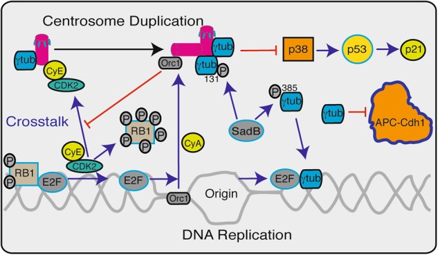
Schematic representation of the different ways in which γ-tubulin (γtub) and the centrosomes control the G1-to-S transition and S phase progression. The blue and black arrows indicate centrosome activation and duplication, respectively, and the red lines indicate inhibition. At the G1–S transition, SadB mediates the phosphorylation of γ-tubulin on Ser131 and Ser385, and this action regulates the recruitment of γ-tubulin to the growing centrosome and leads to the nuclear accumulation of γ-tubulin. The centrosomes inhibit the activation of the p38 mitogen-activated protein kinase (p38)-p53-p21 signal pathway. The centrosomal localization of cyclin E (CyE)–cyclin-dependent kinase (Cdk2) is required for the initiation of DNA replication. Once DNA replication is initiated, the origin replication complex subunit 1 (Orc1) translocates from the origin of replication to the centriole in a cyclin A (CyA)-dependent manner, where it prevents the CyE-dependent reduplication of the centrosomes. To progress through the S phase, the CyE–Cdk2 complex phosphorylates the protein retinoblastoma 1 (RB1), and this initiates the transcriptional activities of E2 promoter binding factors (E2Fs). The activities of the E2Fs trigger the initiation of centrosome duplication and DNA replication, and the accumulation of γ-tubulin in the nuclear compartment turns off the transcriptional activities of E2Fs. Additionally, γ-tubulin inactivates the anaphase-promoting complex/cyclosomeCdh1 at the G1-to-S transition
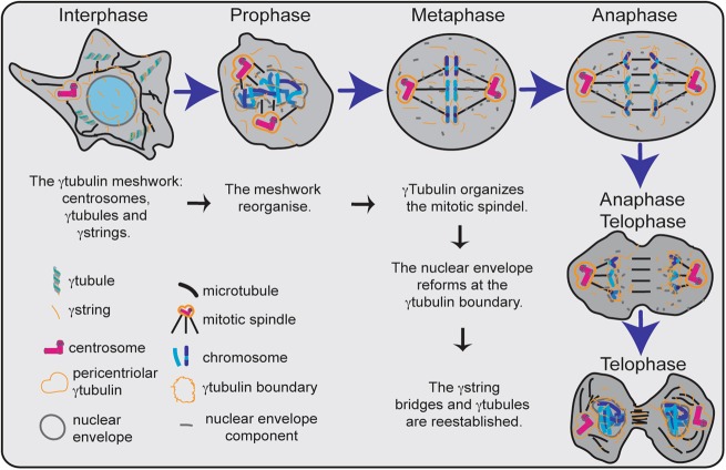
Hypothetical representation of the functions performed by the γ-tubulin meshwork during interphase and mitosis. In interphase, the meshwork is composed of centrosomes, γ-tubules, and γ-strings. The nuclear and the cytosolic pool of γ-tubulin are connected with γ-strings across the nuclear envelope. γ-tubules regulate the concentration of the cytosolic pool of γ-tubulin. The γ-tubulin meshwork changes in a cell-cycle-dependent manner. In prophase, the nuclear envelope is dispersed. γ-tubulin accumulates in the pericentriolar region of the centrosomes and assists in formation of the mitotic spindle. The number of γ-tubules is reduced in mitotic cells (prophase, metaphase, anaphase, and telophase). During mitosis, the centrosomes coordinate the segregation of chromatids between the newborn cells through orientation and organization of the mitotic spindle. The dispersed components of the nuclear envelope localize to the mitotic spindle and cell periphery. In anaphase/telophase, the components of the nuclear envelope nucleate at the γ-tubulin boundary. At the end of mitosis, the nuclear envelope is formed, and the γ-string bridges are reestablished
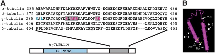
The C terminus of γ-tubulin contains the DNA-binding helix-loop-helix motif. a Sequence alignment of the C-terminal helix (H11)-loop-helix (H12) region of human γ-tubulin 1 (residues 385–436; corresponding to helix numbers 11 and 12 in the γ-tubulin protein) and α- (residues 385–436), β- (residues 375–426), ε- (residues 406–455), and δ-tubulin (residues 404–451). Bold letters indicate identical residues. The bipartite nuclear localization signal (NLS) of γ-tubulin is highlighted in gray, and the magenta letters represent residues included in the NLS. Ser385 and Leu387 in γ-tubulin are labeled in blue. b The known three-dimensional structure of the C-terminal helix-loop-helix region of human γ-tubulin revealed with the three-dimensional structure viewer Cn3D. In the structure, the SadB putative phosphorylation sites at Ser385 and Leu387 are depicted in yellow. The phosphorylation of Ser385 leads to accumulation of γ-tubulin in the nuclear compartment. Mutations in Leu387 have been found in patients suffering from lissencephaly and microcephaly
References
Publication types
LinkOut - more resources
Full Text Sources
Other Literature Sources
