

Retrosplenial cortex and its role in spatial cognition
- PMID: 30221204
- PMCID: PMC6095108
- DOI: 10.1177/2398212818757098
Retrosplenial cortex and its role in spatial cognition
Abstract
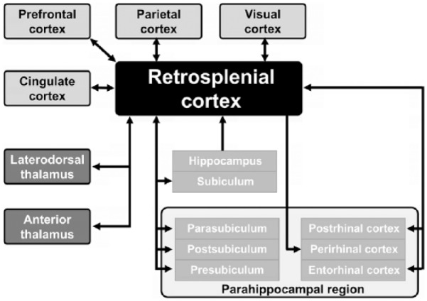
Retrosplenial cortex is a region within the posterior neocortical system, heavily interconnected with an array of brain networks, both cortical and subcortical, that is, engaged by a myriad of cognitive tasks. Although there is no consensus as to its precise function, evidence from both human and animal studies clearly points to a role in spatial cognition. However, the spatial processing impairments that follow retrosplenial cortex damage are not straightforward to characterise, leading to difficulties in defining the exact nature of its role. In this article, we review this literature and classify the types of ideas that have been put forward into three broad, somewhat overlapping classes: (1) learning of landmark location, stability and permanence; (2) integration between spatial reference frames; and (3) consolidation and retrieval of spatial knowledge (schemas). We evaluate these models and suggest ways to test them, before briefly discussing whether the spatial function may be a subset of a more general function in episodic memory.
Keywords: Learning; cingulate cortex; default mode network; electrophysiology; hippocampal formation; immediate-early genes; memory; neuroimaging; primate; thalamus.
Conflict of interest statement
Declaration of conflicting interests: The author(s) declared no potential conflicts of interest with respect to the research, authorship and/or publication of this article.
Figures
Similar articles
-
Cues, context, and long-term memory: the role of the retrosplenial cortex in spatial cognition.Front Hum Neurosci. 2014 Aug 5;8:586. doi: 10.3389/fnhum.2014.00586. eCollection 2014. Front Hum Neurosci. 2014. PMID: 25140141 Free PMC article. Review.
-
A Corticocortical Circuit Directly Links Retrosplenial Cortex to M2 in the Mouse.J Neurosci. 2016 Sep 7;36(36):9365-74. doi: 10.1523/JNEUROSCI.1099-16.2016. J Neurosci. 2016. PMID: 27605612 Free PMC article.
-
Representation of visual landmarks in retrosplenial cortex.Elife. 2020 Mar 10;9:e51458. doi: 10.7554/eLife.51458. Elife. 2020. PMID: 32154781 Free PMC article.
-
The role of the medial prefrontal cortex in cognition, ageing and dementia.Brain Commun. 2021 Jun 11;3(3):fcab125. doi: 10.1093/braincomms/fcab125. eCollection 2021 Jul. Brain Commun. 2021. PMID: 34222873 Free PMC article. Review.
-
Retrosplenial Cortex Indexes Stability beyond the Spatial Domain.J Neurosci. 2018 Feb 7;38(6):1472-1481. doi: 10.1523/JNEUROSCI.2602-17.2017. Epub 2018 Jan 8. J Neurosci. 2018. PMID: 29311139 Free PMC article.
Cited by
-
Region- and neuronal-subtype-specific expression of Na,K-ATPase alpha and beta subunit isoforms in the mouse brain.J Comp Neurol. 2020 Nov 1;528(16):2654-2678. doi: 10.1002/cne.24924. Epub 2020 Apr 28. J Comp Neurol. 2020. PMID: 32301109 Free PMC article.
-
FOS mapping reveals two complementary circuits for spatial navigation in mouse.Sci Rep. 2024 Sep 11;14(1):21252. doi: 10.1038/s41598-024-72272-8. Sci Rep. 2024. PMID: 39261637 Free PMC article.
-
Neonatal ethanol exposure triggers apoptosis in the murine retrosplenial cortex: Role of inhibition of NMDA receptor-driven action potential firing.Neuropharmacology. 2020 Jan 1;162:107837. doi: 10.1016/j.neuropharm.2019.107837. Epub 2019 Nov 2. Neuropharmacology. 2020. PMID: 31689422 Free PMC article.
-
Retrosplenial cortex in spatial memory: focus on immediate early genes mapping.Mol Brain. 2021 Dec 4;14(1):172. doi: 10.1186/s13041-021-00880-w. Mol Brain. 2021. PMID: 34863215 Free PMC article. Review.
-
Context value updating and multidimensional neuronal encoding in the retrosplenial cortex.Nat Commun. 2021 Oct 18;12(1):6045. doi: 10.1038/s41467-021-26301-z. Nat Commun. 2021. PMID: 34663792 Free PMC article.
References
-
- Aggleton JP, Hunt PR, Rawlins JN. (1986) The effects of hippocampal lesions upon spatial and non-spatial tests of working memory. Behavioural Brain Research 19(2): 133–146. - PubMed
-
- Aggleton JP, Hunt PR, Nagle S, et al. (1996) The effects of selective lesions within the anterior thalamic nuclei on spatial memory in the rat. Behavioural Brain Research 81(1–2): 189–198. - PubMed
-
- Alexander AS, Nitz DA. (2015) Retrosplenial cortex maps the conjunction of internal and external spaces. Nature Neuroscience 18(8): 1143–1151. - PubMed
-
- Alexander AS, Nitz DA. (2017) Spatially periodic activation patterns of retrosplenial cortex encode route sub-spaces and distance travelled. Current Biology 27(11): 1551–1560. - PubMed
Publication types
Associated data
Grants and funding
LinkOut - more resources
Full Text Sources
Other Literature Sources
