. 2018 Jul 31;3(7):7663-7672.
doi: 10.1021/acsomega.8b00785. Epub 2018 Jul 11.

Controlled Dye Aggregation in Sodium Dodecylsulfate-Stabilized Poly(methylmethacrylate) Nanoparticles as Fluorescent Imaging Probes

Affiliations

Controlled Dye Aggregation in Sodium Dodecylsulfate-Stabilized Poly(methylmethacrylate) Nanoparticles as Fluorescent Imaging Probes

Samarth Bhargava et al. ACS Omega. .

Abstract

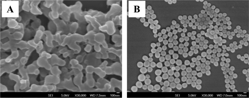
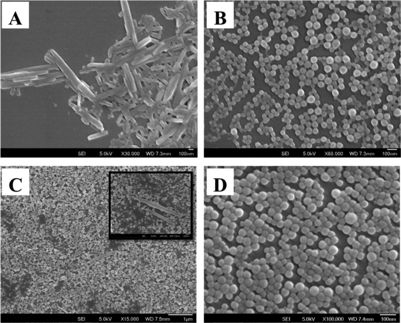
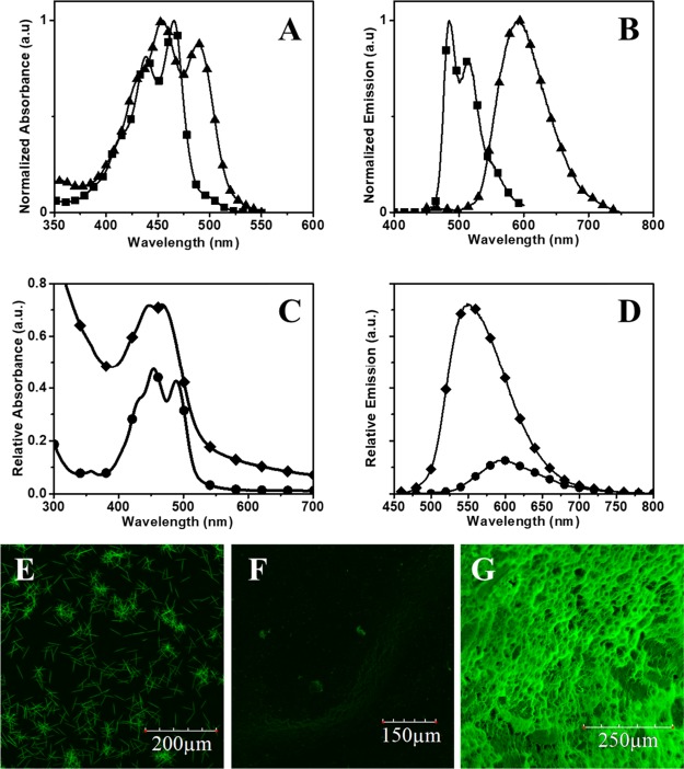
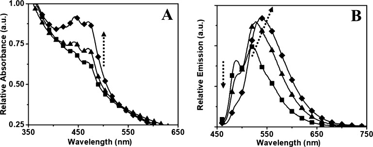
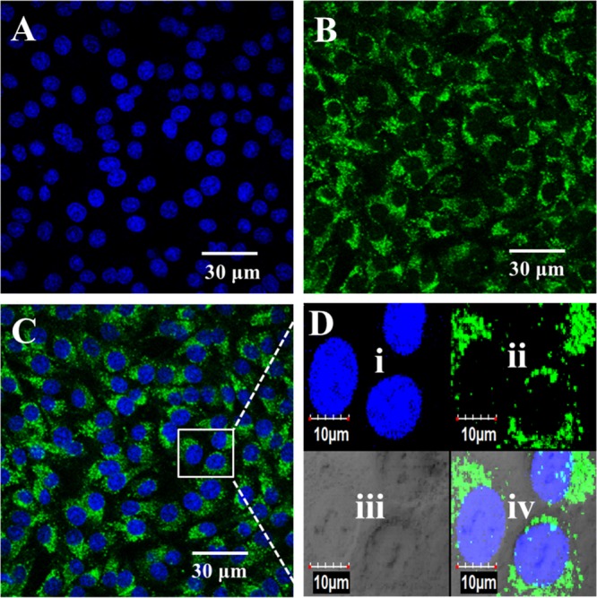
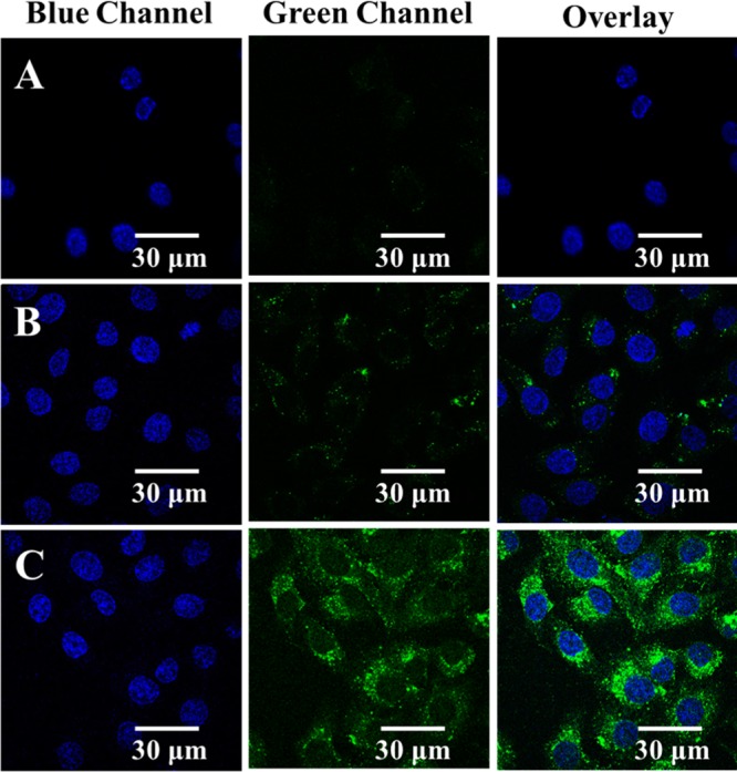
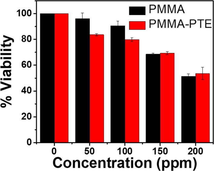
Polymer nanoparticles are used extensively in biomedical applications. Poly(methylmethacrylate) (PMMA) nanoparticles obtained via nanoprecipitation were unstable and flocculate or precipitate from solution within a few hours. A simple method to improve the stability of the particles using surfactants at low concentrations was carried out to produce PMMA nanoparticles with long-term stability in water (>6 months). The increased stability was attributed to the incorporation of surfactants inside the polymer particles during nanoprecipitation. The same methodology was also adopted to encapsulate a highly fluorescent hydrophobic perylene tetraester inside the polymer nanoparticles with good stability in water. Because of the presence of the anionic sodium dodecyl sulfate, the particles showed a negative zeta potential of -34.7 mV and an average size of 150 nm. Similarly, the dye-encapsulated polymer nanoparticles showed a zeta potential of -35.1 mV and an average particle size of 180 nm. By varying the concentration of encapsulated dyes inside the polymer nanoparticles, dye aggregation could be controlled, and the fluorescence profiles of the nanoparticles were altered. To understand the uptake and toxicity of the polymer nanoparticles, baby hamster kidney cells were chosen as a model system. The polymer nanoparticles were taken up by the cells within 3 h and were nontoxic at concentrations as high as 100 ppm. The confocal micrographs of the cells revealed localized fluorescence from the polymer nanoparticles around the nucleus in the cytoplasm without the penetration of the nuclear envelope.


Conflict of interest statement

The authors declare no competing financial interest.

Figures

Figure 1
Figure 1
SEM image of a polymer solution precipitated from water via slow addition (A) and fast addition (B); 30 000× magnification; scale bar is 100 nm.
Figure 2
Figure 2
SEM micrographs of pure PTE (A), PMMA (B) nanomaterials prepared separately using the nanoprecipitation method. Mixture of PTE and PMMA nanomaterials prepared by adding the two solutions separately (C) with a magnified section of the sample (inset) and coprecipitated PMMA–PTE particles (D) from a previously mixed mixture.
Figure 3
Figure 3
Absorption (A) and emission (B) spectra of the PTE solution (-■-; 12.5 μM in acetone) and particles (-▲-; effective PTE concentration of 12.5 μM) in the absence of polymer matrix. Absorption (C) and emission (D) spectra of the nanoprecipitated pure PTE (-●-; 12.5 μM) and PMMA–PTE (-⧫-; effective PTE concentration of 12.5 μM) particles in aqueous solutions under normalized conditions. Confocal images of PTE crystals (E) obtained from the acetone solution through evaporation; nanoprecipitated PTE (F) and PMMA–PTE (G) nanoparticles.
Figure 4
Figure 4
Absorption (A) and emission (B) spectra of the PMMA–PTE nanoparticles with (-■-) 0.5 wt %, (-▲-) 1 wt %, and (-⧫-) 2 wt % PTE loading (the effective concentration ranging from 3 to 13 μM). The arrows indicate the effects of increasing PTE dye concentration encapsulated inside the particle.
Figure 5
Figure 5
Absorption (A) and emission (B) spectra of high-loading PMMA–PTE nanoparticles with PTE loading varied from (-⧫-) 5 wt %, (-▼-) 10 wt %, (-▲-) 17.5 wt %, and (-●-) 25 wt % (effective PTE concentration ranging from 33 to 165 μM in the final nanoprecipitated solution). All fluorescence measurements were measured with an excitation at 450 nm, path length of 1 cm, and concentration of 400 ppm. The fluorescence of diluted nanoparticle solutions (20 ppm) is shown in the Supporting Information (Figure S4). The arrows indicate the effects of increasing the concentration of the PTE dye.
Figure 6
Figure 6
Interaction of BHK cells with PMMA–PTE nanoparticles at a concentration of 25 ppm. The images represent the nucleus stained with DAPI (A), cellular matrix stained with nanoparticles (B), and an overlay of the two images (C), with the magnified images in the inset showing nanoparticle distribution inside the cells (D). Magnified view (D) of nucleus stained with DAPI (i), cellular matrix stained with nanoparticles (ii), cells observed under DIC mode (iii), and overlay of (i)–(iv). The cells were imaged at a magnification of 60× oil objective using a confocal microscope (Olympus FV1000).
Figure 7
Figure 7
Time-based tracking of nanoparticles (25 ppm) inside the cells. From left to right column: DAPI stain in blue channel, nanoparticle stain in green channel, and the overlay of blue and green channels. Images observed after the polymer particle exposure of the cells for 1 (A), 3 (B), and 6 h (C) shown to compare the relative brightness. All images were recorded at 100× using a confocal microscope (Olympus FV1000).
Figure 8
Figure 8
AlamarBlue assay for the determination of the viability of cells exposed to blank (PMMA) nanoparticles and PMMA–PTE nanoparticles.

References

    1. Zhang C.; Pansare V. J.; Prud’homme R. K.; Priestley R. D. Flash nanoprecipitation of polystyrenenanoparticles. Soft Matter 2012, 8, 86–93. 10.1039/c1sm06182h. - DOI
    1. Geissler A.; Biesalski M.; Heinze T.; Zhang K. Formation of nanostructured cellulose stearoyl esters via nanoprecipitation. J. Mater. Chem. A 2014, 2, 1107–1116. 10.1039/c3ta13937a. - DOI
    1. Liu Y.; Lu Y. C.; Luo G. S. Modified nanoprecipitation method for polysulfone nanoparticles preparation. Soft Matter 2014, 10, 3414–3420. 10.1039/c3sm53003e. - DOI - PubMed
    1. Miao M.; Chen Q.; Zhang C.; Cao X.; Zhou W.; Qiu Q.; An Z. Nanoprecipitation of PMMA Stabilized by Core Cross-Linked Star Polymers. Macromol. Chem. Phys. 2013, 214, 1158–1164. 10.1002/macp.201300234. - DOI
    1. Morales-Cruz M.; Flores-Fernández G. M.; Morales-Cruz M.; Orellano E. A.; Rodriguez-Martinez J. A.; Ruiz M.; Griebenow K. Two-step nanoprecipitation for the production of protein-loaded PLGA nanospheres. Results Pharma Sci. 2012, 2, 79–85. 10.1016/j.rinphs.2012.11.001. - DOI - PMC - PubMed

LinkOut - more resources