. 2017;2(3):1030.
Epub 2017 Mar 21.

Diet Modulates Adipose Tissue Oxidative Stress in a Murine Acute Chagas Model

Affiliations

Diet Modulates Adipose Tissue Oxidative Stress in a Murine Acute Chagas Model

Janeesh Plakkal Ayyappan et al. JSM Atheroscler. 2017.

Abstract

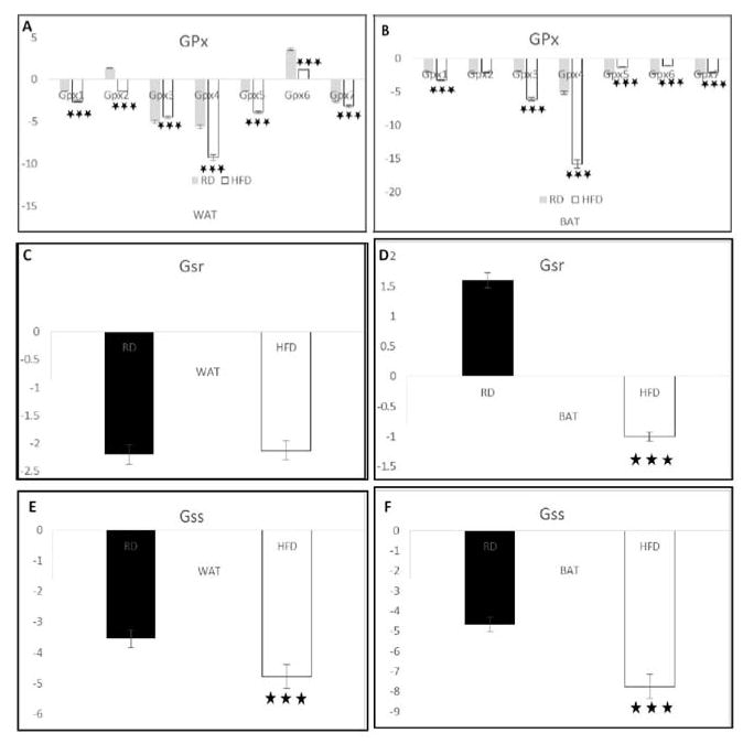
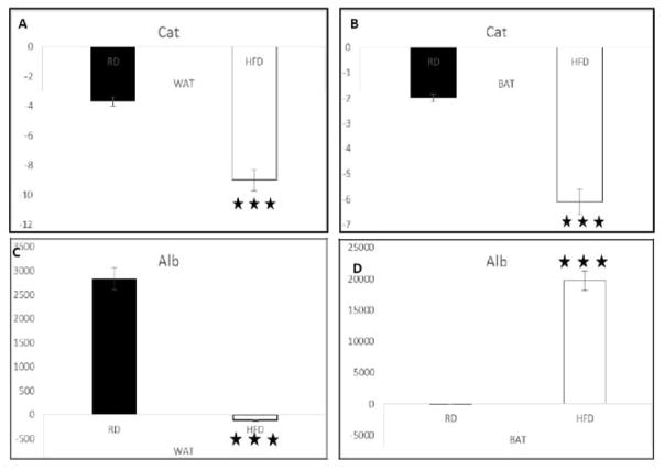
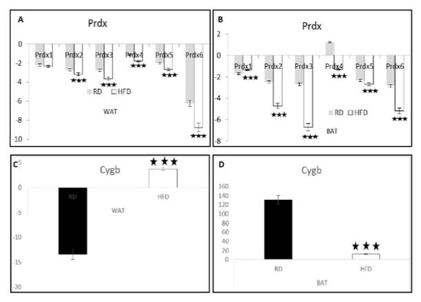
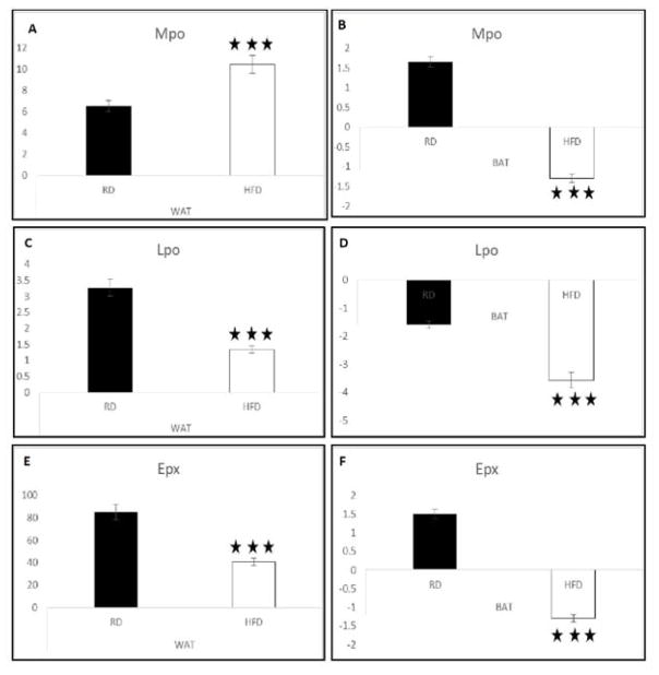
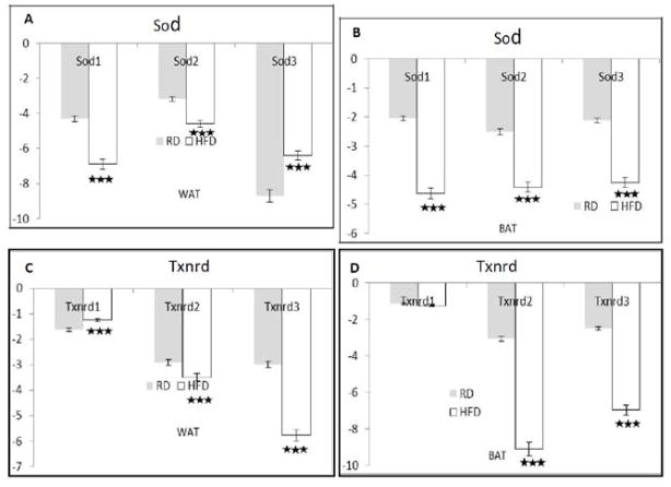
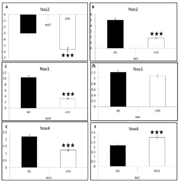
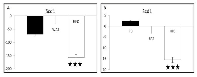
Chagas disease, also known as American trypanosomiasis, is a tropical parasitic disease caused by the protozoan Trypanosoma cruzi. T. cruzi targets adipose tissue, which serves as a reservoir of this parasite. T. cruzi infection of adipose tissue is characterized by increased lipolysis, oxidative stress, and parasitemia. High fat diet (HFD) decreases lipolysis and increases the survival rate in the mice infected with T. cruzi during acute infection. However, the effect of HFD on oxidative stress in adipose tissue has not been examined in detail. In the present study we evaluated the effect of HFD on oxidative stress markers in both white and brown adipose tissues (WAT and BAT) during acute infection. We used qPCR to examine the mRNA expression levels of genes involved in several antioxidant defence systems, such as those acting in ROS metabolism, peroxidases, and relevant oxygen transporter genes. The result of our study showed that HFD regulates the expression levels of oxidative stress genes in adipose tissues and that these effects are often different in WAT and BAT. For instance, while HFD down-regulated the levels of most antioxidant genes in both WAT and BAT, it differentially affected the expression pattern of genes involved in ROS metabolism (e.g. peroxidases) in WAT and BAT tissues of infected mice. Together with our previous studies, these findings show that infection and diet both regulate antioxidant enzymes and other oxidative stress defenses in mouse adipose tissues during acute T. cruzi infection.

Keywords: Adipose tissue; Antioxidant system; High fat diet; Oxidative stress.


Figures

Figure 1
Figure 1. T. cruzi infection and HFD synergistically alter glutathione signalling system in different adipose tissues
The qPCR analysis data suggest that infection reduces mRNA level of genes involved in glutathione system and HFD further decreases the activation of glutathione antioxidant system in the adipose tissues during acute T. cruzi infection. P ≤ 0.005, represented by black stars. Bars represent mean values of the data with Standard Error of the mean (SEM) as vertical lines. RD - regular diet; HFD-high fat diet.
Figure 2
Figure 2. Diet regulates mRNA levels of catalase and albumin in the adipose tissues of T. cruzi infected CD1 mice
qPCR analysis demonstrated that HFD significantly reduces the mRNA levels of catalase in both the WAT (Figure 2A) and BAT (Figure 2B) compared to RD in acute T. cruzi infection. HFD drastically decreases the mRNA levels of Alb in WAT (Figure 2C) and increases in BAT (Figure 2D) compared to RD fed infected mice. P ≤ 0.005, represented by black stars. Bars represent mean values of the data with Standard Error of the mean (SEM) as vertical lines. RD - regular diet; HFD-high fat diet.
Figure 3
Figure 3. Alteration in the mRNA levels of genes involved in peroxiredoxins and cytoglobin levels in the adipose tissues during acute T. cruzi infection
Study at mRNA level by qPCR analysis demonstrated a significant down regulation of various isoforms of Prdx (Prdx 2, 3, 4, 5, & 6) mRNA levels in both the WAT (Figure 3A) and BAT (Figure 3B) of infected mice fed on a HFD compared to RD, except Prdx1. HFD increases CygB mRNA levels to 10 fold in the WAT and decreases to 100 fold in the BAT compared to RD fed infected mice (Figure 3C and 3D). Bars represent mean values of the data with Standard Error of the mean (SEM) as vertical lines. RD - regular diet; HFD-high fat diet.
Figure 4
Figure 4. HFD changes peroxidase levels in the adipose tissue of T. cruzi infected mice
qPCR analysis of MPO gene showed that it was significantly increased (4-fold) in the WAT and significantly decreased (2.5-fold) in the BAT of infected mice fed on a HFD compared to RD (Figure 4A,4B). HFD significantly down regulated the expression of Lpo in both the WAT (Figure 4C) and BAT (Figure 4D) during acute infection in mice compared to feeding with RD. Epx significantly decreased in both the WAT (Figure 4E) and BAT (Figure 4F) (40- and 3-fold, respectively) of the T. cruzi infected mice fed on a HFD compared to RD. Bars represent mean values of the data with Standard Error of the mean (SEM) as vertical lines. RD - regular diet; HFD-high fat diet.
Figure 5
Figure 5. Modulation of superoxide and thioredoxin system signalling in the adipose tissue with diet during acute T. cruzi infection
HFD significantly decreased the mRNA levels of Sod in both the WAT (Figure 5A) and BAT (Figure 5B) when compared to infected RD fed mice (except Sod3 in the WAT). The levels of Txnrd 2 and 3 significantly decreases in both the WAT and BAT of the infected mice fed on a HFD compared to RD (Figure 5C,5D). Bars represent mean values of the data with Standard Error of the mean (SEM) as vertical lines. RD - regular diet; HFD-high fat diet.
Figure 6
Figure 6. Diets differentially regulate T. cruzi infection induced nitrosative stress in different adipose depots during T. cruzi infection
qPCR study showed down regulation expression of Nox1 and Nox4 approximately 3.5 and 2 folds respectively, in the WAT of the infected mice (Figure 6C) on HFD compared to RD. mRNA levels of Nox1 significantly does not alter in the BAT (Figure 6D) of the infected mice fed on either an HFD or RD. However, Nox4 levels significantly decreases on HFD in WAT (Figure 6E) but it is reverse in the case of BAT (Figure 6F), as it is increased on HFD when compared to RD infected mice. Bars represent mean values of the data with Standard Error of the mean (SEM) as vertical lines. RD - regular diet; HFD-high fat diet.
Figure 7
Figure 7. High fat diet alters the level of mono unsaturated fatty acids by limiting the expression of a key fatty acid synthesizing protein Scd1 in the adipose tissues during acute T. cruzi infection
qPCR analysis showed that HFD highly down regulated the level of Scd1 in both WAT (Figure 7A) and BAT (Figure 7B) when compared to RD infected mice. Bars represent mean values of the data with Standard Error of the mean (SEM) as vertical lines. RD - regular diet; HFD-high fat diet.

References

    1. Montgomery SP, Starr MC, Cantey PT, Edwards MS, Meymandi SK. Neglected parasitic infections in the United States: Chagas disease. Am J Trop Med Hyg. 2014;90:814–818. - PMC - PubMed
    1. Bern C, Kjos S, Yabsley MJ, Montgomery SP. Trypanosoma cruzi and Chagas’ Disease in the United States. Clin Microbiol Rev. 2011;24:655–681. - PMC - PubMed
    1. Gascon J, Bern, Pinazo MJ. Chagas Disease in Spain, the United States and Other Non-Endemic Countries. Acta Trop. 2010;115:22–27. - PubMed
    1. Jackson Y, Pinto A, Pettt S. Chagas Disease in Australia and New Zealand: Risks and Needs for Public Health Interventions. Trop Med Int Health. 2014;19:212–218. - PubMed
    1. Navarro M, Navaza B, Guionnet A, López-Vélez R. Chagas Disease in Spain: Need for Further Public Health Measures. PLoS Negl Trop Dis. 2012;6:e1962. - PMC - PubMed

LinkOut - more resources