. 2018 Sep 17;10(1):24.
doi: 10.1186/s13089-018-0105-4.

Combined lung and brain ultrasonography for an individualized "brain-protective ventilation strategy" in neurocritical care patients with challenging ventilation needs

Affiliations

Combined lung and brain ultrasonography for an individualized "brain-protective ventilation strategy" in neurocritical care patients with challenging ventilation needs

Francesco Corradi et al. Crit Ultrasound J. .

Abstract

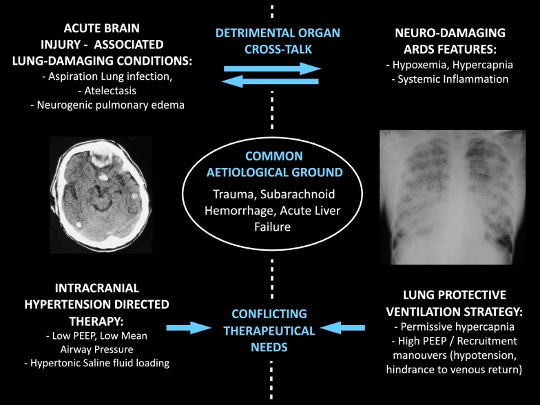
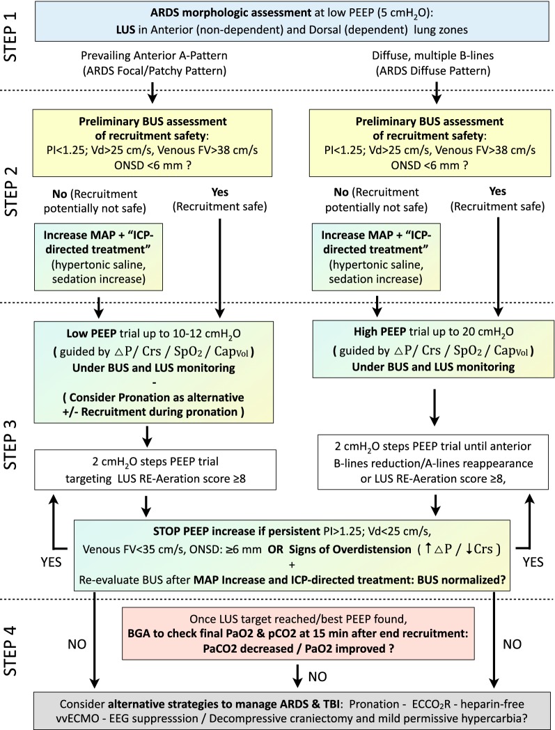
When intracranial hypertension and severe lung damage coexist in the same clinical scenario, their management poses a difficult challenge, especially as concerns mechanical ventilation management. The needs of combined lung and brain protection from secondary damage may conflict, as ventilation strategies commonly used in patients with ARDS are potentially associated with an increased risk of intracranial hypertension. In particular, the use of positive end-expiratory pressure, recruitment maneuvers, prone positioning, and protective lung ventilation can have undesirable effects on cerebral physiology: they may positively or negatively affect intracranial pressure, based on the final repercussions on PaO2 and cerebral perfusion pressure (through changes in cardiac output, mean arterial pressure, venous return, PaO2 and PaCO2), also according to the baseline conditions of cerebral autoregulation. Lung ultrasound (LUS) and brain ultrasound (BUS, as a combination of optic nerve sheath diameter assessment and cerebrovascular Doppler ultrasound) have independently proven their potential in respectively monitoring lung aeration and brain physiology at the bedside. In this narrative review, we describe how the combined use of LUS and BUS on neurocritical patients with demanding mechanical ventilation needs can contribute to ventilation management, with the aim of a tailored "brain-protective ventilation strategy."

Keywords: ARDS; Brain injury; Brain ultrasound; Intracranial hypertension; Lung ultrasound; Mechanical ventilation; Neuro-critical care; Respiratory monitoring.


Figures

Fig. 1
Fig. 1
Potential pathophysiological interactions between severe brain injury and severe lung injury, and their conflicting therapeutical needs. Severe brain injury and severe lung damage may ensue as consequence of the same noxious agent (for example in severe multiple trauma or severe liver failure), but also one be the cause of the other (e.g., neurogenic pulmonary edema in subarachnoid hemorrhage, or lung aspiration and infection in a comatous patient). Indeed, once coexisting, disease of one organ can negatively affect the other, in a harmful organ cross-talk (e.g., hypoxemia can worsen brain damage). Finally, some therapeutical interventions directed at protecting one organ may have detrimental effects on the other (e.g., mechanical ventilation strategies can either reduce systemic mean arterial pressure, decrease cerebral venous return, or cause cerebral vasodilation, thus inducing a worsening of intracranial hypertension and reducing the cerebral perfusion pressure)
Fig. 2
Fig. 2
Invasive and non-invasive intracranial pressure monitoring, respectively, through ICP Bolt and through brain ultrasound (BUS, optic nerve sheath diameter and cerebrovascular Doppler sampling) in a patient with traumatic brain injury and ARDS. A 26-year-old lady was admitted to the intensive care unit after a road traffic accident. She presented with severe traumatic brain injury (TBI) and chest trauma with lung contusions. She was monitored with invasive ICP, intubated and mechanically ventilated. On day 2, she developed hypoxic respiratory failure with bilateral atelectasia. We therefore performed recruitment maneuvers with ABP, CPP, and ICP monitoring and concomitant ONSD and TCD measurements (venous TCD on the SS and arterious TCD on the MCA). Recordings and scanning were performed at baseline (left panels) and during a recruitment maneuver and subsequent increase in PEEP level (right panels). Initially, ICP was below 20 mmHg (mean ONSD = 5.2), with PEEP = 8, with stable arterial blood pressure (ABP) and cerebral perfusion pressure (CPP). After recruitment maneuvers and setting PEEP at 16, ICP spiked up > 20 mmHg, with reflex mean systemic arterial blood pressure (ABP) and cerebral perfusion pressure (CPP) increase. BUS showed consistent increase in middle cerebral artery (MCA) pulsatility index (decrease in diastolic flow, increase in systolic flow), reduction in straight sinus (SS) flow, and increase in optic nerve sheath diameter (ONSD = 7 mm). PaCO2 remained constant during the procedure and the patients experienced no hypotension nor cardiac output decrease. We therefore reduced PEEP levels and noticed that ICP immediately decreased as well as PI, ONSD and the venous flow on the straight sinus. MCA middle cerebral artery, SS straight sinus, ONSD optic nerve sheath diameter, ABP mean systemic arterial blood pressure, ICP intracranial pressure, CPP cerebral perfusion pressure
Fig. 3
Fig. 3
Lung ultrasound–Brain ultrasound (LUS–BUS) combined respiratory and neurological monitoring in patients with traumatic brain injury (TBI) and acute respiratory distress syndrome (ARDS). A four-tiered approach is suggested, in order to decide the best ventilatory strategy and simultaneously monitor the effects on intracranial pressure (ICP) and on cerebrovascular dynamics. The goal is to set the ventilation consistently with a lung-protective strategy without negatively affecting the injured brain. Step 1—scanning of ventral and dorsal chest areas allows to differentiate ARDS with focal/patchy morphology (with less recruitment potential and greater risk of anterior lung overdistention) from ARDS with diffuse, more homogenous, morphology (amenable to successful recruitment at higher PEEP levels). Step 2—once this has been established, the kind of recruitment maneuver suitable for the detected ARDS morphology is preceded by BUS. The detection of signs of intracranial hypertension allows the preemptive institution of medical ICP-directed treatment to reduce the negative impact of the ventilatory maneuvers on the brain. Step 3—the recruiting maneuver is performed [under the guide of driving pressure (ΔP) and static respiratory system compliance (CRS), Volumetric Capnometry, SpO2] while monitoring changes in lung aeration (LUS) and signs of their potential negative impact on ICP and cerebrovascular dynamics and (BUS). Step 4—the final effect of the recruitment maneuver and the chosen PEEP is finally assessed, both in terms of gas exchanges, lung mechanics, and of net effect on the ICP and cerebrovascular dynamics. Should the ventilation target not be reachable nor compatible with brain protection, other respiratory support strategies/ICP treatments should be considered. ARDS acute respiratory distress syndrome, LUS lung ultrasound, BUS brain ultrasound, PEEP positive end expiratory pressure, PI middle cerebral artery pulsatility index, Vd middle cerebral artery diastolic arterial flow velocity, FV flow velocity; ONSD optic nerve sheath diameter, MAP mean systemic arterial pressure, ICP intracranial pressure, BGA blood gas analysis, TBI traumatic brain injury, ECCOR extracorporeal CO2 removal, vvECMO veno-venous extracorporeal membrane oxygenation, EEG electroencephalography, CRS respiratory system compliance, ΔP driving pressure, CapVol volumetric capnometry, SpO2 arterial oxygen saturation

References

    1. Rincon F, Ghosh S, Dey S, Maltenfort M, Vibbert M, Urtecho J, McBride W, Moussouttas M, Bell R, Ratliff JK, Jallo J. Impact of acute lung injury and acute respiratory distress syndrome after traumatic brain injury in the United States. Neurosurgery. 2012;71(4):795–803. doi: 10.1227/NEU.0b013e3182672ae5. - DOI - PubMed
    1. Veeravagu A, Chen YR, Ludwig C, Rincon F, Maltenfort M, Jallo J, Choudhri O, Steinberg GK, Ratliff JK. Acute lung injury in patients with subarachnoid hemorrhage: a nationwide inpatient sample study. World Neurosurg. 2014;82(1–2):e235–e241. doi: 10.1016/j.wneu.2014.02.030. - DOI - PubMed
    1. Damm TW, Kramer DJ. The liver in critical illness. Crit Care Clin. 2016;32(3):425–438. doi: 10.1016/j.ccc.2016.02.002. - DOI - PubMed
    1. Meng L, Gelb AW. Regulation of cerebral autoregulation by carbon dioxide. Anesthesiology. 2015;122(1):196–205. doi: 10.1097/ALN.0000000000000506. - DOI - PubMed
    1. Beuret P, Carton MJ, Nourdine K, Kaaki M, Tramoni G, Ducreux JC. Prone position as prevention of lung injury in comatose patients: a prospective, randomized, controlled study. Intensive Care Med. 2002;28(5):564–569. doi: 10.1007/s00134-002-1266-x. - DOI - PubMed

LinkOut - more resources