. 2019 Feb;234(2):1522-1533.
doi: 10.1002/jcp.27017. Epub 2018 Sep 17.

MiR-24-3p regulates cell proliferation and milk protein synthesis of mammary epithelial cells through menin in dairy cows

Affiliations

MiR-24-3p regulates cell proliferation and milk protein synthesis of mammary epithelial cells through menin in dairy cows

Cao Qiaoqiao et al. J Cell Physiol. 2019 Feb.

Abstract

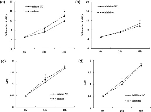
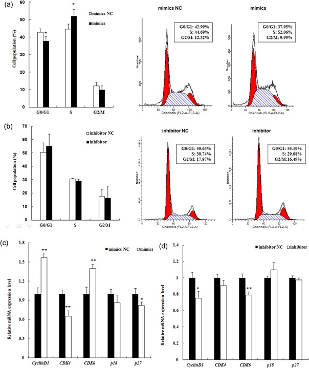
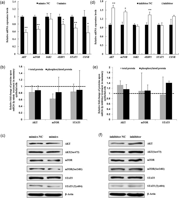
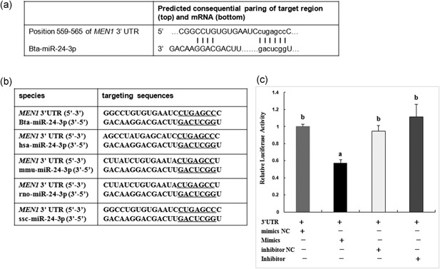
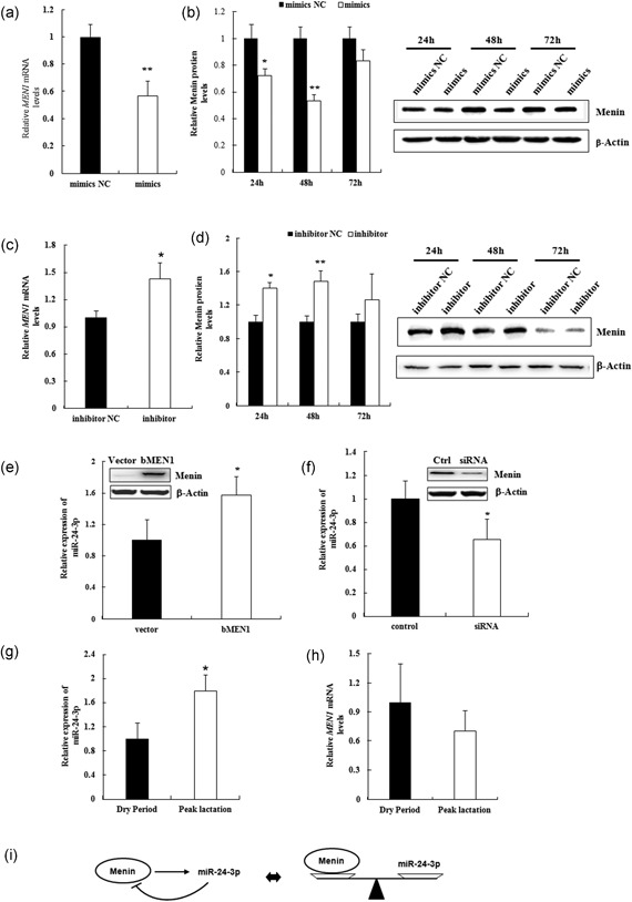
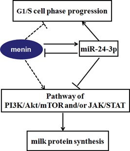
MiR-24-3p, a broadly conserved, small, noncoding RNA, is abundantly expressed in mammary tissue. However, its regulatory role in this tissue remains poorly understood. It was predicted that miR-24-3p targets the 3' untranslated region (3'-UTR) of multiple endocrine neoplasia type 1 (MEN1), an important regulatory factor in mammary tissue. The objective of this study was to investigate the function of miR-24-3p in mammary cells. Using a luciferase assay in mammary epithelial cells (MAC-T), miR-24-3p was confirmed to target the 3'-UTR of MEN1. Furthermore, miR-24-3p negatively regulated the expression of the MEN1 gene and its encoded protein, menin. miR-24-3p enhanced proliferation of MAC-T by promoting G1/S phase progression. MiR-24-3p also regulated the expression of key factors involved in phosphatidylinositol-3-kinase/protein kinase B/mammalian target of rapamycin and Janus kinase/signal transducer and activators of transcription signaling pathways, therefore controlling milk protein synthesis in epithelial cells. Thus, miR-24-3p appears to act on MAC-T by targeting MEN1. The expression of miR-24-3p was controlled by MEN1/menin, indicating a negative feedback loop between miR-24-3p and MEN1/menin. The negatively inhibited expression pattern of miR-24-3p and MEN1 was active in mammary tissues at different lactation stages. The feedback mechanism is a new concept to further understand the lactation cycle of mammary glands and can possibly to be manipulated to improve milk yield and quality.

Keywords: cell proliferation; mammary epithelial cells (MAC-T); miR-24-3p; milk protein synthesis; multiple endocrine neoplasia type 1(MEN1)/menin.


Figures

Figure 1
Figure 1
MiR‐24‐3p promotes the proliferation of mammary epithelial cells. The cell proliferation of mammary epithelial cells (MAC‐T) upon miR‐24‐3p expression modulation were assessed at 0, 24, and 48 hr after transfection of miR‐24‐3p mimics (a) and/or miR‐24‐3p inhibitor (b), as well as their corresponding negative controls (mimics NC and/or inhibitor NC), using cell counting methods after typan blue staining (a,b) and CCK‐8 assay (c,d) measuring the absorbance at 450 nm. Data are represented as the mean ± standard deviation from three independent experiments. *p < 0.05. NC: negative control
Figure 2
Figure 2
MiR‐24‐3p promotes cell cycle progression from G0/G1 to the S phase in mammary epithelial cells. MAC‐T cells were exposed to the miR‐24‐3p mimics (mimics, a) and/or miR‐24‐3p inhibitor (inhibitor, b), as well as their corresponding negative controls (mimics NC and/or inhibitor NC), for 24 hr and assessed for the distribution of cell cycle phases after propidium iodide (PI) staining. The percentages of cells in G0/G1, S, and G2/M phases are shown in (a,b), respectively. Representative FACS images of cell phase analyses illustrated changes of cell cycle in MAC‐T cells upon miR‐24‐3p overexpression (a) and/or low‐expression (b), compared with the negative controls. The cell cycle phase distribution (%) is indicated within each panel. The gene expression of cell cycle regulators specific to G1/S phase, such as cyclinD1, CDK4, CDK6, p18, and p27, were detected in the same miR‐24‐3p mimics (c) and or inhibitor (d) transfected MAC‐T cells using quantitative RT‐PCR. All of the experiments were performed three times for each transfection. **p < 0.01 and *p < 0.05. MAC‐T: mammary epithelial cell; NC: negative control; RT‐PCR: reverse transcription polymerase chain reaction [Color figure can be viewed at wileyonlinelibrary.com]
Figure 3
Figure 3
MiR‐24‐3p modulates the expression of genes that regulate milk protein synthesis in mammary epithelial cells. The expression levels of factors involved in the PI3K/AKT/mTOR (AKT, mTOR, S6K1, and 4E‐BP1) and JAK/STAT (STAT5) pathways that are associated with milk protein synthesis were assessed using quantitative real‐time PCR (qRT‐PCR) in MAC‐T cells at 24 hr after transfection with miR‐24‐3p mimics (mimics)/mimics NC (mimics NC, a) and miR‐24‐3p inhibitor (inhibitor)/inhibitor NC (inhibitor NC, b). Meanwhile, the expression of κ casein (CSNK), one of the key proteins that can be detected in MAC‐T cells, was also assessed in the cells. The data are shown as the relative expression levels normalized to the internal control, β‐actin. *p <  0.05, **p < 0.01. The phosphorylation levels of AKT at Ser473, mTOR at Ser2481, STAT5 at Tyr694 were detected simultaneously. The data are shown as the relative expression levels normalized to the loading controls, β‐actin. The horizontal dashed line represents the normalized level of their corresponding negative controls (b,d). Representative WB images of the expression of AKT, AKT phosphorylated at Ser473, mTOR, mTOR phosphorylated at Ser2481, STAT5 and STAT5 phosphorylated at Tyr694 are shown (c,e). AKT: protein kinase B; JAK: Janus kinase; MAC‐T: mammary epithelial cell; mTOR: mammalian target of rapamycin; NC: negative control; PCR: polymerase chain reaction; PI3K: phosphatidylinositol‐3‐kinase; STAT: signal transducer and activators of transcription
Figure 4
Figure 4
MiR‐24‐3p targets the 3′‐UTR of MEN1 mRNA. The bovine miR‐24‐3p (bta‐miR‐24‐3p) were predicted to interact with the 3′‐UTR region of MEN1 mRNA at position 559–565 using TargetScan 6.2 and RNAhybrid 2.2 software (a). MiR‐24‐3p, broadly conserved in different species, possibly also interacts with 3′‐UTR regions of MEN1 in human (has‐), mouse (mmu‐), rat (rno‐) and pig (ssc‐), with exact the same seed nucleotide sequence (underlined, b). The 3′‐UTR region (789 bp) of bovine MEN1 gene were construed into luciferase‐expressing plasmid (3′‐UTR). The plasmid was cotransfected with miR‐24‐3p mimics (mimics) and/or miR‐24‐3p inhibitor (inhibitor) into mammary epithelial cells, as well as their corresponding negative controls (mimics NC and/or inhibitor NC). The beta‐galactosidase reporter plasmid (β‐gal) was simultaneously transfected for each every transfection, serving as internal control. Significant suppressed luciferase activity was shown in the cotransfection group of 3′‐UTR plasmid and miR‐24‐3p mimics, indicating bta‐miR‐24‐3p targeted the 3′‐UTR of bovine MEN1. All of the experiments were performed three times for each transfection. Each experiment was performed in triplicates. Different letters (a,b) indicate significant difference (*p < 0.05). 3′‐UTR: 3′ untranslated region; MEN1: multiple endocrine tumor type 1; mRNA: messenger RNA; NC: negative control
Figure 5
Figure 5
MiR‐24‐3p and MEN1 act in a negative feedback model. (a–d) Mammary epithelial cells were transfected with miR‐24‐3p mimics/mimics NC (mimics/mimics NC; a and c) and/or miR‐24‐3p inhibitor/inhibitor NC (inhibitor/ inhibitor NC; b,d), and cells were harvested for RNA and protein extraction at 24, 48, and 72 hr after transfection. The expression of bovine MEN1 mRNA at 24 hr (a,c) and protein menin in a time course (b,d) were assessed using qRT‐PCR and western blot technology, respectively. Representative WB images of the expression of protein menin at 24, 28, and 72 hr after transfection of indicated miR‐24‐3p are shown. The data are shown as the relative expression levels normalized to the internal control, β‐actin. * p < 0.05. (e,f) Mammary epithelial cells were transfected with pEGFP‐C2‐bMEN1 (bMEN1)/pEGFP‐C2‐Vector (vector) for MEN1 overexpression system (e) and/or bovine MEN1 specific siRNA/nonspecific negative control siRNA (control) for MEN1 low‐expression system (f). Cells were harvested for RNA extraction and then the expression detection of miR‐24‐3p at 24 hr after transfection. The data are shown as the relative expression levels normalized to the internal control, small nuclear (sn) RNA U6. (g,h) The expression of miR‐24‐3p (g) and MEN1 (h) were measured in mammary tissues of dairy cows at dry period stage (n = 3) and peak lactation stage (n = 3). The data are shown as the relative expression levels normalized to the internal controls, small nuclear RNA U6 for miR‐24‐3p expression (g) and β‐actin for MEN1 expression (h). *p < 0.05. (i) MiR‐24‐3p can negatively modulate the expression level of menin in mammary epithelial cells, whereas menin is a positive regulator of miR‐24‐3p expression. MiR‐24‐3p and menin were suggestive of forming a “negative feedback loop” in mammary epithelial cells, dynamically maintaining the metabolic balance in mammary glands. MEN1: multiple endocrine tumor type 1; mRNA: messenger RNA; NC: negative control; qRT‐PCR: quantitative real‐time polymerase chain reaction; siRNA: small interfering RNA
Figure 6
Figure 6
MiR‐24‐3p regulates cell proliferation and milk protein synthesis in mammary epithelial cells through cooperatively acting with MEN1/menin. MiR‐24‐3p positively regulates the proliferation of mammary epithelial cells, promoting the G1/S cell phase progression. While, MEN1/menin was found, in our previous study (Li et al., 2017), negatively control the proliferation of at G1/S phase progression, causing cell growth arrest. This is contributed to the negative feedback controlling model between miR‐24‐3p and MEN1/menin. The dynamic balance model between miR‐24‐3p and MEN1/menin also make an opposite impacts on milk protein synthesis of mammary epithelial cells through PI3K/AKT/mTOR and/or JAK/STAT signaling pathway. The solid line represents fluxes found in the current study; the dotted line represents previously found effects or fluxes (Li et al., 2017). The arrows indicate the positive regulation, and the blunt‐ended ones indicate the negative regulation. AKT: protein kinase B; JAK: Janus kinase; MEN1: multiple endocrine tumor type 1; mTOR: mammalian target of rapamycin; PI3K: phosphatidylinositol‐3‐kinase; STAT: signal transducer and activators of transcription [Color figure can be viewed at wileyonlinelibrary.com]

Similar articles

Cited by

References

    1. Appuhamy, J. A. , Nayananjalie, W. A. , England, E. M. , Gerrard, D. E. , Akers, R. M. , & Hanigan, M. D. (2014). Effects of AMP‐activated protein kinase (AMPK) signaling and essential amino acids on mammalian target of rapamycin (mTOR) signaling and protein synthesis rates in mammary cells. Journal of Dairy Science, 97, 419–429. - PubMed
    1. Avril‐Sassen, S. , Goldstein, L. D. , Stingl, J. , Blenkiron, C. , Le Quesne, J. , Spiteri, I. , … Caldas, C. (2009). Characterisationof microRNA expression in post‐natal mouse mammary gland development. BMC Genom, 10, 548. - PMC - PubMed
    1. Bartel, D. P. (2004). MicroRNAs: Genomics, biogenesis, mechanism, and function. Cell, 116, 281–297. - PubMed
    1. Bertolino, P. , Tong, W. M. , Galendo, D. , Wang, Z. Q. , & Zhang, C. X. (2003). Heterozygous MEN1 mutant mice develop a range of endocrine tumors mimicking multiple endocrine neoplasia type 1. Molecular Endocrinology, 17(9), 1880–1892. - PubMed
    1. Bionaz, M. , Periasamy, K. , Rodriguez‐Zas, S. L. , Everts, R. E. , Lewin, H. A. , Hurley, W. L. , & Loor, J. J. (2012). Old and new stories: Revelations from functional analysis of the bovine mammary transcriptome during the lactation cycle. PLoS One, 7(3), e33268. - PMC - PubMed

Publication types

LinkOut - more resources