

Repeatability of Dixon magnetic resonance imaging and magnetic resonance spectroscopy for quantitative muscle fat assessments in the thigh
- PMID: 30221479
- PMCID: PMC6240750
- DOI: 10.1002/jcsm.12343
Repeatability of Dixon magnetic resonance imaging and magnetic resonance spectroscopy for quantitative muscle fat assessments in the thigh
Abstract
Background: Changes in muscle fat composition as for example observed in sarcopenia or muscular dystrophy affect physical performance and muscular function, like strength and power. The purpose of the present study is to measure the repeatability of Dixon magnetic resonance imaging (MRI) for assessing muscle volume and fat in the thigh. Furthermore, repeatability of magnetic resonance spectroscopy (MRS) for assessing muscle fat is determined.
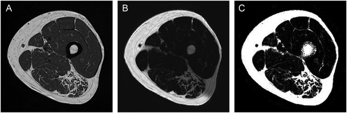
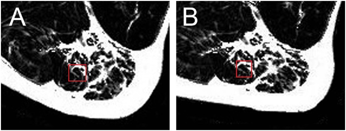
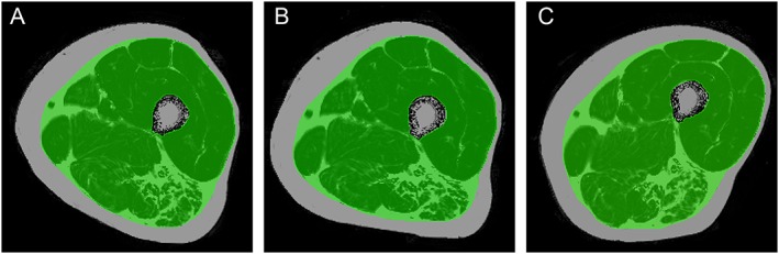
Methods: A prototype 6-point Dixon MRI method was used to measure muscle volume and muscle proton density fat fraction (PDFF) in the left thigh. PDFF was measured in musculus semitendinosus of the left thigh with a T2-corrected multi-echo MRS method. For the determination of short-term repeatability (consecutive examinations), the root mean square coefficients of variation of Dixon MRI and MRS data of 23 young and healthy (29 ± 5 years) and 24 elderly men with sarcopenia (78 ± 5 years) were calculated. For the estimation of the long-term repeatability (13 weeks between examinations), the root mean square coefficients of variation of MRI data of seven young and healthy (31 ± 7 years) and 23 elderly sarcopenic men (76 ± 5 years) were calculated. Long-term repeatability of MRS was not determined.
Results: Short-term errors of Dixon MRI volume measurement were between 1.2% and 1.5%, between 2.1% and 1.6% for Dixon MRI PDFF measurement, and between 9.0% and 15.3% for MRS. Because of the high short-term repeatability errors of MRS, long-term errors were not determined. Long-term errors of MRI volume measurement were between 1.9% and 4.0% and of Dixon MRI PDFF measurement between 2.1% and 4.2%.
Conclusions: The high degree of repeatability of volume and PDFF Dixon MRI supports its use to predict future mobility impairment and measures the success of therapeutic interventions, for example, in sarcopenia in aging populations and muscular dystrophy. Because of possible inhomogeneity of fat infiltration in muscle tissue, the application of MRS for PDFF measurements in muscle is more problematic because this may result in high repeatability errors. In addition, the tissue composition within the MRS voxel may not be representative for the whole muscle.
Keywords: Fat quantification; Magnetic resonance imaging; Magnetic resonance spectroscopy; Muscle; Repeatability; Sarcopenia.
© 2018 The Authors. Journal of Cachexia, Sarcopenia and Muscle published by John Wiley & Sons Ltd on behalf of the Society on Sarcopenia, Cachexia and Wasting Disorders.
Figures
References
-
- Kallman DA, Plato CC, Tobin JD. The role of muscle loss in the age‐related decline of grip strength. J Gerontol 1990;45:M82–M88. - PubMed
-
- Visser M, Goodpaster BH, Kritchevsky SB, Newman AB, Nevitt M, Rubin SM, et al. Muscle mass, muscle strength, and muscle fat infiltration as predictors of incident mobility limitations in well‐functioning older persons. J Gerontol 2005;60:324–333. - PubMed
MeSH terms
LinkOut - more resources
Full Text Sources
Other Literature Sources
Medical
