. 2018 Nov;18(5):4428-4438.
doi: 10.3892/mmr.2018.9485. Epub 2018 Sep 14.

FKBP11 protects intestinal epithelial cells against inflammation‑induced apoptosis via the JNK‑caspase pathway in Crohn's disease

Affiliations

FKBP11 protects intestinal epithelial cells against inflammation‑induced apoptosis via the JNK‑caspase pathway in Crohn's disease

Xiaotong Wang et al. Mol Med Rep. 2018 Nov.

Erratum in

Abstract

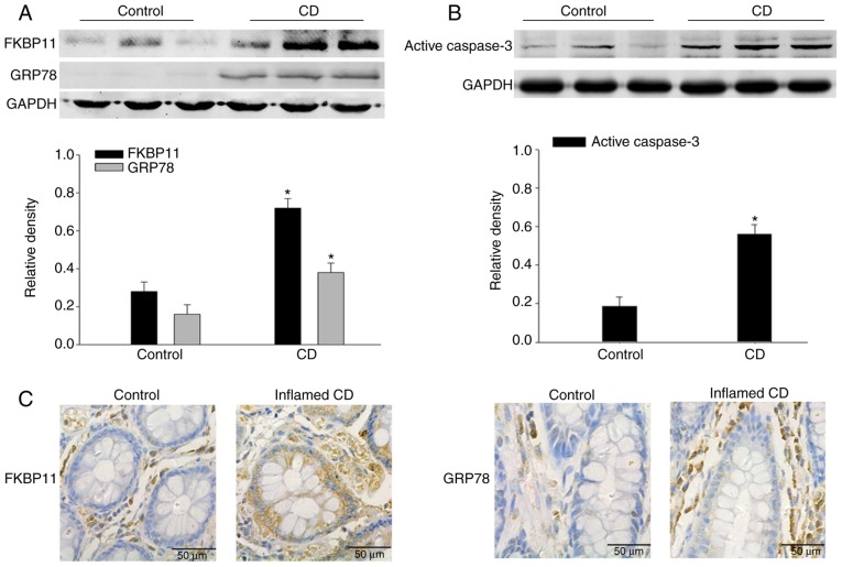
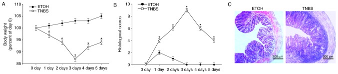
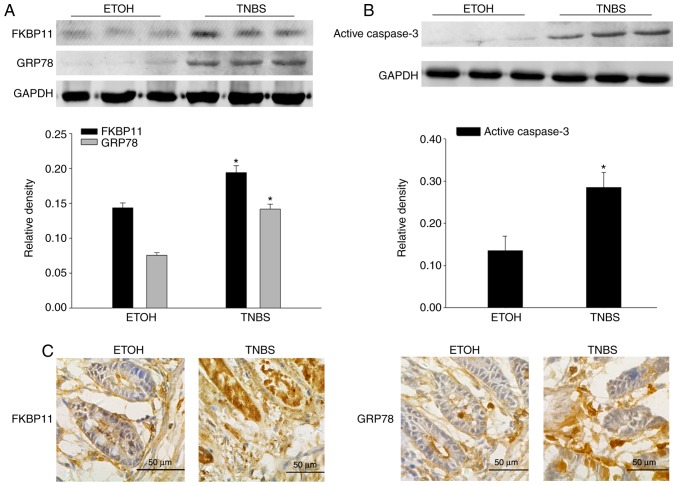
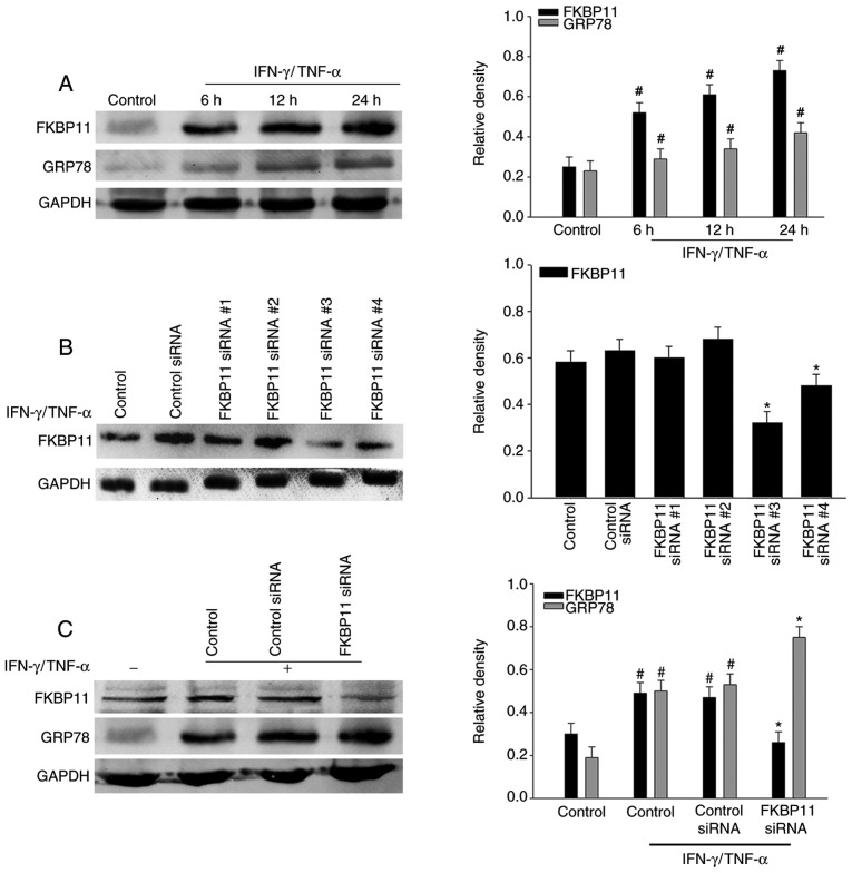
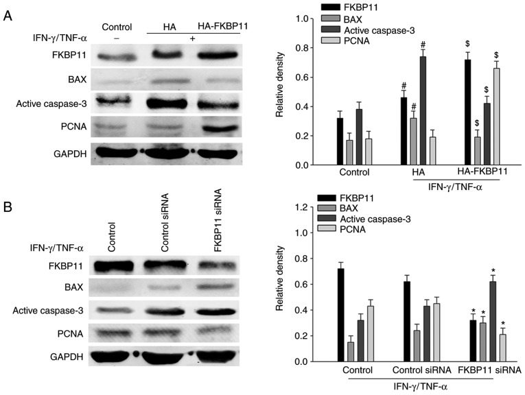
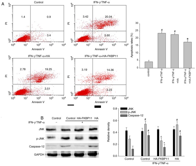
Endoplasmic reticulum (ER) stress in intestinal epithelial cells (IECs) has an important role in the pathogenesis of Crohn's disease (CD). FK506 binding protein 11 (FKBP11), a member of the peptidyl‑prolyl cis‑trans isomerase family, is involved in the unfolded protein response (UPR) and is closely associated with inflammation. Previous bioinformatics analysis revealed a potential association between FKBP11 and human CD. Thus, the present study aimed to investigate the potential significance of FKBP11 in IEC homeostasis and CD. In the present study, increased expression of FKBP11 was detected in the intestinal inflammatory tissues of patients with CD. Furthermore, the results of the present study revealed that overexpression of FKBP11 was accompanied by increased expression levels of the ER stress marker 78 kDa glucose‑regulated protein in the colon tissues of a 2, 4, 6‑trinitrobenzenesulphonic acid‑induced mouse colitis model. Using interferon‑γ (IFN‑γ)/tumor necrosis factor‑α (TNF‑α)‑stimulated IECs as an ER stress and apoptosis cell model, the associated of FKBP11 with ER stress and apoptosis levels was confirmed in IECs. Overexpression of FKBP11 was revealed to significantly attenuate the elevated expression of pro‑apoptotic proteins (Bcl2 associated X apoptosis regulator, caspase‑12 and active caspase‑3), suppress the phosphorylation of c‑Jun N‑terminal kinase (JNK), and decrease apoptosis of IFN‑γ/TNF‑α stimulated IECs. Knockdown of FKBP11 by transfection with small interfering RNA further validated the aforementioned results. In conclusion, these results suggest that the UPR protein FKBP11 may protect IECs against IFN‑γ/TNF‑α induced apoptosis by inhibiting the ER stress‑associated JNK/caspase apoptotic pathway in CD.


Figures

Figure 1.
Figure 1.
Expression of FKBP11 and GRP78 are increased in intestinal tissues of patients with CD. Western blot analyses of (A) FKBP11 and GRP78 expressions levels, and (B) active caspase-3 expression levels in the intestinal tissues of patients with active CD and control tissues. GAPDH was used as a loading control. Bar graphs demonstrate the semi-quantitative analysis of FKBP11, GRP78 and active caspase-3 protein expression levels vs. GAPDH. (C) Immunohistochemistry analysis of FKBP11 and GRP78 expressions in mucosal biopsies tissues obtained from inflamed tissues with active CD and control tissues. Scale bar, 50 µm. Data are presented as mean ± standard error (n=3). *P<0.05 vs. control. CD, Crohn's Disease; FKBP11, FK506 binding protein 11; GRP78, 78 kDa glucose-regulated protein.
Figure 2.
Figure 2.
Indicators confirm success of the TNBS-induced colitis model. (A) Mice body weight changes following administration of TNBS or ETOH at 0, 1, 2, 3, 4 and 5 days time intervals post-treatment. (B) Histological scoring of the H&E staining results at 0, 1, 2, 3, 4 and 5 days time intervals post-treatment with either TNBS or ETOH. (C) Representative light microscopy images of H&E stained colonic tissues from mice following administration of TNBS or ETOH (scale bar, 200 µm). Arrows indicate areas of inflammatory cell infiltration and epithelial cell depletion. Data are presented as mean ± standard error (n=3). *P<0.05 vs. ETOH. H&E, hematoxylin and eosin; ETOH, ethanol; TNBS, 2, 4, 6-trinitrobenzenesulphonic acid.
Figure 3.
Figure 3.
FKBP11 and GRP78 expression levels are increased in TNBS-induced colitis. Western blot analyses revealed that the expression levels of (A) FKBP11 and GRP78, and (B) active caspase-3 in TNBS induced colitis are significantly enhanced 3 days post-treatment compared with the ETOH group. Bar graphs demonstrate the quantitative analysis of FKBP11, GRP78 and active caspase-3 vs. GAPDH. (C) Immunohistochemistry analysis of FKBP11 and GRP78 expression in colonic mucosa of mice treated with TNBS for 3 days (scale bar, 50 µm). Data are presented as mean ± standard error (n=3). *P<0.05 vs. ETOH. ETOH, ethanol; TNBS, 2, 4, 6-trinitrobenzenesulphonic acid; FKBP11, FK506 binding protein 11; GRP78, 78 kDa glucose-regulated protein.
Figure 4.
Figure 4.
Association of FKBP11 expression levels with ER stress in IFN-γ/TNF-α-treated HT-29 cells. (A) HT-29 cells were treated with IFN-γ (2.5 ng/ml) and TNF-α (50 ng/ml) for different time intervals (0, 6, 12 and 24 h) to construct a cell model of ER stress. Western blot analyses revealed a significant upregulation of FKBP11 and GRP78 protein levels in IFN-γ/TNF-α-treated cells in a time-dependent manner. (B) FKBP11 expression following transfection with FKBP11 siRNA in HT-29 cells was revealed by western blot analyses, and transfection with FKBP11siRNA#3 was revealed to exhibit the most significant downregulation of FKBP11 expression. (C) HT-29 cells were transfected with control siRNA or FKBP11siRNA for 48 h, and then treated with IFN-γ/TNF-α for 24 h. Western blot analyses revealed that FKBP11 and GRP78 expression levels increased following IFN-γ/TNF-α treated cells compared with the untreated control, and GRP78 expression was significantly increased following transfection of FKBP11siRNA compared with control siRNA. Bar graphs reveal the densities of FKBP11 or GRP78 protein levels vs. GAPDH. Data are presented as mean ± standard error (n=3). *P<0.05 vs. control siRNA; #P<0.05 vs. IFN-γ/TNF-α untreated control. IFN-γ, interferon-γ; TNF-α, tumor necrosis factor-α; FKBP11, FK506 binding protein 11; GRP78, 78 kDa glucose-regulated protein; siRNA, small interfering RNA.
Figure 5.
Figure 5.
FKBP11 is involved in IFN-γ/TNF-α-induced apoptosis in HT-29 cells. (A) HT-29 cells were transfected with either the FKBP11 overexpressing plasmid pCMV-HA-FKBP11, blank control plasmid pCMV-HA, controls siRNA or FKBP11siRNA for 48 h and then treated with IFN-γ (2.5 ng/ml) and TNF-α (50 ng/ml) for 24 h. Western blot analyses detected FKBP11, BAX and active caspase-3 expression levels following treatment with IFN-γ/TNF-α compared with the untreated control and control plasmid (HA). #P<0.05 vs. IFN-γ/TNF-α untreated control; $P<0.05 vs. control plasmid pCMV-HA. (B) Western blot analyses detected FKBP11, BAX, active caspase-3 and PCNA following transfection with FKBP11 siRNA and treatment with IFN-γ (2.5 ng/ml) and TNF-α (50 ng/ml) for 24 h. Data are presented as mean ± standard error (n=3). *P<0.05 vs. control siRNA. HA, hemagglutinin; FKBP11, FK506 binding protein 11; siRNA, small interfering RNA; GRP78, 78 kDa glucose-regulated protein; IFN-γ, interferon-γ; TNF-α, tumor necrosis factor-α; BAX, Bcl2 associated X apoptosis regulator; PCNA, proliferating cell nuclear antigen.
Figure 6.
Figure 6.
FKBP11 protects HT-29 cells against IFN-γ/TNF-α induced apoptosis by inhibiting the JNK/caspase signaling pathway. (A) Cellular apoptosis and (B) western blot analyses of p-JNK and caspase-12 following post-treatment with IFN-γ/TNF-α and overexpression of FKBP11 in HT-29 cells. Bar graphs present the quantitative analysis of apoptosis rates and the densities of p-JNK, JNK and caspase-12 proteins vs. GAPDH. Data are presented as mean ± standard error of the mean (n=3). #P<0.05 vs. IFN-γ/TNF-α untreated control; *P<0.05 vs. IFN-γ/TNF-α treated control. PI, propidium iodide; IFN-γ, interferon-γ; TNF-α, tumor necrosis factor-α; HA, hemagglutinin; FKBP11, FK506 binding protein 11; JNK, c-Jun N-terminal kinase; p-, phospho-.

References

    1. Cao SS. Endoplasmic reticulum stress and unfolded protein response in inflammatory bowel disease. Inflamm Bowel Dis. 2015;21:636–644. doi: 10.1097/MIB.0000000000000238. - DOI - PubMed
    1. Tawfik A, Flanagan PK, Campbell BJ. Escherichia coli-host macrophage interactions in the pathogenesis of inflammatory bowel disease. World J Gastroenterol. 2014;20:8751–8763. - PMC - PubMed
    1. Cheng L, Huang MF, Mei PF, Bo WH, Deng CS. The clinical, endoscopic and pathologic features of Crohn's disease in the differentiation from intestinal tuberculosis. Zhonghua Nei Ke Za Zhi. 2013;52:940–944. (In Chinese) - PubMed
    1. Galeone C, Pelucchi C, Barbera G, Citterio C, La Vecchia C, Franchi A. Crohn's disease in Italy: A critical review of the literature using different data sources. Dig Liver Dis. 2017;49:459–466. doi: 10.1016/j.dld.2016.12.033. - DOI - PubMed
    1. Kim ES, Chen M, Lee J, Lee CK, Kim YS. Diagnosis of inflammatory bowel disease in Asia: The results of a multinational web-based survey in the 2(nd) Asian Organization for Crohn's and Colitis (AOCC) meeting in Seoul. Intest Res. 2016;14:224–230. doi: 10.5217/ir.2016.14.3.224. - DOI - PMC - PubMed

MeSH terms