. 2018 Oct 2;57(39):5759-5767.
doi: 10.1021/acs.biochem.8b00810. Epub 2018 Sep 17.

The Spastic Paraplegia-Associated Phospholipase DDHD1 Is a Primary Brain Phosphatidylinositol Lipase

Affiliations

The Spastic Paraplegia-Associated Phospholipase DDHD1 Is a Primary Brain Phosphatidylinositol Lipase

Jordon M Inloes et al. Biochemistry. .

Abstract

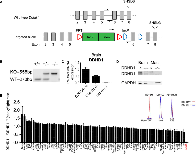
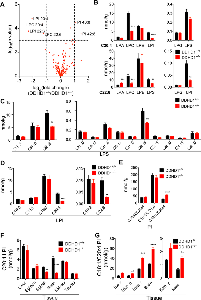
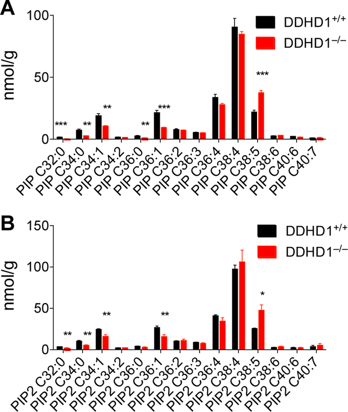
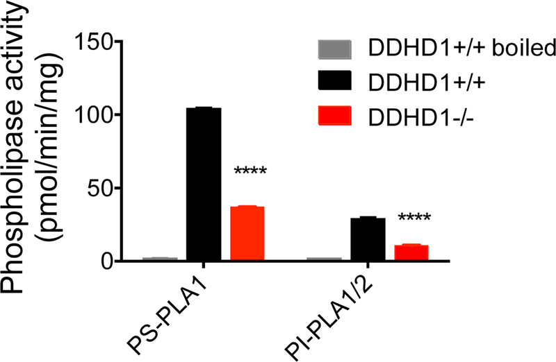
Deleterious mutations in the serine hydrolase DDHD domain containing 1 (DDHD1) cause the SPG28 subtype of the neurological disease hereditary spastic paraplegia (HSP), which is characterized by axonal neuropathy and gait impairments. DDHD1 has been shown to display PLA1-type phospholipase activity with a preference for phosphatidic acid. However, the endogenous lipid pathways regulated by DDHD1 in vivo remain poorly understood. Here we use a combination of untargeted and targeted metabolomics to compare the lipid content of brain tissue from DDHD1+/+ and DDHD1-/- mice, revealing that DDHD1 inactivation causes a substantial decrease in the level of polyunsaturated lysophosphatidylinositol (LPI) lipids and a corresponding increase in the level of phosphatidylinositol (PI) lipids. Levels of other phospholipids were mostly unchanged, with the exception of decreases in the levels of select polyunsaturated lysophosphatidylserine (LPS) and lysophosphatidylcholine lipids and a striking remodeling of PI phosphates (e.g., PIP and PIP2) in DDHD1-/- brain tissue. Biochemical assays confirmed that DDHD1 hydrolyzes PI/PS to LPI/LPS with sn-1 selectivity and accounts for a substantial fraction of the PI/PS lipase activity in mouse brain tissue. These data indicate that DDHD1 is a principal regulator of bioactive LPI and other lysophospholipids, as well as PI phosphates, in the mammalian nervous system, pointing to a potential role for these lipid pathways in HSP.


Figures

Figure 1.
Figure 1.
Generation and initial characterization of DDHD1–/– mice. (A) Map of whole body knockout allele of the Ddhd1 gene with conditional potential. Homologous recombination between Ddhd1 and 5’ and 3’ homology arms of a targeting vector resulted in the insertion of a lacZ/neo insert between exons 5 and 6 of Ddhd1. LoxP sites are introduced flanking exon 6 of Ddhd1. The catalytic serine of DDHD1 is downstream of the insertion. Half arrows represent annealing sites of genotyping primers. Map is not drawn to scale. (B) Genotyping of tail DNA of mice with various Ddhd1 genotypes. The targeted allele (knockout, or KO) generates a larger 558 bp product whereas the wild-type (WT) allele produces a 270 bp product. (C) Quantitative RT-PCR of mouse brain cDNA reveals a profound reduction of Ddhd1 mRNA in DDHD1–/– mice. N = 4, Error bars represent mean ± SEM. (D) Western blot of brain and macrophage lysates from DDHD1+/+ and DDHD1–/– mice. An immunoreactive band at the predicted molecular weight of DDHD1 is absent in DDHD1–/– tissues and cells (mac, macrophages). (E) Quantitative MS-based ABPP of serine hydrolase activities in brain tissue shows complete and selective loss of DDHD1 activity in DDHD1–/– mice. Error bars represent mean ± SEM of median isotopic ratios for tryptic peptides from each indicated serine hydrolase (data are from two independent experiments).
Figure 2.
Figure 2.
Metabolomic analysis identifies phospholipid changes in tissues from DDHD1–/– mice. A) Volcano plot showing relative abundance of metabolites (x-axis) versus significance of the observed changes (y-axis) in brain tissue from DDHD1–/– and DDHD1+/+ mice as measured by untargeted metabolomics. Structural assignments for significantly changing metabolites (P<0.001) with log2 transformed fold changes of <−1 or >1 are shown next to data points. For integrated peak areas of all extracted metabolites see (Table S1). (B) Targeted LC-MS-based quantification of the indicated lysophospholipids containing C20:4 or C22:6 acyl chains in DDHD1+/+ or DDHD1–/– brains. (C-E) Targeted LC-M-based quantification of the indicated LPS (C) and LPI (D) and PI (E) species in DDHD1+/+ and DDHD1–/– brains. (F, G) Targeted LC-M-based quantification of C20:4 LPI (F) and C18:1/C20:4 PI (G) in the indicated tissues from DDHD1+/+ and DDHD1–/– mice. For additional lysophospholipid and PI data, see Figures S5 and S6, respectively. For overall lipid data, see Table S2. N = 4, error bars represent mean ± SEM. ** P<0.01, *** P<0.001, **** P<0.0001, DDHD1+/+ vs. DDHD1–/–.
Figure 3.
Figure 3.. Altered PI phosphate content of brain tissue from DDHD1–/– mice.
(A, B) Targeted LC-MS-based quantification of the indicated PIP (A) and PIP2 (B) species in brain tissue from DDHD1+/+ and DDHD1–/– mice. Data are normalized to PIP or PIP2 internal standards as described in the Supplemental Methods section. N = 4, error bars represent averages ± SEM. * P<0.05,** P<0.01,*** P<0.001, DDHD1+/+ vs. DDHD1–/–.
Figure 4.
Figure 4.
DDHD1 makes a major contribution to brain phospholipase A (PLA1) activity with PI and PS substrates. Soluble brain tissue lysates from DDHD1+/+ or DDHD1–/– mice were assessed for hydrolysis of C16:0/C18:1 PS or C16:0/C16:0 PI substrates as described in Methods. Heat-denatured (boiled) DDHD1+/+ soluble lysates served as an inactivated control sample. N = 3, error bars represent mean ± SEM. **** P<0.0001, DDHD1+/+ vs. DDHD1–/–.

References

    1. Bisogno T; Howell F; Williams G; Minassi A; Cascio MG; Ligresti A; Matias I; Schiano-Moriello A; Paul P; Williams E-J; Gangadharan U; Hobbs C; Di Marzo V; Doherty P Cloning of the First Sn1-DAG Lipases Points to the Spatial and Temporal Regulation of Endocannabinoid Signaling in the Brain. J. Cell Biol 2003, 163 (3), 463–468. - PMC - PubMed
    1. Yung YC; Stoddard NC; Chun J LPA Receptor Signaling: Pharmacology, Physiology, and Pathophysiology. J. Lipid Res 2014, 55 (7), 1192–1214. - PMC - PubMed
    1. Yamashita A; Oka S; Tanikawa T; Hayashi Y; Nemoto-Sasaki Y; Sugiura T The Actions and Metabolism of Lysophosphatidylinositol, an Endogenous Agonist for GPR55. Prostaglandins Other Lipid Mediat. 2013, 107, 103–116. - PubMed
    1. Higgs HN; Han MH; Johnson GE; Glomset JA Cloning of a Phosphatidic Acid-Preferring Phospholipase A1 From Bovine Testis. J. Biol. Chem 1998, 273 (10), 5468–5477. - PubMed
    1. Higgs HN; Glomset JA Purification and Properties of a Phosphatidic Acid-Preferring Phospholipase A1 From Bovine Testis. Examination of the Molecular Basis of Its Activation. J. Biol. Chem 1996, 271 (18), 10874–10883. - PubMed

Publication types