. 2018 Nov;12(6):1169-1177.
doi: 10.1177/1932296818798347. Epub 2018 Sep 15.

Noninvasive Monitoring of Blood Glucose Using Color-Coded Photoplethysmographic Images of the Illuminated Fingertip Within the Visible and Near-Infrared Range: Opportunities and Questions

Affiliations

Noninvasive Monitoring of Blood Glucose Using Color-Coded Photoplethysmographic Images of the Illuminated Fingertip Within the Visible and Near-Infrared Range: Opportunities and Questions

Thorsten Vahlsing et al. J Diabetes Sci Technol. 2018 Nov.

Abstract

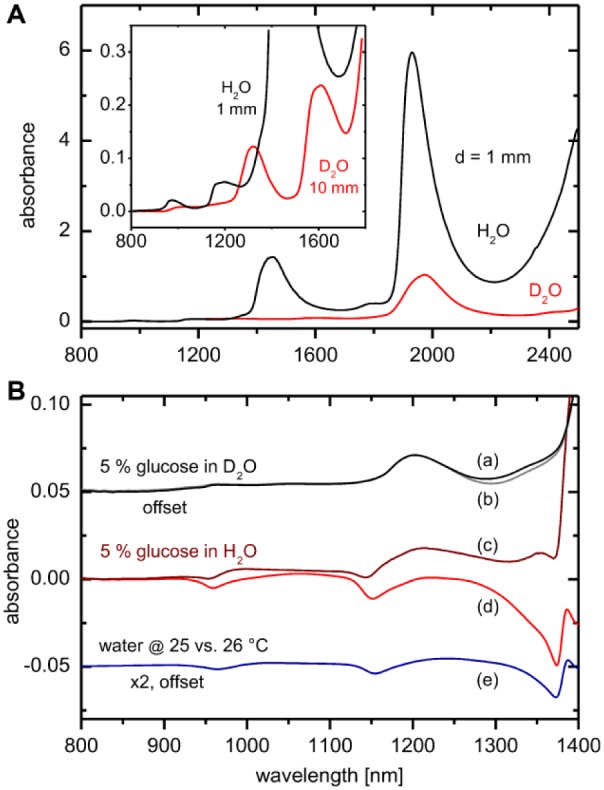
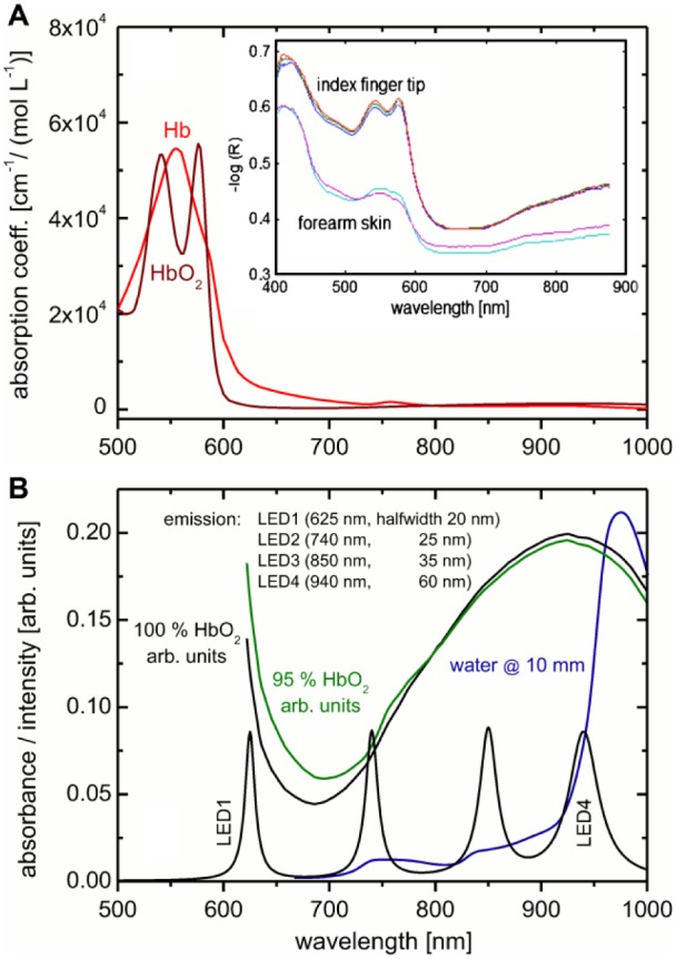
Noninvasive blood glucose assays have been promised for many years and various molecular spectroscopy-based methods of skin are candidates for achieving this goal. Due to the small spectral signatures of the glucose used for direct physical detection, moreover hidden among a largely variable background, broad spectral intervals are usually required to provide the mandatory analytical selectivity, but no such device has so far reached the accuracy that is required for self-monitoring of blood glucose (SMBG). A recently presented device as described in this journal, based on photoplethysmographic fingertip images for measuring glucose in a nonspecific indirect manner, is especially evaluated for providing reliable blood glucose concentration predictions.

Keywords: color sensing; noninvasive glucose sensing; plethysmographic skin imaging; skin tissue spectroscopy; visible/near-infrared spectroscopy.


Conflict of interest statement

Declaration of Conflicting Interests: The author(s) declared no potential conflicts of interest with respect to the research, authorship, and/or publication of this article.

Figures

Figure 1.
Figure 1.
(A) Absorption spectra of H2O and D2O at various optical pathlengths and 25°C within the VIS/NIR spectral range. (B) Glucose absorption spectra at 25°C with D2O as solvent with compensation of displaced deuterated water (a); solution spectrum measured versus a solvent filled cell (b). Glucose spectrum measured in normal water with compensation of displaced water (c) and as measured versus a solvent filled cell (d). Temperature effect on the water absorption spectrum is shown with trace (e). Water displacement factors were calculated by using solution density data from Haynes.
Figure 2.
Figure 2.
(A) Absorptivity data for oxy- and deoxy-hemoglobin (downloaded from https://omlc.org/spectra/hemoglobin/); the inset shows two diffuse reflection spectra of fingertip and forearm skin as measured with a fiber optic probe. (B) Spectra of hemoglobin at different oxygenation rates; included is the absorbance spectrum of water at 10 mm pathlength. Also shown is the illumination irradiance (intensity normalized) for four LEDs with indicated halfwidths from Hamamatsu Catalog at selected center wavelengths, as mentioned by Segman.
Figure 3.
Figure 3.
(A) Subsecond recording of fingertip diffuse reflection spectra using an integrating sphere. (B) Average spectrum including the scaled pulsatile spectrum at heartbeat frequency as obtained from Fourier analysis of the wavelength-dependent time series vectors; shown as inset is the power spectrum of the time series data at 580 nm wavelength up to the fundamental frequency of the transformed PPG waveform.
Figure 4.
Figure 4.
Spectral sensitivity of a camera sensor with Color Filter Array (CFA) modified from Park and Kang, original image by C. Park and M. G. Kang, licensed under CC-BY (http://creativecommons.org/licenses/by/4.0/).

References

    1. Hönes J, Müller P, Surridge N. The technology behind glucose meters: test strips. Diabetes Technol Ther. 2008;10(S1). doi:10.1089/dia.2008.0005. - DOI
    1. Freckmann G, Baumstark A, Schmid C, Pleus S, Link M, Haug C. Evaluation of 12 blood glucose monitoring systems for self-testing: system accuracy and measurement reproducibility. Diabetes Technol Ther. 2014;16(2):113-122. - PubMed
    1. Jungheim K, Koschinsky T. Glucose monitoring at the arm: risky delays of hypoglycemia and hyperglycemia detection. Diabetes Care. 2002;25(6):956-960. doi:10.2337/diacare.25.6.956. - DOI - PubMed
    1. Acciaroli G, Vettoretti M, Facchinetti A, Sparacino G. Calibration of minimally invasive continuous glucose monitoring sensors: state-of-the-art and current perspectives. Biosensors. 2018;8(1):E24. doi:10.3390/bios8010024. - DOI - PMC - PubMed
    1. Corabian P, Chojecki D. IHE Report: Exploratory Brief on Glucose Monitoring Technologies. Edmonton, AB: Institute of Health Economics; 2017. Available at: https://www.ihe.ca/publications/exploratory-brief-on-glucose-monitoring-.... - PubMed

Publication types

MeSH terms