Review
. 2018 Sep 15:4:101-122.
doi: 10.1146/annurev-vision-091517-034018.

Retinal Vasculature in Development and Diseases

Affiliations
Review

Retinal Vasculature in Development and Diseases

Ye Sun et al. Annu Rev Vis Sci. .

Retraction in

Abstract

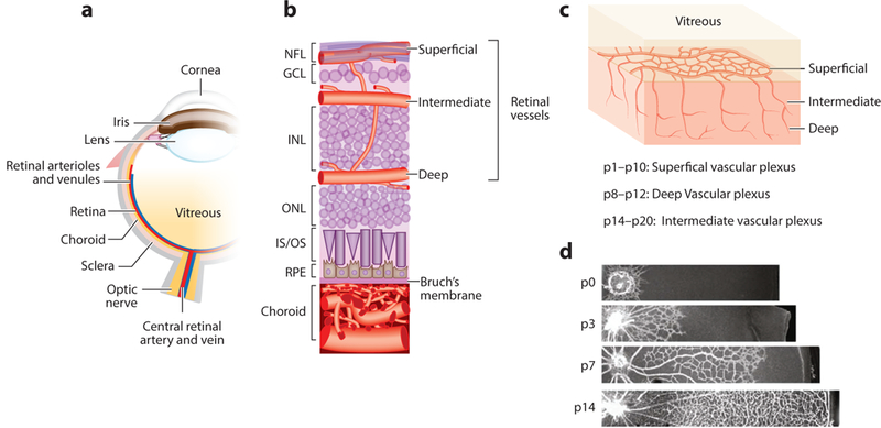
The retina is one of the most metabolically active tissues in the body, consuming high levels of oxygen and nutrients. A well-organized ocular vascular system adapts to meet the metabolic requirements of the retina to ensure visual function. Pathological conditions affect growth of the blood vessels in the eye. Understanding the neuronal biological processes that govern retinal vascular development is of interest for translational researchers and clinicians to develop preventive and interventional therapeutics for vascular eye diseases that address early drivers of abnormal vascular growth. This review summarizes the current knowledge of the cellular and molecular processes governing both physiological and pathological retinal vascular development, which is dependent on the interaction among retinal cell populations, including neurons, glia, immune cells, and vascular endothelial cells. We also review animal models currently used for studying retinal vascular development.

Keywords: AMD; DR; ROP; animal model; development; retina; vasculature.


Figures

Figure 1:
Figure 1:
Schematic diagram of the ocular vasculature. (a) Cross-sectional image of an eye. (b) An enlarged cross-sectional view of the retinal and choroidal vasculature. Three interconnected layers of retinal vessels are embedded among inner retinal neurons: The superficial retinal vasculature lies in the NFL; the intermediate and deep retinal vascular networks lie along each side of the INL. The choroidal vessels are located beneath the RPE and Bruch’s membrane and supply oxygen and nutrients to the outer portion of the retina, which includes primarily photoreceptors in the outer nuclear layer. (c) Retinal vascular development begins with the formation of the superficial vascular plexus from pi to p10, then the deep vascular plexus from p8 to p12, and finally, the intermediate vascular plexus from p14 to p20. (d) Retinal superficial vascular plexus at different developmental stages p0, p3, p7, and p14. Panels a and b adapted from Liu et al. (2017). Panels c and d adapted from Joyal et al. (2018). Abbreviations: GCL, ganglion cell layer; INL, inner nuclear layer; IS/OS, inner segment/outer segment of photoreceptor; NFL, nerve fiber layer; ONL, outer nuclear layer; P, postnatal day; RPE, retinal pigment epithelium.
Figure 2:
Figure 2:
Neuronal and glial cell regulation of pathological neovascularization in the eye via STAT3 and SOCS3 signaling pathways. SOCS3 deficiency in neuronal or glial cells promotes pathological retinal angiogenesis in retinopathy via titrating VEGF signaling controlled by activated transcription factor STAT3. A red arrow indicates upregulation, and a green arrow indicates downregulation. Abbreviations: IL-6, interleukin-6; JAK, Janus kinase; SOCS3, suppressor of cytokine signaling 3; STAT3, Signal transducer and activator of transcription 3; VEGF, vascular endothelial growth factor. Figure adapted from Sun & Smith (2015), with permission from Atlas of Science.
Figure 3:
Figure 3:
Photoreceptor regulation of pathological neovascularization in the eye via control of inflammatory signals in photoreceptors. Targeting inflammatory regulator c-Fos in photoreceptors to control inflammatory proteins may provide a novel approach to suppress pathological ocular neovascularization, which leads to blindness. Abbreviation: RPE, retinal pigment epithelium.
Figure 4:
Figure 4:
Regulation of lipid-metabolism-driven inflammation of pathological neovascularization in the eye via the RORα-SOCS3 pathway. RORα senses lipid metabolites and regulates target gene expression through binding to RORE elements in their promoter region. RORα regulates pathological neovascularization (NV) in the eye via control of tissue inflammation and macrophage polarization. Additional abbreviations: RORa retinoic-acid-receptor-related orphan receptor alpha; RORE, RORα responsive element; SOCS3, suppressor of cytokine signaling 3.

References

    1. Ahmed K, Tunaru S, Tang C, Müller M, Gille A, et al. 2010. An autocrine lactate loop mediates insulin-dependent inhibition of lipolysis through GPR81. CellMetab. 11:311–19 - PubMed
    1. Aiello LP, Pierce EA, Foley ED, Takagi H, Chen H, et al. 1995. Suppression of retinal neovascularization in vivo by inhibition of vascular endothelial growth factor (VEGF) using soluble VEGF-receptor chimeric proteins. PNAS 92:10457–61 - PMC - PubMed
    1. Akula JD, Hansen RM, Martinez-Perez ME, Fulton AB. 2007. Rod photoreceptor function predicts blood vessel abnormality in retinopathy of prematurity. Investig. Ophthalmol Vis. Sci. 48:4351–59 - PubMed
    1. Al-Latayfeh M, Silva PS, Sun JK, Aiello LP. 2012. Antiangiogenic therapy for ischemic retinopathies. Cold Spring Harb. Perspect. Med. 2:a006411. - PMC - PubMed
    1. Anand-Apte B, Hollyfield JG. 2011. Developmental anatomy of the retinal and choroidal vasculature In The Retina and Its Disorders, ed. Besharse J, pp. 9–15. Oxford, UK: Academic

Publication types

LinkOut - more resources