. 2018 Sep 17;13(9):e0202574.
doi: 10.1371/journal.pone.0202574. eCollection 2018.

Satellite cells delivered in their niche efficiently generate functional myotubes in three-dimensional cell culture

Affiliations

Satellite cells delivered in their niche efficiently generate functional myotubes in three-dimensional cell culture

Johanna Prüller et al. PLoS One. .

Abstract

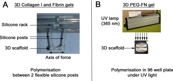
Biophysical/biochemical cues from the environment contribute to regulation of the regenerative capacity of resident skeletal muscle stem cells called satellites cells. This can be observed in vitro, where muscle cell behaviour is influenced by the particular culture substrates and whether culture is performed in a 2D or 3D environment, with changes including morphology, nuclear shape and cytoskeletal organization. To create a 3D skeletal muscle model we compared collagen I, Fibrin or PEG-Fibrinogen with different sources of murine and human myogenic cells. To generate tension in the 3D scaffold, biomaterials were polymerised between two flexible silicone posts to mimic tendons. This 3D culture system has multiple advantages including being simple, fast to set up and inexpensive, so providing an accessible tool to investigate myogenesis in a 3D environment. Immortalised human and murine myoblast lines, and primary murine satellite cells showed varying degrees of myogenic differentiation when cultured in these biomaterials, with C2 myoblasts in particular forming large multinucleated myotubes in collagen I or Fibrin. However, murine satellite cells retained in their niche on a muscle fibre and embedded in 3D collagen I or Fibrin gels generated aligned, multinucleated and contractile myotubes.


Conflict of interest statement

IM and TE are co-founders of EHT Technologies GmbH, Hamburg, a UKE spin-off commercializing the materials for the making and analysis of engineered heart tissue. This does not alter our adherence to PLOS ONE policies on sharing data and materials. JP, PSZ and NF declare no competing interests.

Figures

Fig 1
Fig 1. Experimental setup to generate 3D gels.
Collagen and Fibrin gels were polymerized between two silicone posts that mimic tendons (A). The biomaterial was mixed with muscle cells or muscle fibres. A pair of flexible silicone posts supported by a silicone rack was immersed in the gel. The gel containing the cells polymerises between the two silicone posts. After polymerisation, the silicone rack holding the silicone posts was used to transfer the gel into a 24-well tissue culture dish. The axis of force created by the silicone posts promotes cellular alignment. PEG-Fibrinogen could not to polymerised between the two silicone posts and was instead polymerised in a 96 well plate using UV light (B). Once polymerised, the gel was transferred to a 24 well plate, but undergoes no uniform, directed static strain.
Fig 2
Fig 2. All 3D biomaterial scaffolds promote robust myotube formation of murine C2C12 myoblasts in vitro.
Immortalized C2C12 myoblasts, embedded in Collagen I (450 000 cells / 100 μl: A and D) or Fibrin (450 000 cells / 100 μl: B and E) gels were polymerised between two silicone posts. PEG-Fibrinogen (300 000 cells / 100 μl: C and F) gels were polymerised in a 96 well plate and cultured without uniform static tension. Gels were cultured in proliferation medium for 2 days (A-C) and then switched to differentiation medium for 3 days (D-F). 3D scaffolds were fixed and immunolabelled for β-Tubulin (A-C) or myosin heavy chain (MyHC) (D-F) and nuclei counterstained with DAPI. In Collagen I (A) and Fibrin (B) gels, proliferating myoblasts were often aligned along the line of force created by the silicone posts and gel compaction was observed. In PEG-Fibrinogen gels (C), C2C12 myoblasts present randomly orientated membrane projections and no gel compaction was observed. After 3 days in differentiation medium, large parallel myotubes (arrowed) were observed in Collagen I (D) and Fibrin (E) gels. Thinner and randomly orientated myotubes were present in PEG-Fibrinogen scaffold (F). Scale bar represents 100 μm. Representative images from three or more independent experiments.
Fig 3
Fig 3. Myogenic progression of immortalized human myoblasts in different 3D biomaterial scaffolds in vitro.
Immortalized human myoblasts C25Cl48 were embedded in collagen I (500 000 cells / 100 μl: A and D), Fibrin (500 000 cells / 100 μl: B and E) or PEG-Fibrinogen (500 000 cells / 100 μl: C and F), cultured in proliferation medium for 2 days (A-C) and then switched to differentiation medium for 6 days (D-F). After 2 days in proliferation medium, cells were pulsed with the thymidine analogue 5-Ethynyl-2’deoxyuridine (EdU) for 2 hours and then fixed, before EdU incorporation was visualised and myoblasts immunolabelled for β-Tubulin and nuclei counterstained with DAPI. After 6 days in differentiation medium, cells were fixed and immunolabelled for myosin heavy chain (MyHC) to visualise myotubes and counterstained with DAPI. Proliferating EdU positive (arrow-heads) and β-Tubulin myoblasts were aligned in collagen I (A) and Fibrin (B) gels but randomly orientated in PEG-Fibrinogen scaffold (C). After 6 days in differentiation medium many MyHC positive cells were present but only a few small myotubes were detected in collagen I (D) or Fibrin (E) gels. In PEG-Fibrinogen scaffold (C), small myotubes were randomly orientated. Scale bar represents 100 μm. Representative images from 3 or more independent experiments. Expression of the sarcomeric proteins Myosin Heavy chain (MYH2, 3, 8) (G), Actinin α3 (ACTN3) (H) and Tropomyosin 1 (TPM1) (I) and creatine kinase (CKM) (J) after 7 days of differentiation were analysed by RT-qPCR. Expression was normalized to the house keeping gene TBP. Data are mean±SEM from 3 independent gels where an asterisk denotes a significant difference (p<0.05) from the Collagen gel using an unpaired two-tailed Student's t-test.
Fig 4
Fig 4. Limited myogenic differentiation of expanded primary murine satellite cells in 3D biomaterial scaffolds.
In vitro expanded primary satellite cell-derived myoblasts were embedded in collagen I (500, 000 cells/100 μl: A and D), Fibrin (500, 000 cells/100 μl: B and E) or PEG-Fibrinogen (500, 000 cells/100 μl: C and F) and were cultured in proliferation medium for 4 days (A-C) and then switched to differentiation medium for 10 days (D-F). Cellular morphology of proliferating myoblasts (A-C) and myotubes (D-F) were visualized by immunolabelling for β-tubulin and Desmin respectively, and nuclei counterstained with DAPI. After 4 days in proliferation medium (A, B and C) most of the satellite cells had a rounded shape (arrow heads) and only few were elongated (arrows). Even after 10 days in differentiation medium, no myotubes were observed in collagen I (D) or PEG-Fibrinogen scaffolds (F) and only a few thin myotubes were present in Fibrin gel (arrow in E). Scale bar represents 100 μm. Representative images from 3 independent experiments. Expression of the sarcomeric proteins myosin heavy chain (Myh1) (G), tropomyosin 1 (Tpm1) (H) and actinin α3 (Actn3) (I) and creatine kinase (Ckm) (J) were analysed by RT-qPCR (G-J). Expression was normalized to the house keeping gene Tbp. Data are mean±SEM from satellite cells isolated from 3 mice where an asterisk denotes a significant difference (p<0.05) from Collagen gels using a paired two-tail Student's t-test.
Fig 5
Fig 5. Satellite cells delivered in their niche on a myofibre make contractile myotubes in 3D scaffolds.
Approximately 100 freshly isolated Soleus myofibres were embedded in 100 μl of collagen I (A-D), 100 μl of Fibrin (E-H) or 100 μl PEG-Fibrinogen (I-L) and were cultured in proliferation medium for 1 (A, E and I), 3 (B, F and J) or 6 days (C, G and K). After 10 days in proliferation medium, 3D scaffolds were switched to differentiation medium for 3 days (D, H and L). After 1 day (A, E and I), intact myofibres (arrows) were visible and some hypercontracted myofibres (asterisk) were observed in PEG-Fibrinogen (I). After 3 days (B, F and J), satellite cells (arrow heads) were present at the surface of myofibres and some had migrated into the biomaterial scaffold. After 6 days (C, G and K) satellite cells were still proliferating. After 10 days in proliferation medium and 3 days in differentiation medium (D, H and L) multinucleated, matured and functional myotubes (arrows) were observed. Representative images from repeats using myofibres from 3 mice. Scale bars represent approximately 100 μm. Freshly isolated Soleus myofibres from 3F-nlacZ-E transgenic mice were embedded in collagen I (M), Fibrin (N) or PEG-Fibrinogen (O) gels. Biomaterial scaffolds were incubated in proliferation medium for 10 days, switched to differentiation medium for 3 days and then fixed and stained in X-gal solution to reveal β-galactosidase activity in myonuclei from the 3F-nlacZ-E transgene. Differentiated X-Gal positive myonuclei were detected in myotubes (arrows). More X-Gal positive nuclei and parallel aligned myotubes were observed in Fibrin (N) compared to Collagen I (M). Representative images of 3 independent experiments using myofibres from 3 x 3F-nlacZ-E mice.

Similar articles

Cited by

References

    1. Janssen I, Heymsfield SB, Wang ZM, Ross R. Skeletal muscle mass and distribution in 468 men and women aged 18–88 yr. J Appl Physiol (1985). 2000;89(1):81–8. 10.1152/jappl.2000.89.1.81 . - DOI - PubMed
    1. Gillies AR, Lieber RL. Structure and function of the skeletal muscle extracellular matrix. Muscle Nerve. 2011;44(3):318–31. 10.1002/mus.22094 ; PubMed Central PMCID: PMC3177172. - DOI - PMC - PubMed
    1. Relaix F, Zammit PS. Satellite cells are essential for skeletal muscle regeneration: the cell on the edge returns centre stage. Development. 2012;139(16):2845–56. 10.1242/dev.069088 . - DOI - PubMed
    1. Mauro A. Satellite cell of skeletal muscle fibers. J Biophys Biochem Cytol. 1961;9:493–5. ; PubMed Central PMCID: PMC2225012. - PMC - PubMed
    1. Dumont NA, Bentzinger CF, Sincennes MC, Rudnicki MA. Satellite Cells and Skeletal Muscle Regeneration. Comprehensive Physiology. 2015;5(3):1027–59. 10.1002/cphy.c140068 . - DOI - PubMed

Publication types