

Refining muscle geometry and wrapping in the TLEM 2 model for improved hip contact force prediction
- PMID: 30222777
- PMCID: PMC6141086
- DOI: 10.1371/journal.pone.0204109
Refining muscle geometry and wrapping in the TLEM 2 model for improved hip contact force prediction
Abstract
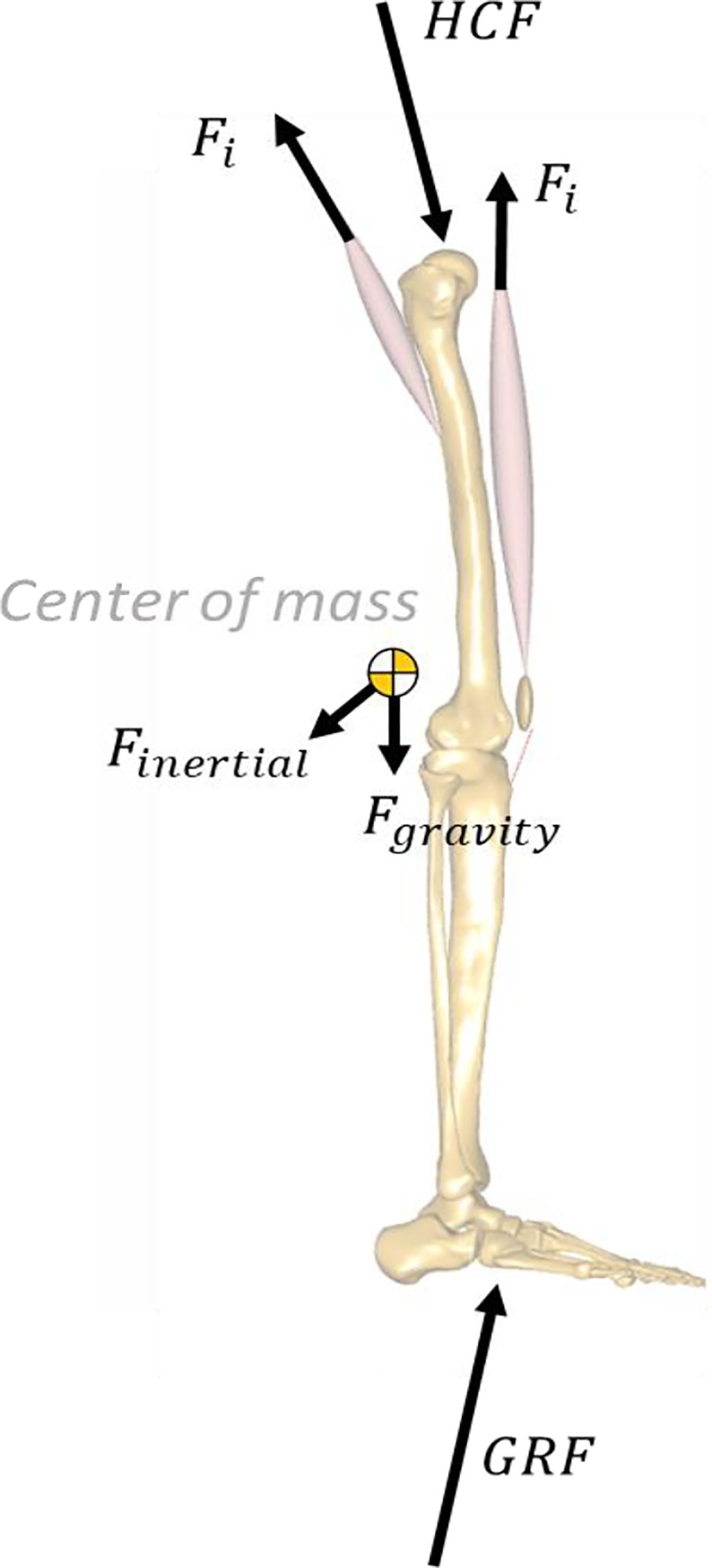
Musculoskeletal models represent a powerful tool to gain knowledge on the internal forces acting at the joint level in a non-invasive way. However, these models can present some errors associated with the level of detail in their geometrical representation. For this reason, a thorough validation is necessary to prove the reliability of their predictions. This study documents the development of a generic musculoskeletal model and proposes a working logic and simulation techniques for identifying specific model features in need of refinement; as well as providing a quantitative validation for the prediction of hip contact forces (HCF). The model, implemented in the AnyBody Modeling System and based on the cadaveric dataset TLEM 2.0, was scaled to match the anthropometry of a patient fitted with an instrumented hip implant and to reproduce gait kinematics based on motion capture data. The relative contribution of individual muscle elements to the HCF and joint moments was analyzed to identify critical geometries, which were then compared to muscle magnetic resonance imaging (MRI) scans and, in case of inconsistencies, were modified to better match the volumetric scans. The predicted HCF showed good agreement with the overall trend and timing of the measured HCF from the instrumented prosthesis. The average root mean square error (RMSE), calculated for the total HCF was found to be 0.298*BW. Refining the geometries of the muscles thus identified reduced RMSE on HCF magnitudes by 17% (from 0.359*BW to 0.298*BW) over the whole gait cycle. The detailed study of individual muscle contributions to the HCF succeeded in identifying muscles with incorrect anatomy, which would have been difficult to intuitively identify otherwise. Despite a certain residual over-prediction of the final hip contact forces in the stance phase, a satisfactory level of geometrical accuracy of muscle paths has been achieved with the refinement of this model.
Conflict of interest statement
Morten E. Lund, Kasper P. Rasmussen, and Anantharaman Gopalakrishnan are employed by AnyBody Technology A/S, which maintains and sells the modelling system used for the musculoskeletal models developed in the project. The funding for their salaries in this work comes from the EU grant mentioned in the financial disclosure. The remaining authors report no financial relationships with any organisations that might have an interest in the submitted work. None of the authors have any personal or non-financial competing interest in relation to this work. None of the competing interests alter our adherence to PLOS ONE policies on sharing data and materials.
Figures


Similar articles
-
Subject-specific geometrical detail rather than cost function formulation affects hip loading calculation.Comput Methods Biomech Biomed Engin. 2016 Nov;19(14):1475-88. doi: 10.1080/10255842.2016.1154547. Epub 2016 Mar 1. Comput Methods Biomech Biomed Engin. 2016. PMID: 26930478
-
Evaluation of predicted knee-joint muscle forces during gait using an instrumented knee implant.J Orthop Res. 2009 Oct;27(10):1326-31. doi: 10.1002/jor.20876. J Orthop Res. 2009. PMID: 19396858
-
A subject-specific musculoskeletal modeling framework to predict in vivo mechanics of total knee arthroplasty.J Biomech Eng. 2015 Feb 1;137(2):020904. doi: 10.1115/1.4029258. Epub 2015 Jan 26. J Biomech Eng. 2015. PMID: 25429519
-
Contributions to the understanding of gait control.Dan Med J. 2014 Apr;61(4):B4823. Dan Med J. 2014. PMID: 24814597 Review.
-
Multibody dynamics-based musculoskeletal modeling for gait analysis: a systematic review.J Neuroeng Rehabil. 2024 Oct 5;21(1):178. doi: 10.1186/s12984-024-01458-y. J Neuroeng Rehabil. 2024. PMID: 39369227 Free PMC article.
Cited by
-
Comparison of kinematic parameters of children gait obtained by inverse and direct models.PLoS One. 2022 Jun 24;17(6):e0270423. doi: 10.1371/journal.pone.0270423. eCollection 2022. PLoS One. 2022. PMID: 35749351 Free PMC article.
-
Virtual reality skateboarding training for balance and functional performance in degenerative lumbar spine disease.J Neuroeng Rehabil. 2024 May 9;21(1):74. doi: 10.1186/s12984-024-01357-2. J Neuroeng Rehabil. 2024. PMID: 38724981 Free PMC article.
-
Increased Femoral Anteversion Does Not Lead to Increased Joint Forces During Gait in a Cohort of Adolescent Patients.Front Bioeng Biotechnol. 2022 Jun 6;10:914990. doi: 10.3389/fbioe.2022.914990. eCollection 2022. Front Bioeng Biotechnol. 2022. PMID: 35733525 Free PMC article.
-
Reduced Muscle Strength Can Alter the Impact of Gait Modifications on Knee Cartilage Mechanics.J Orthop Res. 2025 Sep;43(9):1566-1580. doi: 10.1002/jor.70007. Epub 2025 Jun 27. J Orthop Res. 2025. PMID: 40576011 Free PMC article.
-
A review of musculoskeletal modelling of human locomotion.Interface Focus. 2021 Aug 13;11(5):20200060. doi: 10.1098/rsfs.2020.0060. eCollection 2021 Oct 6. Interface Focus. 2021. PMID: 34938430 Free PMC article. Review.
References
-
- Gao L, Fisher J, Jin Z. Effect of walking patterns on the elastohydrodynamic lubrication of metal-on-metal total hip replacements. Proc Inst Mech Eng Part J J Eng Tribol. 2011;225: 515–525. 10.1177/1350650110396802 - DOI
-
- Jonkers I, Sauwen N, Lenaerts G, Mulier M, Van der Perre G, Jaecques S. Relation between subject-specific hip joint loading, stress distribution in the proximal femur and bone mineral density changes after total hip replacement. J Biomech. 2008;41: 3405–3413. 10.1016/j.jbiomech.2008.09.011 - DOI - PubMed
Publication types
MeSH terms
LinkOut - more resources
Full Text Sources
Other Literature Sources
