. 2018 Sep 17;14(9):e1007664.
doi: 10.1371/journal.pgen.1007664. eCollection 2018 Sep.

Disrupted structure and aberrant function of CHIP mediates the loss of motor and cognitive function in preclinical models of SCAR16

Affiliations

Disrupted structure and aberrant function of CHIP mediates the loss of motor and cognitive function in preclinical models of SCAR16

Chang-He Shi et al. PLoS Genet. .

Abstract

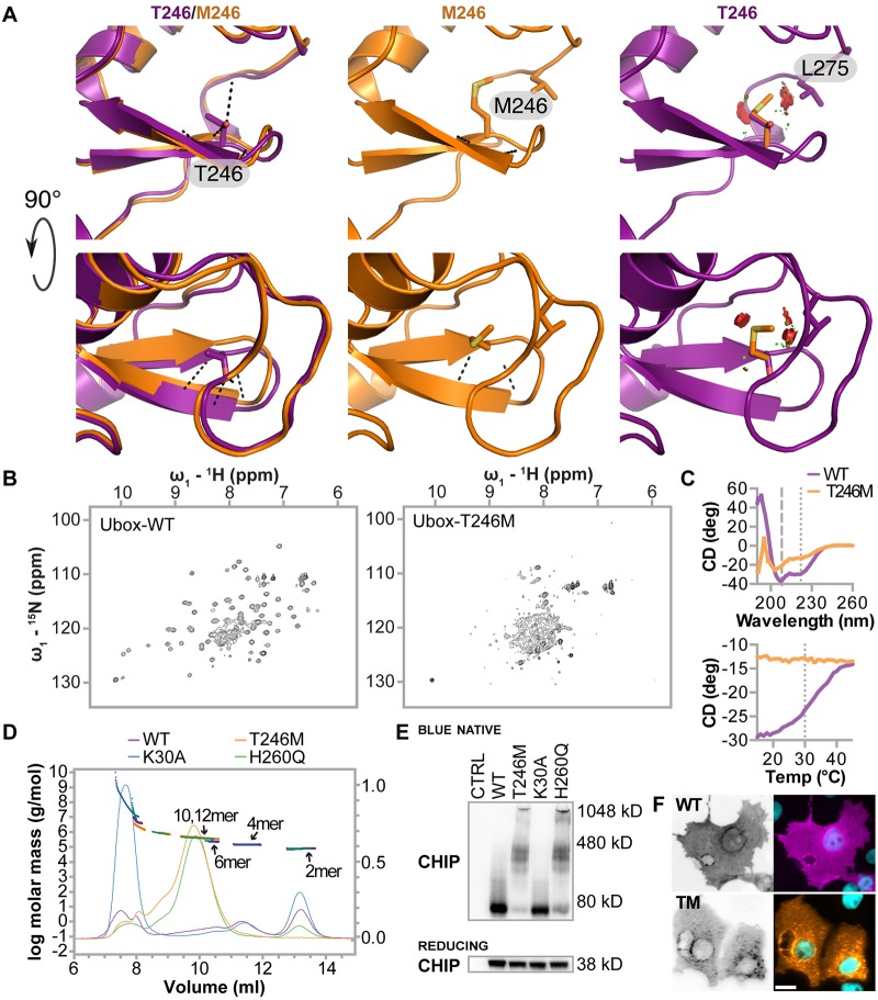
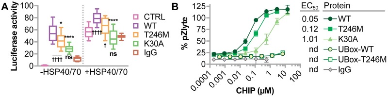
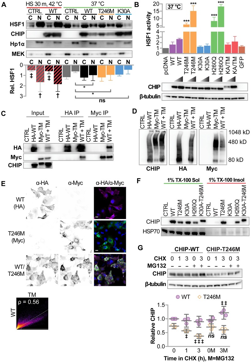
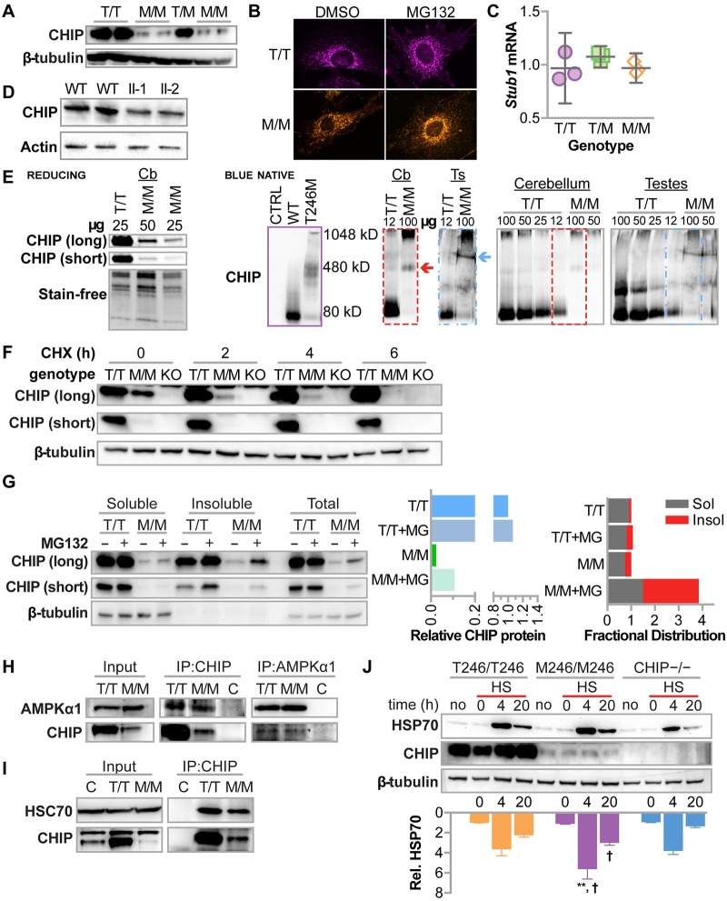
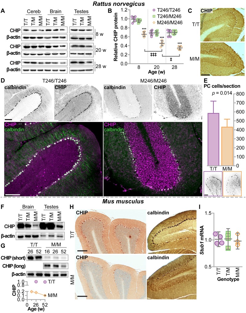
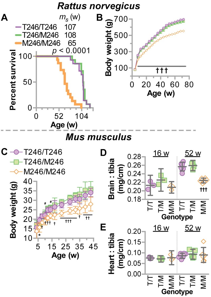
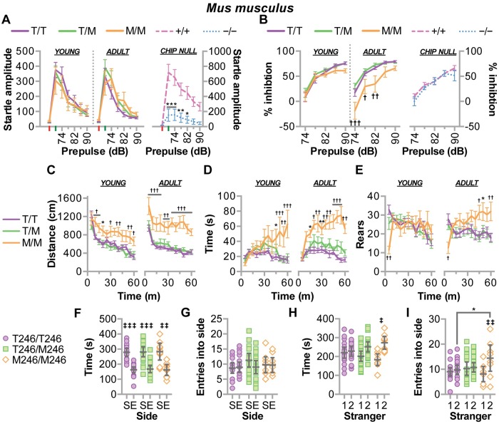
CHIP (carboxyl terminus of heat shock 70-interacting protein) has long been recognized as an active member of the cellular protein quality control system given the ability of CHIP to function as both a co-chaperone and ubiquitin ligase. We discovered a genetic disease, now known as spinocerebellar autosomal recessive 16 (SCAR16), resulting from a coding mutation that caused a loss of CHIP ubiquitin ligase function. The initial mutation describing SCAR16 was a missense mutation in the ubiquitin ligase domain of CHIP (p.T246M). Using multiple biophysical and cellular approaches, we demonstrated that T246M mutation results in structural disorganization and misfolding of the CHIP U-box domain, promoting oligomerization, and increased proteasome-dependent turnover. CHIP-T246M has no ligase activity, but maintains interactions with chaperones and chaperone-related functions. To establish preclinical models of SCAR16, we engineered T246M at the endogenous locus in both mice and rats. Animals homozygous for T246M had both cognitive and motor cerebellar dysfunction distinct from those observed in the CHIP null animal model, as well as deficits in learning and memory, reflective of the cognitive deficits reported in SCAR16 patients. We conclude that the T246M mutation is not equivalent to the total loss of CHIP, supporting the concept that disease-causing CHIP mutations have different biophysical and functional repercussions on CHIP function that may directly correlate to the spectrum of clinical phenotypes observed in SCAR16 patients. Our findings both further expand our basic understanding of CHIP biology and provide meaningful mechanistic insight underlying the molecular drivers of SCAR16 disease pathology, which may be used to inform the development of novel therapeutics for this devastating disease.


Conflict of interest statement

The authors have declared that no competing interests exist.

Figures

Fig 1
Fig 1. CHIP-T246M disrupts the structure of the U-box and promotes the formation of soluble oligomers.
(A) The structure of the U-box β-hairpin harboring residue 246 is shown for CHIP-WT (purple) and a Rosetta-relaxed model of CHIP-T246M (orange). The left panels show the CHIP-WT and CHIP-T246M backbones and the side chain of T246. The middle panels show the CHIP-T246M model. The right panels show T246M and the clashes (red discs) that would occur without adjustment of the U-box backbone structure. The lower panels shows identical structures rotated 90° about the x-axis from the view in the upper panels. Hydrogen bonds between residue 246 and neighboring residues are shown as black dashed lines for the left and middle panels. (B) 600-MHz 15N-1H transverse relaxation-optimized spectroscopy-HSQC spectra collected at 293 K for 2H,15N-labeled WT (left) and T246M (right) CHIP U box (218–303). (C, upper) Circular dichroism spectroscopy data collected for the U-box of WT (purple) and T246M (orange). (C, lower) Melting point determinations for the U-box of WT and T246M CHIP at 222nm. (D) Size distribution of full-length proteins of either WT (purple), T246M (orange), K30A (blue) and H260Q (green) CHIP determined by size-exclusion chromatography and multi-angle lights scattering. The molecular mass of oligomeric species of each protein are indicated (left Y-axis, thick lines) and the Rayleigh ratio chromatographs represent the amount of light scattering (right Y-axis, thin lines). (E) COS-7 cells were co-transfected with the indicated vectors (CTRL = pcDNA3, WT = pcDNA3-mycCHIP, T246M = pcDNA3-myc CHIP-T246M, K30A = pcDNA3-mycCHIP-K30A, H260Q = pcDNA3-mycCHIP-H260Q). Cells were collected on ice and total protein collected and freshly separated by BN PAGE under native conditions or SDS-PAGE under reducing conditions and immunoblotted with an anti-myc (CHIP). Locations of molecular weight standards in kilodaltons (kD) are indicated. (F) COS-7 cells were co-transfected with the indicated transgenes. 24 hours post transfection cells were fixed and immunostained for myc-CHIP expression (scale bar = 20 microns).
Fig 2
Fig 2. CHIP-T246M has chaperone activity in cell-free assays.
(A) Relative luciferase activity on heat-denatured luciferase after one hour incubation with the indicated CHIP proteins (CTRL = no protein, IgG = immunoglobulin G), either in the absence (left) or presence of HSP40 and HSP70, represented by dot plot and summarized by the mean ± 95% CI, N = 4 experimental replicates: *, **** correspond to p < 0.05, and 0.0001 via Tukey’s post hoc test compared to CHIP-WT, †, †††† correspond to p < 0.05, and 0.0001 via Tukey’s post hoc test compared to IgG; ns indicates p > 0.05 compared to IgG. (B) AMPK kinase activity towards the Z’LYTE peptide substrate in the presence of increasing amounts of the indicated recombinant CHIP proteins or IgG protein control represented by scatter plot and summarized by the mean ± 95% CI, N = 3 experimental replicates. The half maximal effective concentration (EC50) for each protein is included.
Fig 3
Fig 3. Cellular characterization of CHIP-T246M revealed enhanced activation of HSF1, changes in solubility, and increased turnover.
(A) Immunoblot analysis of HSF1, CHIP, HP1α (nuclear marker) and MEK (cytosolic marker) in cytosolic (C) and nuclear (N) fractions from COS-7 cells transiently transfected with the indicated vectors (CTRL = pcDNA3, WT = pcDNA3-mycCHIP, T246M = pcDNA3-mycCHIP-T246M, K30A = pcDNA3-mycCHIP-K30A) treated with or without heat shock as indicated. Densitometry of relative HSF1 protein is represented by the bar graph and 95% CI, N = 3 biological replicates; effect of heat shock: ‡ and † correspond to p < 0.01 and 0.0001 via Sidak’s post hoc comparison test to cytosolic or nuclear HSF1 in control conditions at 37 °C; effect of CHIP transgene: * p < 0.05 via Dunnett’s post hoc comparison to nuclear HSF1 levels in control conditions. (B) Bar graph of HSF1 transcription activity represented by the mean and 95% CI normalized to control vector (pcDNA) conditions in COS-7 cells transiently transfected with increasing amount if DNA using the indicated vectors (K30A-T246M = pcDNA3-mycCHIP-K30A-T246M, GFP = green fluorescent protein), N = 4 biological replicates, *** p < 0.0001 via Dunnet’s multiple comparison test to WT conditions. Immunoblot analysis of the myc-tag and β-tubulin confirmed transgene expression. (C) COS-7 cells were co-transfected with the indicated vectors (transgenes, HA-WT = pcDNA3-HA-CHIP, Myc-TM = pcDNA3-mycCHIP-T246M, WT + TM = HA-WT and Myc-TM). CHIP protein was immunoprecipitated with Anti-HA or Anti-Myc affinity gel. The inputs and resulting precipitants (IP) were separated by SDS-PAGE and immunoblotted with the indicated antibodies. (D) COS-7 cells were co-transfected with the indicated transgenes and cell lysates were separated by BN-PAGE and immunoblotted with the indicated antibodies. Locations of molecular weight standards in kilodaltons (kD) are indicated. (E) Micrographs of indirect immunofluorescence from COS-7 expressing the indicated transgenes to detect CHIP-WT (left), CHIP-T246M (center), or both (right). DAPI nuclear staining is also included (right panels) and the scale bar represents 20 microns. Co-localization of HA-WT and Myc-TM is represented by scatter plot and the indicated Pearson correlation (ρ). (F) Solubility analysis (in 1% Triton X-100, TX-100) of CHIP proteins, HSP70, and AMPKα in COS-7 cells determined by immunoblot analysis. (G) Immunoblot analysis of CHIP expression (top) in COS-7 cells treated with 50 μg/ml cycloheximide (CHX) for the indicated time (h) in the presence or absence of 20 μM MG132. β-tubulin. Densitometry analysis (bottom) represented by dot plot and summarized by the mean ± 95% CI of relative levels of CHIP protein were normalized to β-tubulin, N = 3 biological replicates, ‡, ‡‡, ‡‡‡ correspond to p < 0.05, 0.01, 0.0001 compared to starting expression levels within each construct via Tukey’s post hoc test.
Fig 4
Fig 4. CHIP-T246M expressed from the endogenous locus results is regulated post-translationally resulting in decreased steady-state protein levels, however, interactions between CHIP and known interactors are not affected.
(A) Immunoblot analysis of CHIP and β-tubulin protein in primary fibroblasts (MEFs) isolated from T246/T246 (T/T), T246/M246 (T/M), or M246/M246 (M/M) mouse embryos (B) Maximum intensity projections of CHIP immunofluorescence in MEFs treated with 0.05% DMSO or MG132. (C) qPCR analysis of Stub1 mRNA levels in MEFs represented by the dot plot and summarized by the mean and 95% confidence intervals. (D) Immunoblot analysis of CHIP and β-actin protein in fibroblasts isolated from control patients (WT) or siblings that are homozygous for CHIP-T246M (II-1 and II-2). (E) Immunoblot analysis of CHIP under reducing SDS-PAGE (left) or blue native PAGE (right) of varying amounts of protein extracts (μg) from cerebellum (Cbl) or testes (Ts) from mice with the indicated genotypes. Protein loading on the reducing blot was confirmed via stain-free technology. As a reference, migration of recombinant CHIP protein is provided (purple). The dashed boxes identify the regions used for longer exposure. (F) Immunoblot analysis of CHIP and β-tubulin in soluble cell lysates from either T/T or M/M, fibroblasts, or fibroblasts isolated from CHIP−/− embryos (KO) after exposure to cycloheximide (CHX) indicated in hours (h). Two exposures of CHIP immunoblots are provided to help visualize M/M conditions. (G) Immunoblot analysis of CHIP and β-actin in either the soluble or insoluble fraction of lysates or whole cell lysates from T/T or M/M MEFs treated with 20 μM MG132 or 0.05% DMSO control for 4 hours. (H) Immunoblot analysis of AMPKα1 and CHIP in cell lysates from T/T or M/M MEFs either before (input) or after immunoprecipitation (IP) of the indicated protein. Control samples (C) contained a mixture of 50% T/T and T/M and were immunoprecipitated with either rabbit IgG or goat IgG as controls for the CHIP and AMPKα1 antibodies, respectively. (I) Immunoblot analysis of HSC70 and CHIP in cell lysates from T/T or M/M MEFs either before (input) or after immunoprecipitation (IP) of CHIP. Control samples (C) contained a mixture of 50% T/T and T/M and were immunoprecipitated with rabbit IgG to control for the CHIP antibody. (J) Immunoblot analysis of HSP70 and CHIP in MEFs with the indicated genotypes that were treated without heat shock (no) or with heat shock (HS) followed by the indicated recovery time. Densitometry of relative HSP70 protein is represented by the bar graph and 95% CI, N = 3; **, † correspond to p < 0.01 or 0.05 via Tukey’s post hoc test comparing M/M to either T/T or CHIP−/−, respectively, at the indicated time point.
Fig 5
Fig 5. Rodents engineered with CHIP-T246M had decreased expression of CHIP in the brain and testes, Purkinje cell degeneration, increased mortality, and selective tissue atrophy.
(A) Immunoblot analysis of CHIP and β-actin in the cerebellum (cereb), brain, and testes of rats with either T246/T246 (T/T), T246/M246 (T/M), or M246/M246 (M/M) genotypes at harvested at different ages. (B) Densitometry analysis of relative CHIP protein represented by dot plot and summarized by the mean ± 95% CI: *** p < 0.001 compared to T/T or T/M; ‡, ‡‡‡ correspond to p < 0.05, or 0.001 compared to previous time point via Tukey’s post hoc test. (C) Representative micrographs of immunohistochemical detection of CHIP expression in rat cerebellums, 32 weeks of age, scale bar = 100 microns. (D) Indirect immunofluorescence of either calbindin or CHIP expression and the false color overlap (co-expression) in cerebellums of rats with the indicated genotypes, 32 weeks of age, scale bar = 100 microns. (E) Average number of Purkinje cells per section from rats at 32 weeks of age, summarized by the mean and 95% CI, N = 3 animals per genotype. Immunoblot analysis of CHIP in (F) brain and testes (26 weeks of age) or (G) cerebellum of mice of varying age, with the indicated genotypes. Densitometry analysis of relative CHIP protein levels, N = 3, * p < 0.05 via t-test in comparing CHIP in M/M mice at 52 versus 26 weeks normalized to stain-free protein levels. (H) Representative micrographs of immunohistochemical detection of either CHIP or calbindin expression in mouse cerebellums with the indicated genotypes, 52 weeks of age, scale bar = 200 microns. (I) qPCR analysis of Stub1 mRNA levels in four different tissues (heart, liver, brain, testes) isolated from mice with the indicated genotypes, N = 3 mice per tissue. Relative mRNA levels are represented by the dot plot and summarized by the mean and 95% confidence intervals.
Fig 6
Fig 6. Decreased mortality and weight in CHIP-T246M rodents.
(A) Survival analysis of rats with the indicated genotypes (Mantel-Cox test) with the median survival (ms) indicated in weeks (w), N = 12 animals per genotype. Total body weight of (B) rats or (C) mice with the indicated genotypes over age represented by scatter plot and summarized by the mean ± 95% CI. For rats, N = 10 (per genotype); for mice, N = 30, 29, and 18 for T/T, T/M, and M/M, respectively. Tukey’s post hoc test: * p < 0.05 M/M vs. T/T; †, ††, ††† correspond to p < 0.05, 0.01, 0.001 comparing M/M to T/T and T/M; # p < 0.05 M/M vs. T/M. (D) brain weight or (E) heart weight normalized to tibia length from mice with indicated genotypes represented by dot plot and summarized by the mean ± 95% CI: ††† corresponds to p < 0.001 comparing M/M to T/T and T/M.
Fig 7
Fig 7. CHIP-T246M in rodents resulted in progressive ataxia and altered gait.
Changes in (A) rotatrod performance, (B) support base, (C) stride, and (D) support ratio over time in rats with the indicated genotypes represented by line plot and summarized by the mean ± SEM (N = 6 animals/genotype), Tukey’s post hoc test: a, b, c, and ††† correspond to p < 0.05, 0.01, 0.001, and 0.0001 comparing M/M to T/T and T/M. (E) Graphical representation of gait parameters with foot locations indicated (RH = right hand, RF = right foot, LH = left hand, LF = left foot). (F) Rotarod analysis during the initial learning phase (left) or over time (right) represented by a line graph of the change in latency to fall over the first three trials, including a re-test (R) two days later, summarized by the mean ± SEM (N = 10–20 animals/genotype), Dunnett’s post hoc test: #, ## correspond to p < 0.05, 0.01 within T/T and T/M mice compared to trial 1; ††† p < 0.001 within all three genotypes compared to trial 1; 0.001 compared to previous time point; Tukey’s post hoc test: *, ** correspond to p < 0.05, 0.01 comparing M/M vs. T/T. (G) The initial rotarod performance in CHIP null mice (−/−) or wild-type mice (+/+) over the first three trials, including a re-test (R) two days later, summarized by the mean ± SEM (N = 10 animals/genotype), Tukey’s post hoc test: ‡ corresponds to p < 0.05 compare to trial 1; *, ** correspond to p < 0.05, 0.01 comparing genotypes. (H) Composite ataxia score of mice with the indicated genotypes including wild-type (+/+), heterozygous (+/−), and homozygous (−/−) knockouts of the CHIP allele represented by scatterplot and summarized by the mean ± SEM (N = 12 animals/genotype). Results of the linear regression analysis are depicted by the line and equation provided and results of the mixed model analysis provided. In comparing slopes, only M/M mice had a different slope at α = 0.05.
Fig 8
Fig 8. CHIP-T246M resulted in age-dependent deficits in sensorimotor skills, increased anxiety, and changes in sociability in mice.
(A) The amplitude of startle response (red and green are no stimulus and acoustic startle stimulus alone trials, respectively) and (B) the amount of prepulse inhibition over increasing levels of sound, decibels (dB) in mice of the indicated genotypes. (C) The distance traveled, (D) time spent in the center, and (E) the number of rearing movements in open field test over time. Tests (A-E) were performed on mice with the indicated genotypes at 11–13 w of age (young) and repeated at 33 w of age (adult) and are represented by line plot and summarized by the mean ± SEM (N = 10–20 animals/genotype), Tukey’s post hoc test: *, ** correspond to p < 0.05, 0.01 comparing M/M to T/T (or +/+ to −/−); †, ††, ††† correspond to p < 0.05, 0.01, 0.001 comparing M/M to T/T and T/M; ‡ corresponds to p < 0.001 compared to the initial stimulus within the genotype (learning), ns = p > 0.05. (F) Time spent and (G) number of entries in either the side with stranger 1 (S) compared to an empty cage (E). (H) Time spent and (I) number of entries in either the side with previous stranger 1 (1) or novel stranger 2 (2). Tests F-I were performed on mice with the indicated genotypes represented by dot plot and summarized by the mean ± 95% CI (N = 10–20 animals/genotype), Tukey’s post hoc test: ‡, ‡‡, ‡‡‡ correspond to p < 0.05, 0.01, 0.001 within genotype; * p < 0.05 M/M vs. T/T.
Fig 9
Fig 9. CHIP-T246M rodents developed cognitive deficits.
In rats, (A) the average latency to find the platform over four consecutive training days in the Morris Water maze task is represented by line plot and summarized by the mean ± SEM. (B) The amount of time spent in the target quadrant when the platform was removed after the last trial is represented by dot plot and summarized by the mean ± 95% CI. Tests A-B were administered on rats with the indicated genotypes at 16 w of age (young) and repeated at 36 w of age (adult), N = 6 animals/genotype, Tukey’s post hoc test: ** corresponds to p < 0.01 comparing M/M vs. T/T; †, ††, ††† correspond to p < 0.05, 0.01, 0.001 comparing M/M to T/T and T/M; ‡ corresponds to p < 0.05 within genotype. Conditioned fear testing was measured in mice by calculating the percentage of time freezing (no movement) during either (C) context or (D) cue-dependent learning during the training day (D1), and over 5 minute periods either a day after training (left) or two weeks later (right). Tests C-D are represented by line plot and summarized by the mean ± SEM (N = 10–20 animals/genotype), Tukey’s post hoc test: †, ††, ††† correspond to p < 0.05, 0.01, 0.001 comparing M/M to T/T and T/M; ‡ corresponds to p < 0.001 compared to the initial stimulus within the genotype (learning), ns = p > 0.05.
Fig 10
Fig 10. Changes in the cerebellar proteome due to CHIP-T246M.
(A) Heatmap (left) of cerebellar proteins determined to be differentially expressed in M246/M246 relative to T246/T246 rats and immunoblot analysis (right) of selected proteins in either cerebellums or whole brain in rats with the indicated genotype, GAPDH was used as a loading control. (B) Densitometry analysis of selected proteins from either cerebellum (Cbl) or brain (Brn) from rats of the indicated genotypes at different ages, represented by line plot and summarized by the mean ± SEM (N = 2 animals per tissue per genotype per timepoint). 3-way ANOVA analysis identified interactions between age and genotype for all five proteins (p < 0.0001) and no interaction between age, genotype, and tissue type (p range 0.14–0.91). Tukey’s post hoc test: **, ***, **** correspond to p < 0.01, 0.001, 0.0001 comparing M/M to T/T and T/M in both tissues. (C) Protein interactions analysis identified both the interaction clusters and functional enrichment of the differentially expressed proteins identified due to CHIP-T246M. The interaction clusters are connected with solid lines and additionally indicated by colored protein labels. Functional enrichment analysis is labeled by the colored spheres.

Similar articles

Cited by

References

    1. Ballinger CA, Connell P, Wu Y, Hu Z, Thompson LJ, Yin LY, et al. Identification of CHIP, a novel tetratricopeptide repeat-containing protein that interacts with heat shock proteins and negatively regulates chaperone functions. Mol Cell Biol. 1999;19: 4535–4545. - PMC - PubMed
    1. Kampinga HH, Kanon B, Salomons FA, Kabakov AE, Patterson C. Overexpression of the cochaperone CHIP enhances Hsp70-dependent folding activity in mammalian cells. Mol Cell Biol. 2003;23: 4948–4958. 10.1128/MCB.23.14.4948-4958.2003 - DOI - PMC - PubMed
    1. Jiang J, Ballinger CA, Wu Y, Dai Q, Cyr DM, Hohfeld J, et al. CHIP is a U-box-dependent E3 ubiquitin ligase: identification of Hsc70 as a target for ubiquitylation. J Biol Chem. 2001;276: 42938–44. 10.1074/jbc.M101968200 - DOI - PubMed
    1. Al-Ramahi I, Lam YC, Chen HK, de Gouyon B, Zhang M, Perez AM, et al. CHIP protects from the neurotoxicity of expanded and wild-type ataxin-1 and promotes their ubiquitination and degradation. J Biol Chem. 2006;281: 26714–26724. 10.1074/jbc.M601603200 - DOI - PubMed
    1. Dickey CA, Patterson C, Dickson D, Petrucelli L. Brain CHIP: removing the culprits in neurodegenerative disease. Trends Mol Med. 2007;13: 32–38. 10.1016/j.molmed.2006.11.003 - DOI - PubMed

Publication types

MeSH terms