Review
. 2018 Sep 14;19(9):2765.
doi: 10.3390/ijms19092765.

TOP2B: The First Thirty Years

Affiliations
Review

TOP2B: The First Thirty Years

Caroline A Austin et al. Int J Mol Sci. .

Abstract

Type II DNA topoisomerases (EC 5.99.1.3) are enzymes that catalyse topological changes in DNA in an ATP dependent manner. Strand passage reactions involve passing one double stranded DNA duplex (transported helix) through a transient enzyme-bridged break in another (gated helix). This activity is required for a range of cellular processes including transcription. Vertebrates have two isoforms: topoisomerase IIα and β. Topoisomerase IIβ was first reported in 1987. Here we review the research on DNA topoisomerase IIβ over the 30 years since its discovery.

Keywords: Cell cycle; TOP2; Topoisomerase II; cell biology; review; transcription.


Conflict of interest statement

The authors declare no conflict of interest.

Figures

Figure 1
Figure 1
Schematic representation of the domain arrangement of human topoisomerase IIβ. The ATPase domain is shown in red, the breakage-reunion domain in green and the C-terminal domain in blue. The positions of conserved motifs are marked above. The line underneath shows the location of site-directed mutations reported by West et al. [7] (above) and mutations selected for drug resistance reported in Leontiou et al. [8] (below). The locations of inter-domain hinge regions elucidated by limited proteolysis reported in Austin et al. [9] are also shown. At the bottom of Figure 1, the locations of the initial partial cDNA clones encoding portions of TOP2B are shown [5]; SP11, and CAA5, clone CAA6, confirming that CAA5 and SP11 were part of the same gene, are also shown [10,11].
Figure 2
Figure 2
Elutriation of K562 cells separates the cells according to size. (A) FACS analysis (Flowjo) of each elutriation fraction showing the percentage of G1, S or G2/M phase cells. (B) Immunofluorescence analysis of each elutriation fraction showing the percentage of each fraction in lateS/G2/M determined by CENPF immunofluorescence and the percentage of mitotic cells determined by DAPI staining. (C,D) TOP2A and TOP2B protein level per cell was determined by quantitative immunofluorescence [77], the data shown represent the mean ± SEM of median values obtained from three replicate experiments. Statistical analysis was carried out using one way ANOVA (* p < 0.05; *** p < 0.0001).
Figure 3
Figure 3
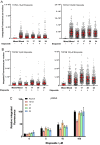
TOP2 covalent complex levels determined by trapped in agarose DNA immunostaining (TARDIS) and γH2AX integrated fluorescence. Quantification of etoposide-induced TOP2A and TOP2B covalent-DNA complexes, (A,B, respectively). Cells from each elutriated fraction were treated with etoposide (10 or 100 μM) and TOP2-DNA covalent complexes were detected and quantified by TARDIS analysis [73,74,75,77]. The data are shown as scattergrams, derived from a single representative experiment. Bars represent the median and interquartile range. (C) Cells from each elutriated fraction were treated with etoposide (5, 10 or 100 μM) or solvent (DMSO) and analysed by γH2AX immunofluorescence; the level of γH2AX integrated fluorescence was determined by quantitative immunofluorescence [77]. The data are shown as means of the medians obtained from three replicate experiments.
Figure 4
Figure 4
Drug sensitivity of the starting mixed population and the elutriated fractions. Cells from each elutriated fraction were treated with etoposide (0–100 μM) for 1 h. Cells were then replated in fresh medium and were counted after 24, 48 and 96 h. Population doublings were calculated according to the formula PD = Log10(cell count/initial cell count)/Log10 (2). Data represent the mean ± SEM derived from duplicate elutriation experiments.

References

    1. Drake F.H., Zimmerman J.P., McCabe F.L., Bartus H.F., Per S.R., Sullivan D.M., Ross W.E., Mattern M.R., Johnson R.K., Crooke S.T. Purification of topoisomerase II from amsacrine-resistant P388 leukemia cells. Evidence for two forms of the enzyme. J. Biol. Chem. 1987;262:16739–16747. - PubMed
    1. Per S.R., Mattern M.R., Mirabelli C.K., Drake F.H., Johnson R.K., Crooke S.T. Characterization of a subline of P388 leukemia resistant to amsacrine: Evidence of altered topoisomerase II function. Mol. Pharmacol. 1987;32:17–25. - PubMed
    1. Holden J.A., Rolfson D.H., Wittwer C.T. Human DNA topoisomerase II: Evaluation of enzyme activity in normal and neoplastic tissues. Biochemistry. 1990;29:2127–2134. doi: 10.1021/bi00460a024. - DOI - PubMed
    1. Roca J., Mezquita C. DNA topoisomerase II activity in nonreplicating, transcriptionally inactive, chicken late spermatids. EMBO J. 1989;8:1855–1860. doi: 10.1002/j.1460-2075.1989.tb03581.x. - DOI - PMC - PubMed
    1. Chung T.D., Drake F.H., Tan K.B., Per S.R., Crooke S.T., Mirabelli C.K. Characterization and immunological identification of cDNA clones encoding two human DNA topoisomerase II isozymes. Proc. Natl. Acad. Sci. USA. 1989;86:9431–9435. doi: 10.1073/pnas.86.23.9431. - DOI - PMC - PubMed

MeSH terms

Grants and funding

LinkOut - more resources