. 2018 Sep 14;10(9):1304.
doi: 10.3390/nu10091304.

Cardioprotective Effects of Nanoemulsions Loaded with Anti-Inflammatory Nutraceuticals against Doxorubicin-Induced Cardiotoxicity

Affiliations

Cardioprotective Effects of Nanoemulsions Loaded with Anti-Inflammatory Nutraceuticals against Doxorubicin-Induced Cardiotoxicity

Vincenzo Quagliariello et al. Nutrients. .

Abstract

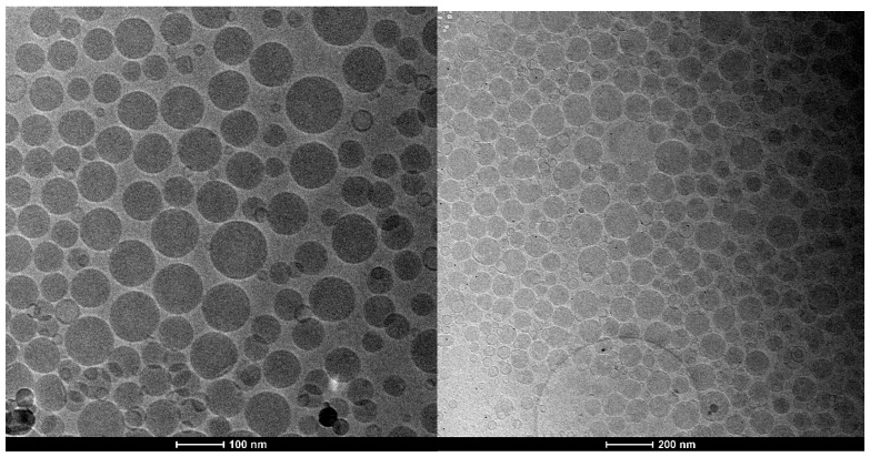
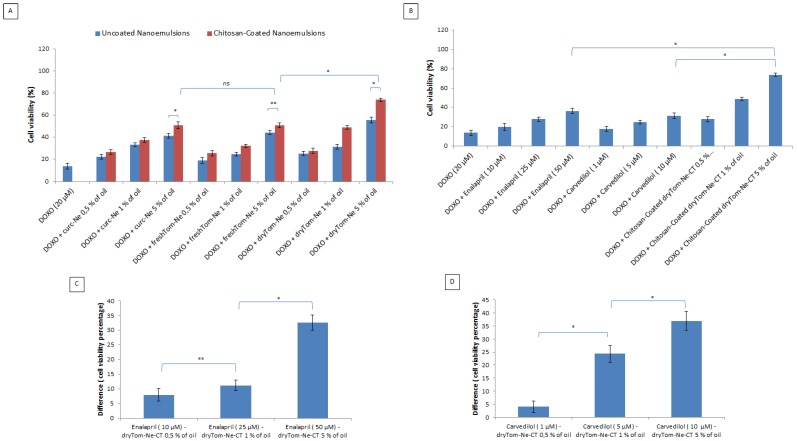
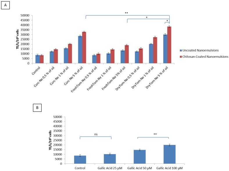
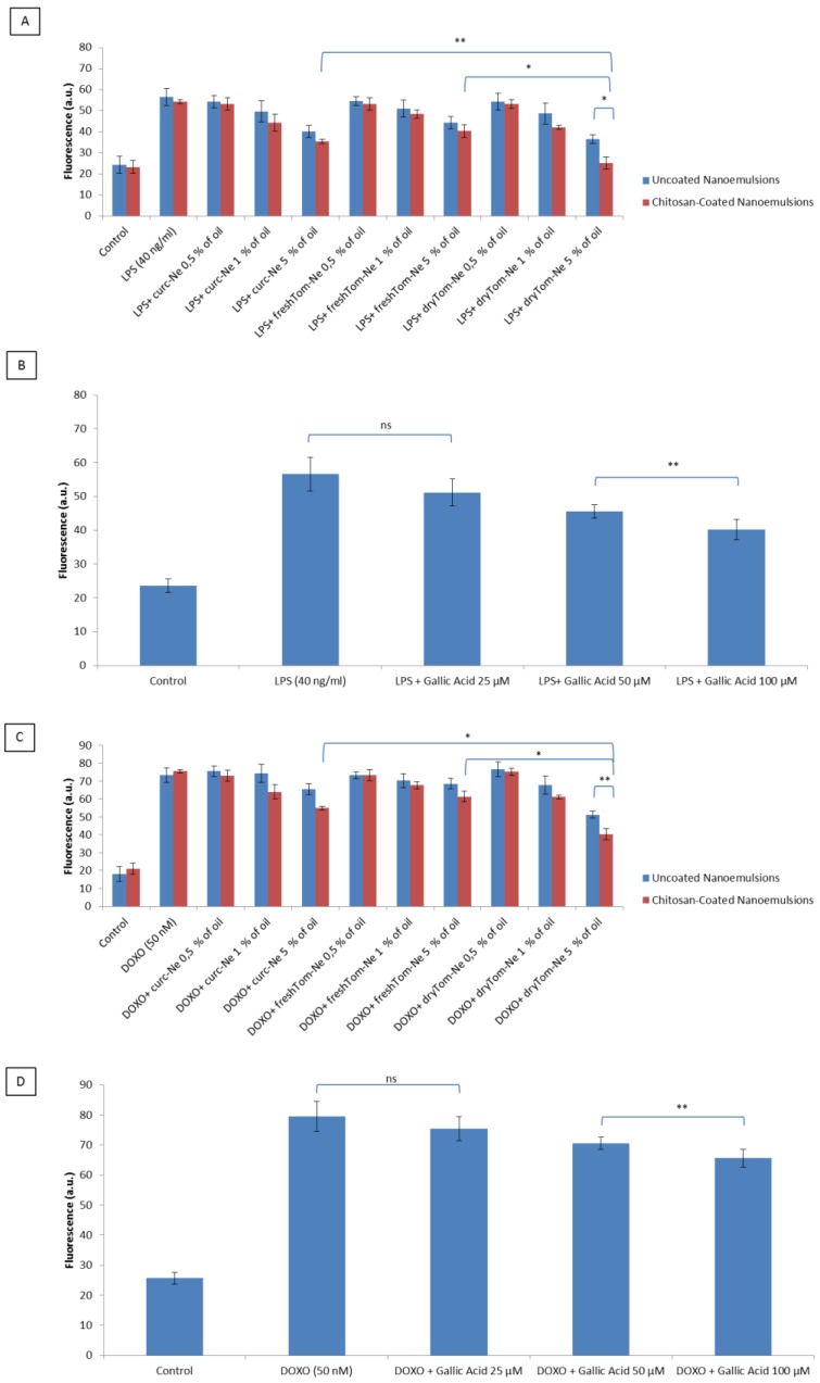
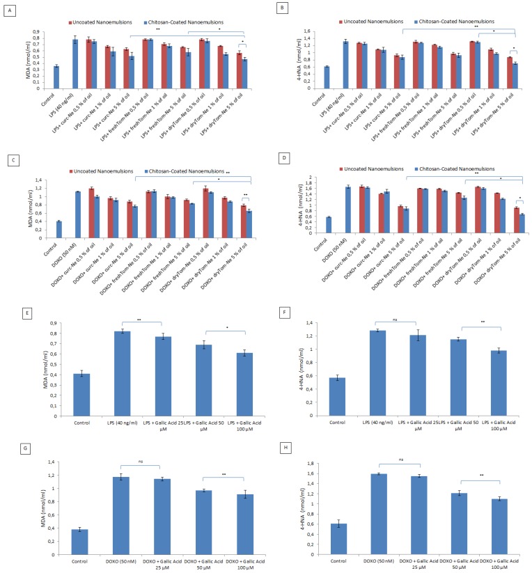
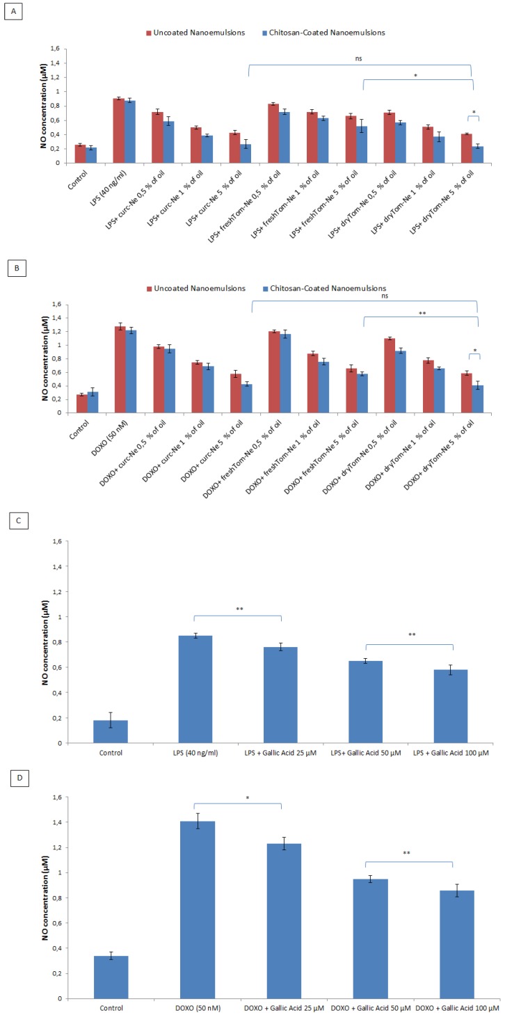
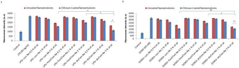
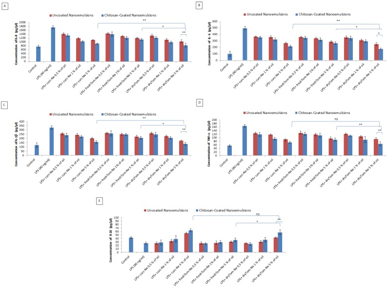
Doxorubicin is a highly active antineoplastic agent, but its clinical use is limited because of its cardiotoxicity. Although nutraceuticals endowed with anti-inflammatory properties exert cardioprotective activity, their bioavailability and stability are inconsistent. In an attempt to address this issue, we evaluated whether bioavailable nanoemulsions loaded with nutraceuticals (curcumin and fresh and dry tomato extracts rich in lycopene) protect cardiomyoblasts (H9C2 cells) from doxorubicin-induced toxicity. Nanoemulsions were produced with a high-pressure homogenizer. H9C2 cells were incubated with nanoemulsions loaded with different nutraceuticals alone or in combination with doxorubicin. Cell viability was evaluated with a modified MTT method. The levels of the lipid peroxidation products malondialdehyde (MDA) and 4-hydroxy-2-butanone (4-HNA), and of the cardiotoxic-related interleukins IL-6, IL-8, IL-1β and IL-10, tumor necrosis factor-alpha (TNF-α), and nitric oxide were analyzed in cardiomyoblasts. The hydrodynamic size of nanoemulsions was around 100 nm. Cell viability enhancement was 35⁻40% higher in cardiomyoblasts treated with nanoemulsion + doxorubicin than in cardiomyoblasts treated with doxorubicin alone. Nanoemulsions also protected against oxidative stress as witnessed by a reduction of MDA and 4-HNA. Notably, nanoemulsions inhibited the release of IL-6, IL-8, IL-1β, TNF-α and nitric oxide by around 35⁻40% and increased IL-10 production by 25⁻27% versus cells not treated with emulsions. Of the nutraceuticals evaluated, lycopene-rich nanoemulsions had the best cardioprotective profile. In conclusion, nanoemulsions loaded with the nutraceuticals described herein protect against cardiotoxicity, by reducing inflammation and lipid oxidative stress. These results set the stage for studies in preclinical models.

Keywords: cardiology; cytokines; doxorubicin; nanomedicine; nutraceuticals.


Conflict of interest statement

The authors declare no conflict of interest. Any role of the funding sponsors in the design of the study; in the collection, analyses or interpretation of data; in the writing of the manuscript, or in the decision to publish the results.

Figures

Figure 1
Figure 1
Cryo-TEM projection images of dryTom-Ne (left) and dryTom-Ne-CT (right).
Figure 2
Figure 2
Representative chromatograms for the samples “dry tomato extract” (left) and “fresh tomato extract” (right). Lycopene is peak n. 5.
Figure 3
Figure 3
Cell viability in function of the concentration of nutraceutical-loaded uncoated and chitosan-coated nanoemulsions all tested at concentrations from 0.5 to 5% v/v of oil tested combined with doxorubicin at 20 µM. (A). The viability of cardiomyoblasts incubated with doxorubicin (at 20 µM) in association with Enalapril at 10, 25 and 50 µM and Carvedilol at 1, 5 and 10 µM. (B). Difference in the percentage of cell viability versus control cells, between (C) Enalapril and (D) Carvedilol and the best nanoemulsion (dryTom-Ne-CT) at different concentrations * p < 0.001; ** p < 0.05; ns: not significant.
Figure 4
Figure 4
Left, overall cellular uptake quantification of H9c2 cells (at a density of 5 × 10³ cells/well) from 0.5 up to 24 h of contact with fluorescent nanocarriers at 1% v/v oil. Right, effect of different inhibitors on the internalization of fluorescent nanocarriers (1% v/v oil) in H9c2 cells after 4 h of incubation. The data are normalized against their controls.
Figure 5
Figure 5
Mean antioxidant values (±SEM) of H9c2 cell lysates (TE/L/106 cells). Antioxidant values of cell lysates after challenge with 50 mL of AAPH (4 mM) for 10 min and recovery with PBS (control) or chitosan-coated or uncoated nutraceutical-loaded nanoemulsions (A) or gallic acid (B) at 25, 50 and 100 µM. * p < 0.001; ** p < 0.05.
Figure 6
Figure 6
Detection of intracellular reactive oxygen species by fluorescence (a.u) in the H9c2 cell line (5000 cells/well). Cells were pretreated or not with uncoated and chitosan-coated nutraceutical-loaded nanoemulsions for 4 h before stimulation with 40 ng/mL of lipopolysaccharide (LPS) (A) or 50 nM of doxorubicin (C) for 24 h. Gallic acid was also exposed to cardiomyoblasts as positive control before stimulation with LPS (B) or doxorubicin (D). * p < 0.001; ** p < 0.05.
Figure 7
Figure 7
Cellular quantification of malondialdehyde (MDA) and 4HNA production under pro-inflammatory conditions and doxorubicin exposure. MDA and 4HNA (nmol/mL) production by cardiomyoblasts treated with LPS (A,B) and doxorubicin (C,D) alone or combined with uncoated or chitosan-coated nutraceutical-loaded nanocarriers at concentrations ranging from 0.5 to 5% oil. At the same condition, as positive control, we tested also the effects of gallic acid at 25, 50 and 100 µM (E,F) for LPS treatments; (G,H) for doxorubicin treatments). * p < 0.001; ** p < 0.05; ns: not significant.
Figure 8
Figure 8
Measurement of nitric oxide (NO) production expressed as nitric oxide concentration (µM) in H9c2 cells (5000 cells/well). Cells were pretreated or not with uncoated and chitosan-coated nutraceutical-loaded nanoemulsions at 0.5%, 1% and 5% of oil 4 h before stimulation with 40 ng/mL of LPS (A) or 50 nM of DOXO, (B) for 24 h. Pretreatments were also performed by incubating cells with gallic acid, as positive control, for both LPS (C) and DOXO (D) treatments. * p < 0.001; ** p < 0.05; ns: not significant.
Figure 9
Figure 9
Intracellular calcium fluorescence staining in H9c2 cells (expressed as fluorescence intensity a.u). Cells were pretreated or not with uncoated and chitosan-coated nutraceutical-loaded nanoemulsions at 0.5%, 1% and 5% of oil for 4 h before stimulation with 40 ng/mL of LPS (A) or 50 nM of DOXO, (B) for 24 h. * p < 0.001; ** p < 0.05; ns: not significant.
Figure 10
Figure 10
Anti-inflammatory effects of nutraceutical-loaded nanoemulsionson IL-8 (A), IL-6 (B), IL-1β (C), TNF-α (D) and IL-10 (E) production of cardiomyoblasts (at a density of 1.2 × 10⁵ cells/well). Cells were treated or not with 0.1 mL of a 0.5%, 1% and 5% of oil of chitosan-coated and uncoated nanoemulsions for 5 h before exposure to lipopolysaccharides (40 ng/mL) for 12 h. * p < 0.001, ** p < 0.05; ns: not significant.

References

    1. Fernandez-Chas M., Curtis M.J., Niederer S.A. Mechanism of doxorubicin cardiotoxicity evaluated by integrating multiple molecular effects into a biophysical model. Br. J. Pharmacol. 2018;175:763–781. doi: 10.1111/bph.14104. - DOI - PMC - PubMed
    1. Mele D., Tocchetti C.G., Pagliaro P., Madonna R., Novo G., Pepe A., Zito C., Maurea N., Spallarossa P. Pathophysiology of anthracycline cardiotoxicity. J. Cardiovasc. Med. 2016;17(Suppl. 1):e3–e11. doi: 10.2459/JCM.0000000000000378. - DOI - PubMed
    1. Maurea N., Coppola C., Piscopo G., Galletta F., Riccio G., Esposito E., De Lorenzo C., De Laurentiis M., Spallarossa P., Mercuro G. Pathophysiology of cardiotoxicity from target therapy and angiogenesis inhibitors. J. Cardiovasc. Med. 2016;17(Suppl. 1):e19–e26. doi: 10.2459/JCM.0000000000000377. - DOI - PubMed
    1. Simůnek T., Stérba M., Popelová O., Adamcová M., Hrdina R., Gersl V. Anthracycline-induced cardiotoxicity: Overview of studies examining the roles of oxidative stress and free cellular iron. Pharmacol. Rep. 2009;61:154–171. doi: 10.1016/S1734-1140(09)70018-0. - DOI - PubMed
    1. Wu K., Schwartz S.J., Platz E.A. Variations in plasma lycopene and specific isomers over time in a cohort of U.S. men. J. Nutr. 2003;133:1930–1936. doi: 10.1093/jn/133.6.1930. - DOI - PubMed

MeSH terms

LinkOut - more resources