Review
. 2018 Sep 14;8(3):94.
doi: 10.3390/biom8030094.

Plasma Membrane Lipid Domains as Platforms for Vesicle Biogenesis and Shedding?

Affiliations
Review

Plasma Membrane Lipid Domains as Platforms for Vesicle Biogenesis and Shedding?

Hélène Pollet et al. Biomolecules. .

Abstract

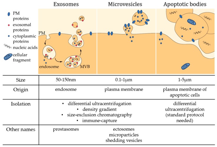
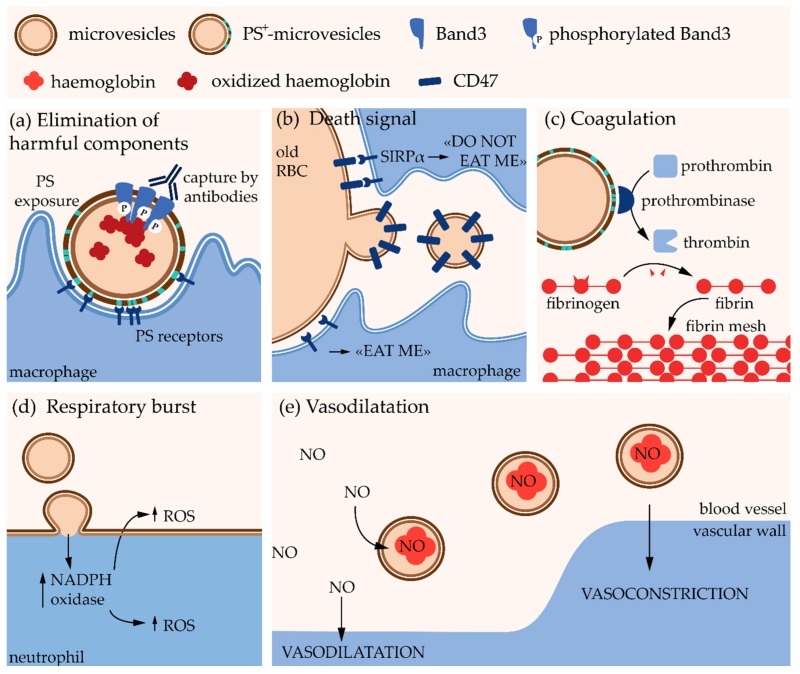
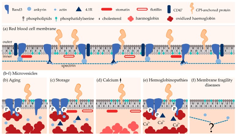
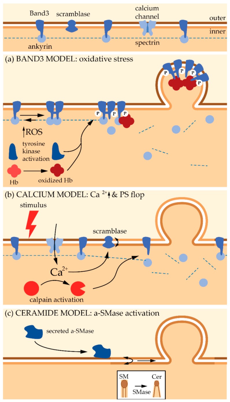
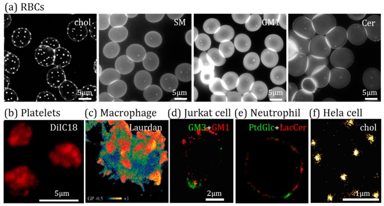
Extracellular vesicles (EVs) contribute to several pathophysiological processes and appear as emerging targets for disease diagnosis and therapy. However, successful translation from bench to bedside requires deeper understanding of EVs, in particular their diversity, composition, biogenesis and shedding mechanisms. In this review, we focus on plasma membrane-derived microvesicles (MVs), far less appreciated than exosomes. We integrate documented mechanisms involved in MV biogenesis and shedding, focusing on the red blood cell as a model. We then provide a perspective for the relevance of plasma membrane lipid composition and biophysical properties in microvesiculation on red blood cells but also platelets, immune and nervous cells as well as tumor cells. Although only a few data are available in this respect, most of them appear to converge to the idea that modulation of plasma membrane lipid content, transversal asymmetry and lateral heterogeneity in lipid domains may play a significant role in the vesiculation process. We suggest that lipid domains may represent platforms for inclusion/exclusion of membrane lipids and proteins into MVs and that MVs could originate from distinct domains during physiological processes and disease evolution.

Keywords: calcium; ceramide; cholesterol; cytoskeleton; lipid domains; microvesicle; oxidative stress; raft; red blood cell; sphingomyelinase.


Conflict of interest statement

The authors declare no conflict of interest.

Figures

Figure 1
Figure 1
Characteristics of the three main classes of extracellular vesicles. MVB, multivesicular body; PM, plasma membrane.
Figure 2
Figure 2
Pathophysiological effects of erythrocyte-derived microvesicles. PS, phosphatidylserine; RBC, red blood cell; SIRPα, signal regulatory protein α; NADPH; reduced form of nicotinamide adenine dinucleotide phosphate; ROS, reactive oxygen species; NO, nitric oxide.
Figure 3
Figure 3
Schematic representation of lipid and protein composition of red blood cell-derived microvesicles. (a) RBC plasma membrane. (bf) RBC-derived microvesicles in (b,c) physiological processes (senescence in vivo and storage at 4 °C), (d) pharmacological Ca2+ boost, and (e,f) pathological situations (hemoglobinopathies and membrane fragility diseases).
Figure 4
Figure 4
Models described in the literature for the biogenesis and shedding of red blood cell-derived microvesicles. Hb: haemoglobin; P: phosphorylation; a-SMase: acid sphingomyelinase.
Figure 5
Figure 5
Visualization of plasma membrane lipid domains. (a) RBCs labeled for chol with Theta-D4-mCherry or with boron-dipyrromethene-sphingomyelin (BODIPY-SM), -GM1 (BODIPY-GM1) or –ceramide (BODIPY-Cer) and analyzed by fluorescence or confocal microscopy. (b) Platelets labeled with DilC18 and analyzed by fluorescence microscopy. (c) Macrophage labeled with Laurdan and vizualized by two-photon microscopy. (d) Jurkat cell labeled for GM3 and GM1 using anti-GM3 serum and cholera toxin B subunit, respectively, and visualized by confocal imaging. (e) Human neutrophil stained for phosphatidylglucoside (PtdGlc) and lactosylceramide (LacCer) and examined by stimulated emission depletion microscopy (STED). (f) HeLa cell labeled for chol with Theta-D4-DRONPA and processed by photoactivated localization microscopy (PALM). Adapted from (a) [225]; (b) [229]; (c) [230]; (d) [231]; (e) [232]; (f) [233].

Similar articles

Cited by

References

    1. Deatherage B.L., Cookson B.T. Membrane vesicle release in bacteria, eukaryotes, and archaea: A conserved yet underappreciated aspect of microbial life. Infect. Immun. 2012;80:1948–1957. doi: 10.1128/IAI.06014-11. - DOI - PMC - PubMed
    1. Robinson D.G., Ding Y., Jiang L. Unconventional protein secretion in plants: A critical assessment. Protoplasma. 2016;253:31–43. doi: 10.1007/s00709-015-0887-1. - DOI - PubMed
    1. Gould S.J., Raposo G. As we wait: Coping with an imperfect nomenclature for extracellular vesicles. J. Extracell. Vesicles. 2013;2 doi: 10.3402/jev.v2i0.20389. - DOI - PMC - PubMed
    1. Raposo G., Stoorvogel W. Extracellular vesicles: Exosomes, microvesicles, and friends. J. Cell Biol. 2013;200:373–383. doi: 10.1083/jcb.201211138. - DOI - PMC - PubMed
    1. Gyorgy B., Szabo T.G., Pasztoi M., Pal Z., Misjak P., Aradi B., Laszlo V., Pallinger E., Pap E., Kittel A., et al. Membrane vesicles, current state-of-the-art: Emerging role of extracellular vesicles. Cell. Mol. Life Sci. 2011;68:2667–2688. doi: 10.1007/s00018-011-0689-3. - DOI - PMC - PubMed

Publication types

LinkOut - more resources