. 2018 Sep 15;9(3):53.
doi: 10.3390/jfb9030053.

Specialized Living Wound Dressing Based on the Self-Assembly Approach of Tissue Engineering

Affiliations

Specialized Living Wound Dressing Based on the Self-Assembly Approach of Tissue Engineering

Laurence Cantin-Warren et al. J Funct Biomater. .

Abstract

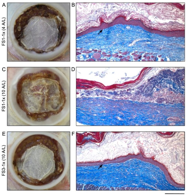
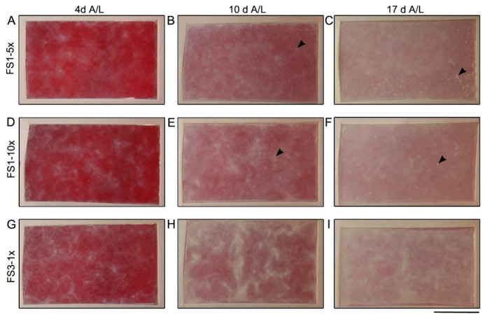
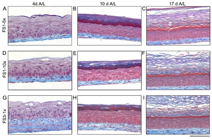
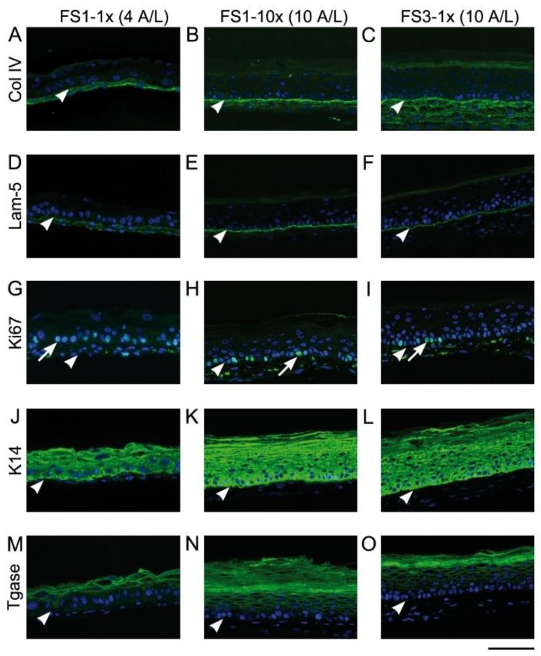
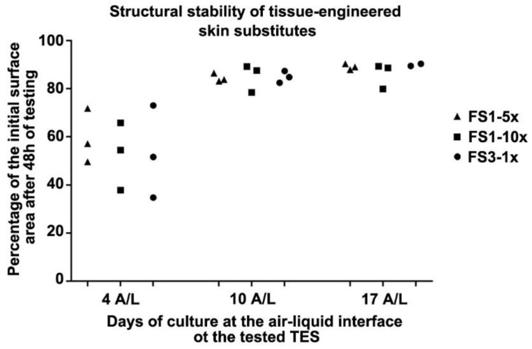
There is a high incidence of failure and recurrence for chronic skin wounds following conventional therapies. To promote healing, the use of skin substitutes containing living cells as wound dressings has been proposed. The aim of this study was to produce a scaffold-free cell-based bilayered tissue-engineered skin substitute (TES) containing living fibroblasts and keratinocytes suitable for use as wound dressing, while considering production time, handling effort during the manufacturing process, and stability of the final product. The self-assembly method, which relies on the ability of mesenchymal cells to secrete and organize connective tissue sheet sustaining keratinocyte growth, was used to produce TESs. Three fibroblast-seeding densities were tested to produce tissue sheets. At day 17, keratinocytes were added onto 1 or 3 (reference method) stacked tissue sheets. Four days later, TESs were subjected either to 4, 10, or 17 days of culture at the air⁻liquid interface (A/L). All resulting TESs were comparable in terms of their histological aspect, protein expression profile and contractile behavior in vitro. However, signs of extracellular matrix (ECM) digestion that progressed over culture time were noted in TESs produced with only one fibroblast-derived tissue sheet. With lower fibroblast density, the ECM of TESs was almost completely digested after 10 days A/L and lost histological integrity after grafting in athymic mice. Increasing the fibroblast seeding density 5 to 10 times solved this problem. We conclude that the proposed method allows for a 25-day production of a living TES, which retains its histological characteristics in vitro for at least two weeks.

Keywords: bilayered skin substitutes; culture techniques; regenerative medicine; skin equivalent; skin ulcer; tissue culture; tissue engineering.


Conflict of interest statement

The authors declare no conflict of interest.

Figures

Figure 1
Figure 1
Macroscopic and histological analysis of tissue-engineered skin substitutes matured in vitro. Representative macroscopic pictures (left panel) and histological staining (right panel) of FS1-1× (AF), and FS3-1× (G,H) cultured for 4 (4 A/L) and 10 (10 A/L) days at the air–liquid interface. A,D,G are top view pictures of the TES in their culture flasks, the red appearance is due to the culture media (red) that is under the TES. The arrowheads point to opaque white dots correlated with keratinocyte infiltrations across and under the reconstructed dermis (E) and arrow points a clear area correlated with ECM digestion zone (F). Note the whitish appearance of the FS3-1× (G) indicating a well differentiated epidermis (H). Histological coloration: Masson’s trichrome. Scale bar: A,D,G, 25 mm; B,C,E,F,H: 100 µm.
Figure 2
Figure 2
Macroscopic and histological analysis of tissue-engineered skin substitutes matured 21 days in vivo. Representative macroscopic (left panel) and histological (right panel) results of FS1-1× (AD) and FS3-1× (E,F) cultured for 4 (4 A/L) and 10 (10 A/L) days at the air–liquid interface before grafting in athymic mice. Arrows point out the fully differentiated epidermis well attached to the underlying dermis. Note that when the air–liquid interface culture period of FS1-1× was prolonged to 10 days before grafting, epidermis was absent in several areas after in vivo maturation (D). Histological coloration: Masson’s trichrome. Scale bar: A,C,D: 10 mm; B,D,F 235 μm.
Figure 3
Figure 3
Macroscopic analysis of tissue-engineered skin substitutes matured in vitro. Top view pictures of FS1-5× (A–C), FS1-10× (D–F), and FS3-1× (G–I) cultured for 4 (4 A/L), 10 (10 A/L), and 17 (17 A/L) days at the air–liquid interface. Arrowheads point opaque dots correlated with keratinocyte infiltrations across and under the reconstructed dermis. The red appearance is due to the culture media (red) that is under the TES. The culture media (red) under the FS1 is more apparent when the stratum corneum of the epidermis is thinner. Scale bar: 25 mm.
Figure 4
Figure 4
Histological analysis of tissue-engineered skin substitutes matured in vitro. Representative histological results of TES produced with FS1-5× (A–C), FS1-10× (D–F), and FS3-1× (G–I) cultured for 4 (4 A/L), 10 (10 A/L) and 17 (17 A/L) days at the air–liquid interface. Histological coloration: Masson’s trichrome. Scale bar: 100 µm.
Figure 5
Figure 5
Analysis of skin marker expression in tissue-engineered skin substitutes matured in vitro. Representative pictures FS1-1× (left panel), FS1-10× (center panel), and FS3-1× (right panel) cultured for 4 (4 A/L) and 10 (10 A/L) days at the air–liquid interface immunolabeled for the detection of type IV collagen (AC), laminin 5 (D–F), Ki67 (GI), keratin 14 (JL), and transglutaminase (MO). Arrowheads point out the level of the dermo-epidermal junction. Arrows point out nuclei expressing the proliferation marker Ki67. Col IV, type IV collagen; K14, keratin 14; Lam-5, laminin 5; Tgase, transglutaminase. Scale bar: 100 µm.
Figure 6
Figure 6
Structural stability of tissue-engineered skin substitutes matured in vitro for various time at the air–liquid interface before measuring surface area after an additional 48 h on agar substrate. Resulting contraction indicated as a percentage of the initial (at t = 0 h on agar) surface area of TES FS1-5× 10d A/L (triangles), FS1-10× (squares), and FS3-1× (circles) cultured for 4 (4 A/L), 10 (10 A/L), and 17 (17 A/L) days at the air–liquid interface remaining after 48 h of in vitro testing on agar.

References

    1. Sen C.K., Gordillo G.M., Roy S., Kirsner R., Lambert L., Hunt T.K., Gottrup F., Gurtner G.C., Longaker M.T. Human skin wounds: A major and snowballing threat to public health and the economy. Wound Repair Regen. 2009;17:763–771. doi: 10.1111/j.1524-475X.2009.00543.x. - DOI - PMC - PubMed
    1. Zhou K., Krug K., Brogan M.S. Physical therapy in wound care: A cost–effectiveness analysis. Med. Baltim. 2015;94 doi: 10.1097/MD.0000000000002202. - DOI - PMC - PubMed
    1. Bosanquet N., Franks P., Moffatt C., Connolly M., Oldroyd M., Brown P., Greenhalgh R., McCollum C. Community leg ulcer clinics: Cost-effectiveness. Health Trends. 1993;25:146–148. - PubMed
    1. Nelson E.A., Adderley U. Venous leg ulcers. [(accessed on 15 January 2016)];2016 Available online: https://www.ncbi.nlm.nih.gov/pubmed/26771825.
    1. Ashby R.L., Gabe R., Ali S., Adderley U., Bland J.M., Cullum N.A., Dumville J.C., Iglesias C.P., Kang’ombe A.R., Soares M.O., et al. Clinical and cost-effectiveness of compression hosiery versus compression bandages in treatment of venous leg ulcers (venous leg ulcer study iv, venus iv): A randomised controlled trial. Lancet. 2014;383:871–879. doi: 10.1016/S0140-6736(13)62368-5. - DOI - PubMed

LinkOut - more resources