. 2018 Jan 1:17:1533033818791794.
doi: 10.1177/1533033818791794.

The Pathological Features of Colorectal Cancer Determine the Detection Performance on Blood ctDNA

Affiliations

The Pathological Features of Colorectal Cancer Determine the Detection Performance on Blood ctDNA

Na He et al. Technol Cancer Res Treat. .

Abstract

Background and aim: Methylated SEPT9 is a novel circulating tumor DNA marker for colorectal cancer, while the effects of various colorectal cancer clinicopathological factors on its detection performance have not been fully evaluated. This study aims to investigate the significance of the clinicopathological factors on methylated SEPT9 performance in a symptomatic endoscopy cohort, with a specific focus on colorectal cancer.

Methods: A total of 1160 participants were recruited in this study, including 300 patients with colorectal cancer, 122 patients with adenoma, 103 patients with hyperplastic polyps, 568 normal participants (no evidence of disease), and 67 patients with other gastrointestinal diseases. Peripheral blood samples of these participants were collected from 3 Chinese hospitals, and the methylated SEPT9 level was measured using the Epi proColon 2.0 assay.

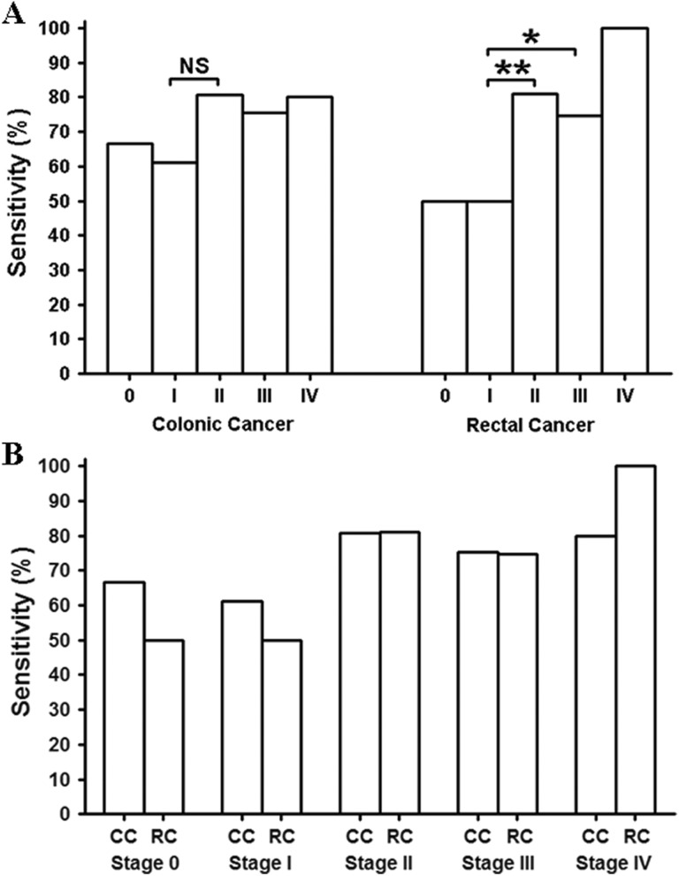
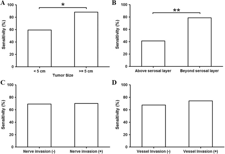
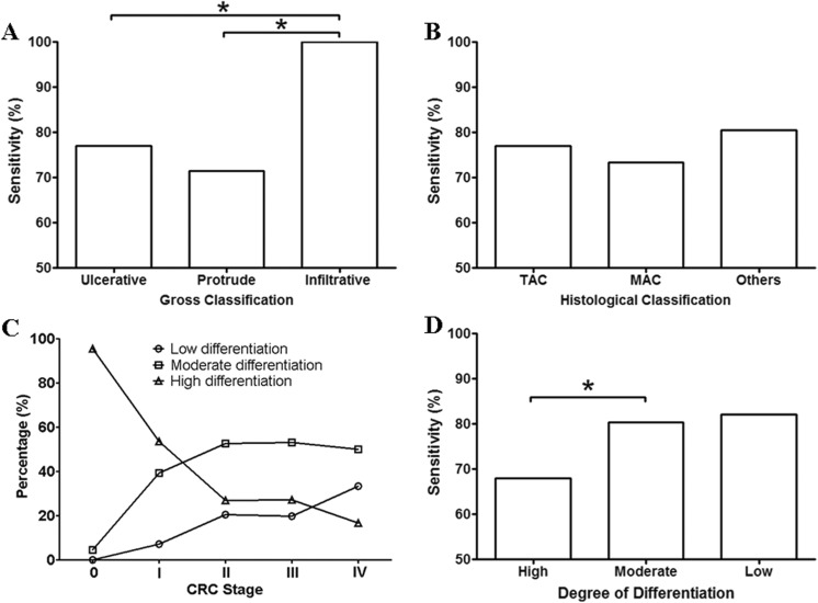
Results: Cancer stage, size, and invasion depth were positively correlated with the detection sensitivity, while no difference in sensitivity was identified among cancers at various locations. Infiltrative colorectal cancer exhibited higher sensitivity than ulcerative and protrude colorectal cancer, while no difference in sensitivity was observed among assessed histological types. The colorectal cancer differentiation showed a clear correlation with the cancer stage, and moderate and poorly differentiated colorectal cancer exhibited higher sensitivity than well-differentiated colorectal cancer. Furthermore, colorectal cancer with distal metastasis (M1) showed higher sensitivity than those without any metastasis, while colorectal cancer with lymph node metastasis (N1 and N2) did not show statistical significance compared to those without it. Finally, local vessel or nerve invasion did not affect the sensitivity.

Conclusion: Factors that reflect the colorectal cancer intrinsic properties, including cancer stage, size, invasion depth, classification, differentiation, and metastasis, exhibited significant effect on the mSEPT9 detection performance.

Keywords: SEPT9; adenoma; colorectal cancer; methylation; polyps; septin 9.


Conflict of interest statement

Declaration of Conflicting Interests: The author(s) declared the following potential conflicts of interest with respect to the research, authorship, and/or publication of this article: Lele Song, Guangpeng Zhou, Jianming Wang, and Xiaoliang Han are current employees of BioChain (Beijing) Science and Technology, Inc. BioChain is a collaborator of Epigenomics AG, a Germany-based company that launched the first commercial SEPT9 assay. Other authors claim no conflict of interests.

Figures

Figure 1.
Figure 1.
The positive detection rate (sensitivity) of the mSEPT9 assay for NED (normal control), HP, adenoma, and CRC stage 0-IV using the 2/3 algorithm. The number of patients for each group was 300, 122, 67, 103, and 568 for CRC, Adenoma, Other GID, HP, and NED, respectively, and was 23, 42, 99, 124, and 12 for Stage 0, I, II, III, and IV CRC, respectively. CRC indicates colorectal cancer; GID, gastrointestinal disease; HP, hyperplastic polyps; NED, no evidence of disease.
Figure 2.
Figure 2.
The sensitivity for CC and RC at each stage. The stage-dependent sensitivity was compared in panel A for CC and RC, respectively, and the sensitivity of each stage of CC or RC was compared in panel B. The number of patients for Stage 0, I, II, III, and IV was 15, 18, 57, 61, and 5, respectively, for CC and was 8, 24, 42, 63 and 7, respectively, for RC. CC indicates colonic cancer; NS, not significant; RC, rectal cancer; *significant difference; **highly significant difference.
Figure 3.
Figure 3.
The detection sensitivity for colorectal cancer (CRC) at ascending, transverse, descending, sigmoid colon and rectum. The number of participants was 60, 20, 17, 49, and 154 for ascending, transverse, descending, sigmoid colon and rectum, respectively.
Figure 4.
Figure 4.
The sensitivity for colorectal cancer (CRC) categorized by size, invasion depth, nerve or vessel invasion. Panel A shows the sensitivity of CRC < 5 cm or ≥5 cm. Panel B shows the sensitivity for CRC not reaching the serosal layer (above serosal layer) or reaching or growing beyond serosal layer (beyond serosal layer). The sensitivity for CRC with or without nerve or vessel invasion was shown in panel C and D, respectively. The number of patients was 193 for tumor size <5 cm, and was 107 for tumor size >5 cm. The number of patients was 70 for tumors above serosal layer, and was 230 for tumors beyond serosal layer. The number of patients was 53 for those without nerve invasion, and was 247 for those with nerve invasion. The number of patients was 189 for those with no vessel invasion, and was 111 for those with vessel invasion. *significant difference; **highly significant difference.
Figure 5.
Figure 5.
The sensitivity for CRC categorized by gross classification, histological classification, and degree of differentiation. Panel A shows the sensitivity of CRC categorized by gross classification, including ulcerative, protrude, and infiltrative CRC. Panel B shows the sensitivity of CRC categorized by histological classification, including the TAC, the MAC, and other types of CRC (others). Panel C shows the relationship between the ratio of low, moderate or high differentiation and the clinical stage of CRC. Panel D shows the detection sensitivity of CRC with high, moderate, and low differentiation. The number of patients was 231, 53, and 16 for ulcerative, protrude, and infiltrate CRC, respectively. The number of patients was 198, 85, and 17 for TAC, MAC, and other types, respectively. The number of patients for Stage 0-IV CRC is shown in Table 1. The number of patients with high, moderate, and low differentiation was 110, 140, and 50, respectively. CRC indicates colorectal cancer; MAC, mucinous adenocarcinoma; TAC, tubular adenocarcinoma, *significant difference (P < .05).
Figure 6.
Figure 6.
Sensitivity of CRC categorized by the grade distal or LN metastasis and vessel or nerve invasion. Panel A shows the sensitivity of CRC with (M1) or without (M0) distal metastasis. Panel B shows the sensitivity of CRC without (N0), or with 1-3 (N1), or with ≥4 (N2; LN) metastasis. The number of patients without distal metastasis (M0) was 288, and 12 had distal metastasis (M1). The number of patients with no LN metastasis (N0), 1 to 3 LN metastasis (N1) and ≥4 LN metastasis was 166, 96, and 38, respectively. CRC indicates colorectal cancer; LN, lymph node; *significant difference (P < .05).

References

    1. Schreuders EH, Ruco A, Rabeneck L, et al. Colorectal cancer screening: a global overview of existing programmes. Gut. 2015;64(10):1637–1649. - PubMed
    1. National Cancer Institute, PDQ, Treatment, Health Professionals. Survival Rate for Colorectal Cancer by Stage. Bethesda, MD: National Cancer Institute; 1999.
    1. Lofton-Day C, Model F, Devos T, et al. DNA methylation biomarkers for blood-based colorectal cancer screening. Clin Chem. 2008;54(2):414–423. - PubMed
    1. Gruètzmann R, Molnar B, Pilarsky C, et al. Sensitive detection of colorectal cancer in peripheral blood by septin 9 DNA methylation assay. PLoS One. 2008;3(11):e3759. - PMC - PubMed
    1. Song L, Li Y. SEPT9: a specific circulating biomarker for colorectal cancer. Adv Clin Chem. 2015;72:171–204. - PubMed

Publication types