

The role of diffusion tensor imaging as an objective tool for the assessment of motor function recovery after paraplegia in a naturally-occurring large animal model of spinal cord injury
- PMID: 30223849
- PMCID: PMC6142343
- DOI: 10.1186/s12967-018-1630-4
The role of diffusion tensor imaging as an objective tool for the assessment of motor function recovery after paraplegia in a naturally-occurring large animal model of spinal cord injury
Abstract
Background: Traumatic spinal cord injury (SCI) results in sensory and motor function impairment and may cause a substantial social and economic burden. For the implementation of novel treatment strategies, parallel development of objective tools evaluating spinal cord (SC) integrity during motor function recovery (MFR) is needed. Diffusion tensor imaging (DTI) enables in vivo microstructural assessment of SCI.
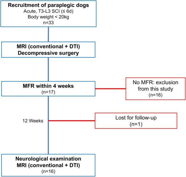
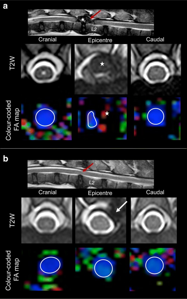
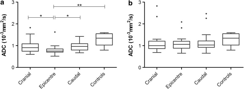
Methods: In the current study, temporal evolvement of DTI metrics during MFR were examined; therefore, values of fractional anisotropy (FA) and apparent diffusion coefficient (ADC) were measured in a population of 17 paraplegic dogs with naturally-occurring acute SCI showing MFR within 4 weeks after surgical decompression and compared to 6 control dogs. MRI scans were performed preoperatively and 12 weeks after MFR was observed. DTI metrics were obtained at the lesion epicentre and one SC segment cranially and caudally. Variance analyses were performed to compare values between evaluated localizations in affected dogs and controls and between time points. Correlations between DTI metrics and clinical scores at follow-up examinations were assessed.
Results: Before surgery, FA values at epicentres were higher than caudally (p = 0.0014) and control values (p = 0.0097); ADC values were lower in the epicentre compared to control values (p = 0.0035) and perilesional (p = 0.0448 cranially and p = 0.0433 caudally). In follow-up examinations, no significant differences could be found between DTI values from dogs showing MFR and control dogs. Lower ADC values at epicentres correlated with neurological deficits at follow-up examinations (r = - 0.705; p = 0.0023).
Conclusions: Findings suggest that a tendency to the return of DTI values to the physiological situation after surgical decompression accompanies MFR after SCI in paraplegic dogs. DTI may represent a useful and objective clinical tool for follow-up studies examining in vivo SC recovery in treatment studies.
Keywords: Canine; DTI; Follow-up studies; Hemilaminectomy; IVDH; Intervertebral disc herniation; MRI; SCI; Translational medicine.
Figures

Similar articles
-
Spontaneous acute and chronic spinal cord injuries in paraplegic dogs: a comparative study of in vivo diffusion tensor imaging.Spinal Cord. 2017 Dec;55(12):1108-1116. doi: 10.1038/sc.2017.83. Epub 2017 Aug 1. Spinal Cord. 2017. PMID: 28762382
-
Comparison of Preoperative Quantitative Magnetic Resonance Imaging and Clinical Assessment of Deep Pain Perception as Prognostic Tools for Early Recovery of Motor Function in Paraplegic Dogs with Intervertebral Disk Herniations.J Vet Intern Med. 2017 May;31(3):842-848. doi: 10.1111/jvim.14715. Epub 2017 Apr 25. J Vet Intern Med. 2017. PMID: 28440586 Free PMC article.
-
The role of diffusion tensor imaging in the diagnosis, prognosis, and assessment of recovery and treatment of spinal cord injury: a systematic review.Neurosurg Focus. 2019 Mar 1;46(3):E7. doi: 10.3171/2019.1.FOCUS18591. Neurosurg Focus. 2019. PMID: 30835681
-
Dynamic diffusion tensor imaging of spinal cord contusion: A canine model.J Neurosci Res. 2018 Jun;96(6):1093-1103. doi: 10.1002/jnr.24222. Epub 2018 Feb 27. J Neurosci Res. 2018. PMID: 29485189
-
The role of diffusion tensor imaging and fractional anisotropy in the evaluation of patients with idiopathic normal pressure hydrocephalus: a literature review.Neurosurg Focus. 2016 Sep;41(3):E12. doi: 10.3171/2016.6.FOCUS16192. Neurosurg Focus. 2016. PMID: 27581308 Review.
Cited by
-
Porcine Model of the Growing Spinal Cord-Changes in Diffusion Tensor Imaging Parameters.Animals (Basel). 2023 Feb 6;13(4):565. doi: 10.3390/ani13040565. Animals (Basel). 2023. PMID: 36830353 Free PMC article.
-
Prognostic Factors in Canine Acute Intervertebral Disc Disease.Front Vet Sci. 2020 Nov 26;7:596059. doi: 10.3389/fvets.2020.596059. eCollection 2020. Front Vet Sci. 2020. PMID: 33324703 Free PMC article. Review.
-
A Pilot Study on the Safety of a Novel Antioxidant Nanoparticle Delivery System and Its Indirect Effects on Cytokine Levels in Four Dogs.Front Vet Sci. 2020 Jul 30;7:447. doi: 10.3389/fvets.2020.00447. eCollection 2020. Front Vet Sci. 2020. PMID: 32851027 Free PMC article.
-
Diffusion tensor imaging predicting neurological repair of spinal cord injury with transplanting collagen/chitosan scaffold binding bFGF.J Mater Sci Mater Med. 2019 Nov 4;30(11):123. doi: 10.1007/s10856-019-6322-y. J Mater Sci Mater Med. 2019. PMID: 31686219
-
Diagnostic Imaging in Intervertebral Disc Disease.Front Vet Sci. 2020 Oct 22;7:588338. doi: 10.3389/fvets.2020.588338. eCollection 2020. Front Vet Sci. 2020. PMID: 33195623 Free PMC article. Review.
References
-
- Krueger H, Noonan VK, Trenaman LM, Joshi P, Rivers CS. The economic burden of traumatic spinal cord injury in Canada. Chronic Dis Inj Can. 2013;33(3):113–122. - PubMed
Publication types
MeSH terms
Grants and funding
LinkOut - more resources
Full Text Sources
Other Literature Sources
Medical
