. 2018 Sep 17;9(1):237.
doi: 10.1186/s13287-018-0979-x.

Mesenchymal stem cells alleviate experimental autoimmune cholangitis through immunosuppression and cytoprotective function mediated by galectin-9

Affiliations

Mesenchymal stem cells alleviate experimental autoimmune cholangitis through immunosuppression and cytoprotective function mediated by galectin-9

Junyu Fan et al. Stem Cell Res Ther. .

Abstract

Background: Mesenchymal stem cells (MSCs) play an anti-inflammatory role by secreting certain bioactive molecules to exert their therapeutic effects for disease treatment. However, the underlying mechanism of MSCs in chronic autoimmune liver diseases-primary biliary cholangitis (PBC), for example-remains to be elucidated.

Methods: Human umbilical cord-derived MSCs (UC-MSCs) were injected intravenously into 2-octynoic acid coupled to bovine serum albumin (2OA-BSA)-induced autoimmune cholangitis mice. Serum levels of biomarkers and autoantibodies, histologic changes in the liver, diverse CD4+ T-cell subsets in different tissues, and chemokine activities were analyzed. Moreover, we investigated galectin-9 (Gal-9) expression and its function in UC-MSCs.

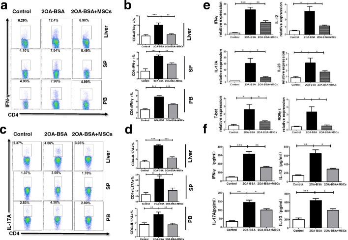
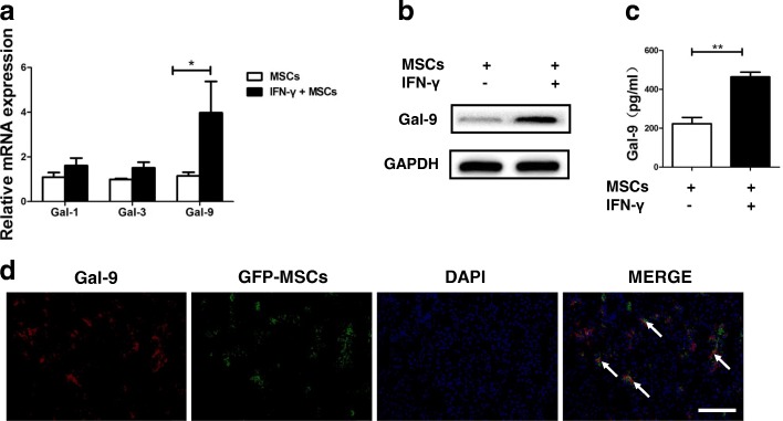
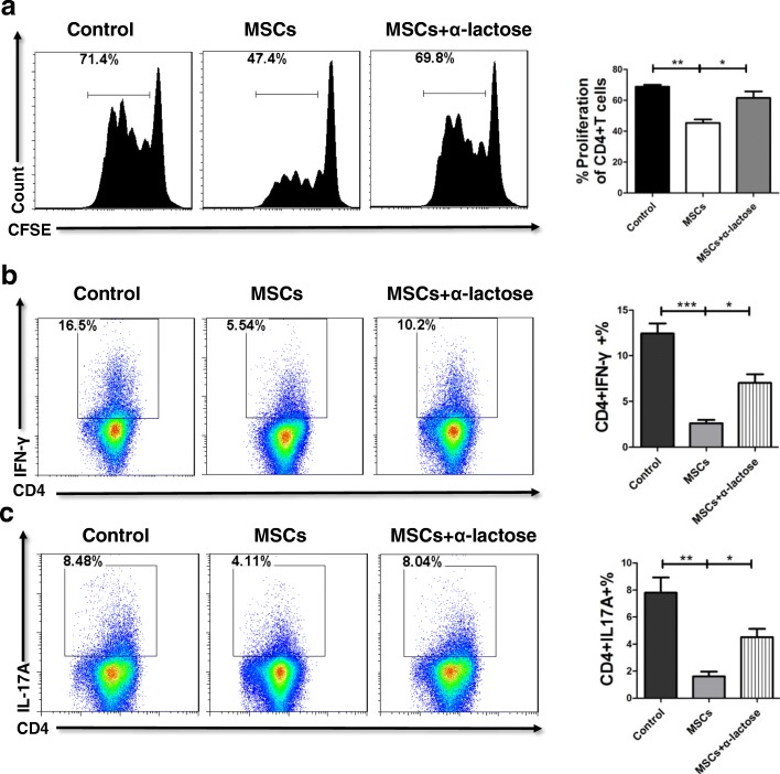
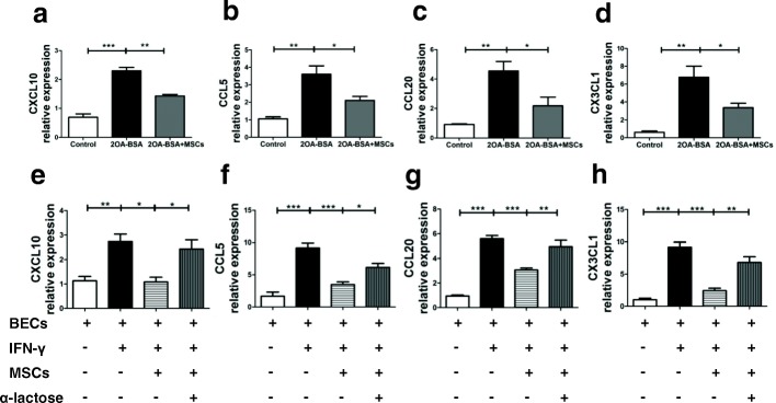
Results: In this study, UC-MSC transplantation (UC-MSCT) significantly ameliorated liver inflammation, primarily by diminishing T helper 1 (Th1) and Th17 responses as well as modifying liver chemokine activities in experimental autoimmune cholangitis mice. Mechanistically, UC-MSCs significantly repressed the proliferation of CD4+ T cells and suppressed the differentiation of Th1 and Th17 cells, which was likely dependent on Gal-9. Furthermore, the signal transducer and activator of transcription (STAT) and c-Jun N-terminal kinase (JNK) signaling pathways were involved in the production of Gal-9 in UC-MSCs.

Conclusions: These results suggest that Gal-9 contributes significantly to UC-MSC-mediated therapeutic effects and improve our understanding of the immunomodulatory mechanisms of MSCs in the treatment of PBC.

Keywords: Galectin-9; Inflammation; Primary biliary cholangitis; Umbilical cord–derived mesenchymal stem cells.


Conflict of interest statement

Ethics approval and consent to participate

The animal-related experiments and the isolation of human umbilical cord mesenchymal stem cells were approved by the ethics committee of the Affiliated Drum Tower Hospital of Nanjing University Medical School. All applicable institutional and national guidelines for the care and use of animals were followed.

Consent for publication

All authors declare their support for the publication and its contents.

Competing interests

The authors declare that they have no competing interests.

Publisher’s Note

Springer Nature remains neutral with regard to jurisdictional claims in published maps and institutional affiliations.

Figures

Fig. 1
Fig. 1
Umbilical cord–derived mesenchymal stem cell transplantation alleviates 2-octynoic acid coupled to bovine serum albumin (2OA-BSA)–induced autoimmune cholangitis. a Schematic illustration of the experimental procedure. b Representative images of liver tissue sections by hematoxylin-and-eosin staining from control, primary biliary cholangitis, or umbilical cord–derived mesenchymal stem cell (UC-MSC)–treated mice, showing liver inflammation and the extent of bile duct damage. c Histological scores were assessed on the basis of lymphocytic infiltration and bile duct damage in the liver. d The serum levels of alanine aminotransferase (ALT), aspartate aminotransferase (AST), alkaline phosphatase (ALP), and glutamyl transpeptidase (GGT) were measured. e The levels of anti-PDC-E2 autoantibodies were measured. Bars represent the mean ± standard error of the mean (SEM); n = 6 per group. *P <0.05, **P <0.01, ***P <0.001
Fig. 2
Fig. 2
Umbilical cord–derived mesenchymal stem cell transplantation inhibits the aberrant T helper 1 (Th1) and Th17 responses in 2-octynoic acid coupled to bovine serum albumin (2OA-BSA)–induced autoimmune cholangitis. a Representative flow cytometric profiles showing the frequencies of Th1 cells in the liver, spleen, and blood. b The frequencies of Th1 cells were quantified. c Representative flow cytometric profiles showing the frequencies of Th17 cells in the liver, spleen, and blood. d The frequencies of Th17 cells were quantified. e Intrahepatic mRNA expressions of interferon-gamma (IFN-γ), interleukin-17A (IL-17A), IL-12, IL-23, T-bet, and RAR-related orphan receptor gamma t (RORγt) were determined by real-time polymerase chain reaction. f Levels of IFN-γ, IL-17A, IL-12, and IL-23 in serum were detected by Luminex. Bars represent the mean ± standard error of the mean (SEM); n = 6 per group. *P <0.05, **P <0.01, ***P <0.001. Abbreviations: PB peripheral blood, SP spleen
Fig. 3
Fig. 3
Galectin-9 (Gal-9) is highly expressed in umbilical cord–derived mesenchymal stem cells (UC-MSCs). a Total mRNA of UC-MSCs was extracted after activation with or without interferon-gamma (IFN-γ) and analyzed by real-time polymerase chain reaction for the expression of Gal-1, Gal-3, and Gal-9. Bars represent the mean ± standard error of the mean (SEM); n = 4 per group. b, c UC-MSCs were treated with or without IFN-γ and analyzed by Western blot and enzyme-linked immunosorbent assay for the expression of Gal-9. Bars represent the mean ± SEM; n = 3 per group. d The fluorescence microscope was employed to detect green fluorescent protein (GFP)-positive UC-MSCs in the livers of primary biliary cholangitis mice. Colocalization (white arrow) of the GFP (green) with Gal-9 (red) staining indicated that UC-MSCs expressed Gal-9. Scale bar = 50 μm. Abbreviation: DAPI 4′,6-diamidino-2-phenylindole
Fig. 4
Fig. 4
Umbilical cord–derived mesenchymal stem cells (UC-MSCs) suppress CD4+ T-cell proliferation as well as T helper 1 (Th1) and Th17 cell differentiation via galectin-9. a Carboxyfluorescein diacetate succinimidyl ester (CFSE)–labeled CD4+ T cells were cultured in the presence of UC-MSCs or UC-MSCs plus α-lactose for 4 days. The proliferation of murine CD4+ T cells was assessed on the basis of the fluorescence intensity of CFSE. Bars represent the mean ± standard error of the mean (SEM); n = 3 per group. b Purified naïve murine CD4+ T cells were cultured under Th1 polarization conditions in the presence of UC-MSCs or UC-MSCs plus α-lactose. Representative flow cytometric profiles that show the frequencies of Th1 cells are presented. The frequencies of Th1 cells were quantified. c Purified naïve murine CD4+ T cells were cultured under Th17 polarization conditions in the presence of UC-MSCs or UC-MSCs plus α-lactose. Representative flow cytometric profiles that show the frequencies of Th17 cells are presented. The frequencies of Th17 cells were quantified. Bars represent the mean ± SEM; n = 4 per group. *P <0.05, **P <0.01, ***P <0.001
Fig. 5
Fig. 5
Umbilical cord–derived mesenchymal stem cell (UC-MSC) transplantation downregulates proinflammatory chemokines to repress inflammatory responses. ad Intrahepatic mRNA expressions of CCL5, CCL20, CXCL10, and CX3CL1 were determined by real-time polymerase chain reaction. Bars represent the mean ± standard error of the mean (SEM); n = 6 per group. eh CCL5, CCL20, CXCL10, and CX3CL1 mRNA expression in unstimulated, interferon-gamma (IFN-γ)–stimulated, UC-MSCs or UC-MSCs plus α-lactose co-cultured with biliary epithelial cells (BECs) demonstrated by real-time polymerase chain reaction. Bars represent the mean ± SEM; n = 4 per group. *P <0.05, **P <0.01, ***P <0.001. Abbreviation: 2OA-BSA 2-octynoic acid coupled to bovine serum albumin
Fig. 6
Fig. 6
Interferon-gamma (IFN-γ)–induced galectin-9 (Gal-9) upregulation of umbilical cord–derived mesenchymal stem cells (UC-MSCs) is mediated through the c-Jun N-terminal kinase (JNK) and signal transducer and activator of transcription (STAT) signaling pathways. a The expression of STAT, p38/mitogen-activated protein kinase (p38/MAPK), extracellular-regulated protein kinase (ERK), JNK, and their phosphorylation forms were detected by Western blot analysis in unstimulated and IFN-γ–stimulated UC-MSCs. b Total mRNA of UC-MSCs was extracted after the activation of IFN-γ with JNK or STAT inhibitors and then analyzed by real-time polymerase chain reaction for the expression of Gal-9. Bars represent the mean ± standard error of the mean (SEM); n = 3 per group. ***P <0.001

References

    1. Griffiths L, Dyson JK, Jones DE. The new epidemiology of primary biliary cirrhosis. Semin Liver Dis. 2014;34:318–328. doi: 10.1055/s-0034-1383730. - DOI - PubMed
    1. Mousa HS, Carbone M, Malinverno F, Ronca V, Gershwin ME, Invernizzi P. Novel therapeutics for primary biliary cholangitis: toward a disease-stage-based approach. Autoimmun Rev. 2016;15:870–876. doi: 10.1016/j.autrev.2016.07.003. - DOI - PubMed
    1. Hirschfield GM, Gershwin ME. The immunobiology and pathophysiology of primary biliary cirrhosis. Annu Rev Pathol. 2013;8:303–330. doi: 10.1146/annurev-pathol-020712-164014. - DOI - PubMed
    1. Hirschfield GM, Mason A, Luketic V, Lindor K, Gordon SC, Mayo M, et al. Efficacy of obeticholic acid in patients with primary biliary cirrhosis and inadequate response to ursodeoxycholic acid. Gastroenterology. 2015;148:751–61 e8. doi: 10.1053/j.gastro.2014.12.005. - DOI - PubMed
    1. Markham A, Keam SJ. Obeticholic acid: first global approval. Drugs. 2016;76:1221–1226. doi: 10.1007/s40265-016-0616-x. - DOI - PubMed

Publication types

MeSH terms