. 2019 Oct;38(5):2145-2154.
doi: 10.1016/j.clnu.2018.08.036. Epub 2018 Sep 1.

Effects of dietary omega-3 fatty acids on bones of healthy mice

Affiliations

Effects of dietary omega-3 fatty acids on bones of healthy mice

Lorenzo Anez-Bustillos et al. Clin Nutr. 2019 Oct.

Abstract

Background & aims: Altering the lipid component in diets may affect the incidence of metabolic bone disease in patients dependent on parenteral nutrition. Consumption of polyunsaturated fatty acids (PUFA) can impact bone health by modulating calcium metabolism, prostaglandin synthesis, lipid oxidation, osteoblast formation, and osteoclastogenesis. The aim of this study was to evaluate the dietary effects of PUFA on murine bone health.

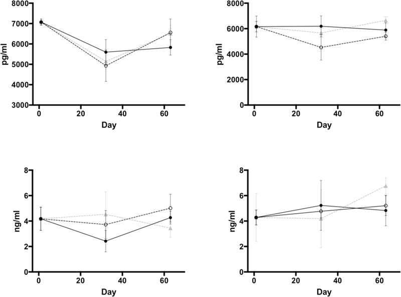
Methods: Three-weeks-old male (n = 30) and female (n = 30) C57BL/6J mice were randomized into one of three dietary groups. The diets differed only in fat composition: soybean oil (SOY), rich in ω-6 PUFA; docosahexaenoic acid alone (DHA), an ω-3 PUFA; and DHA with arachidonic acid, an ω-6 PUFA, at a 20:1 ratio (DHA/ARA). After 9 weeks of dietary treatment, femurs were harvested for micro-computed tomographic analysis and mechanical testing via 3-point bending. Separate mice from each group were used solely for serial blood draws for measurement of biomarkers of bone formation and resorption.

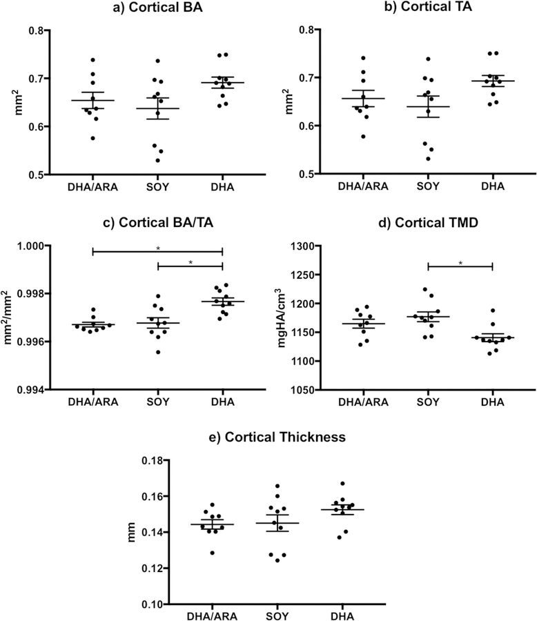
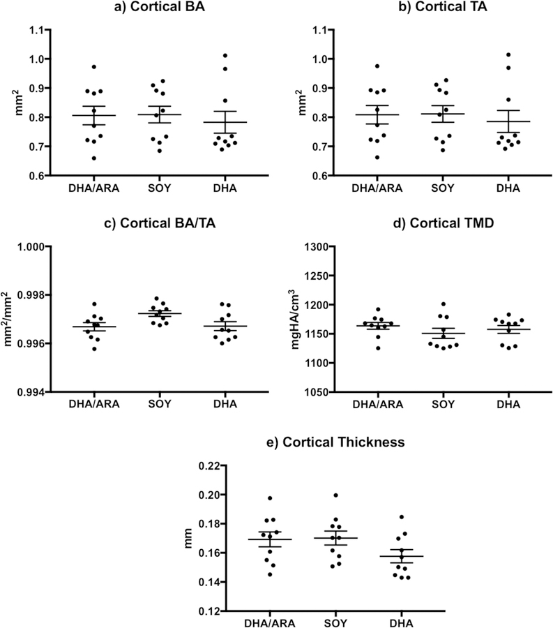
Results: At the microstructural level, although some parameters in cortical bone reached differences that were statistically significant in female mice, these were too small to be considered biologically relevant. Similarly, trabecular bone parameters in male mice were statistically different in some dietary groups, although the biological interpretation of such subtle changes translate into a lack of effect in favor of any of the experimental diets. No differences were noted at the mechanical level and in blood-based biomarkers of bone metabolism across dietary groups within gender.

Conclusions: Subtle differences were noted at the bones' microstructural level, however these are likely the result of random effects that do not translate into changes that are biologically relevant. Similarly, differences were not seen at the mechanical level, nor were they reflected in blood-based biomarkers of bone metabolism. Altogether, dietary consumption of PUFA do not seem to affect bone structure or metabolism in a healthy model of growing mice.

Keywords: Bone; Bone micro-architecture; Bone strength; Polyunsaturated fatty acids.


Figures

Figure 1.
Figure 1.
Weight (grams) for n=89 animals. Shown are males (filled circles) and females (empty circles), with means from a broken-stick mixed model shown for soy, DHA/ARA, and DHA. Data are jittered right/left to prevent male/female overlap.
Figure 2.
Figure 2.
Graphs representing cortical bone indices (mean ± standard error of the mean) measured in female mice (DHA/ARA, n=9; SOY, n=10; DHA, n=10): a) cortical BA (bone area); b) cortical TA (total cross-sectional area); c) cortical BA/TA (bone area fraction); d) cortical TMD (tissue mineral density); and e) cortical thickness. Where indicated, mean value differences between treatment groups are considered statistically significant (* P < 0.05).
Figure 3.
Figure 3.
Graphs representing cortical bone indices (mean ± standard error of the mean) measured in male mice (DHA/ARA, n=10; SOY, n=10; DHA, n=10): a) cortical BA (bone area); b) cortical TA (total cross-sectional area); c) cortical BA/TA (bone area fraction); d) cortical TMD (tissue mineral density); and e) cortical thickness. Where indicated, mean value differences between treatment groups are considered statistically significant (* P < 0.05).
Figure 4.
Figure 4.
Graphs representing trabecular bone indices (mean ± standard error of the mean) measured in female mice (DHA/ARA, n=9; SOY, n=10; DHA, n=10): a) BV/TV (bone volume fraction); b) connectivity density; c) SMI (structure model index); d) trabecular number; e) trabecular thickness; f) trabecular separation; g) trabecular TMD (tissue mineral density). Where indicated, mean value differences between treatment groups are considered statistically significant (* P < 0.05).
Figure 5.
Figure 5.
Graphs representing trabecular bone indices (mean ± standard error of the mean) measured in male mice (DHA/ARA, n=10; SOY, n=10; DHA, n=10): a) BV/TV (bone volume fraction); b) connectivity density; c) SMI (structure model index); d) trabecular number; e) trabecular thickness; f) trabecular separation; g) trabecular TMD (tissue mineral density). Where indicated, mean value differences between treatment groups are considered statistically significant (* P < 0.05).
Figure 6.
Figure 6.
Changes (mean ± standard error of the mean) in levels of P1NP (procollagen type 1 propeptide, top panels) and CTX (C-terminal telopeptide of type I collagen, bottom panels) in female (left-sided panels) and male (right-sided panels) animals. Blood samples collected at baseline (day 0), day 32, and prior to euthanasia (day 63). Filled circles and full lines represent animals in the DHA/ARA group; empty circles and interrupted line belong to animals in the SOY group; and gray triangles and gray interrupted lines belong to the DHA group.

References

    1. Jeejeebhoy KN, Parenteral nutrition in the intensive care unit, Nutr. Rev 70 (2012) 623–630. doi: 10.1111/j.1753-4887.2012.00538.x. - DOI - PubMed
    1. Rangel SJ, Calkins CM, Cowles RA, Barnhart DC, Huang EY, Abdullah F, Arca MJ, Teitelbaum DH, Parenteral nutrition–associated cholestasis: an American Pediatric Surgical Association Outcomes and Clinical Trials Committee systematic review, J. Pediatr. Surg 47 (2012) 225–240. doi: 10.1016/j.jpedsurg.2011.10.007. - DOI - PubMed
    1. Diamanti A, Bizzarri C, Bizzarri C, Basso MS, Gambarara M, Cappa M, Daniele A, Noto C, Castro M, How does long-term parenteral nutrition impact the bone mineral status of children with intestinal failure?, J. Bone Miner. Metab 28 (2010) 351–8. doi: 10.1007/s00774-009-0140-0. - DOI - PubMed
    1. Buchman AL, Moukarzel A, Metabolic bone disease associated with total parenteral nutrition, Clin. Nutr 19 (2000) 217–231. doi: 10.1054/clnu.1999.0083. - DOI - PubMed
    1. Klein GL, Metabolic bone disease of total parenteral nutrition., Nutrition 14 (1998) 149–52. - PubMed

Publication types

Substances

LinkOut - more resources