. 2018 Oct 26;293(43):16899-16911.
doi: 10.1074/jbc.RA118.004138. Epub 2018 Sep 17.

Cox2p of yeast cytochrome oxidase assembles as a stand-alone subunit with the Cox1p and Cox3p modules

Affiliations

Cox2p of yeast cytochrome oxidase assembles as a stand-alone subunit with the Cox1p and Cox3p modules

Leticia Veloso R Franco et al. J Biol Chem. .

Abstract

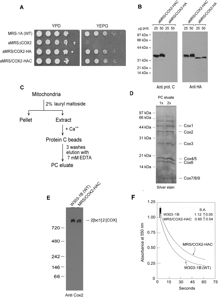
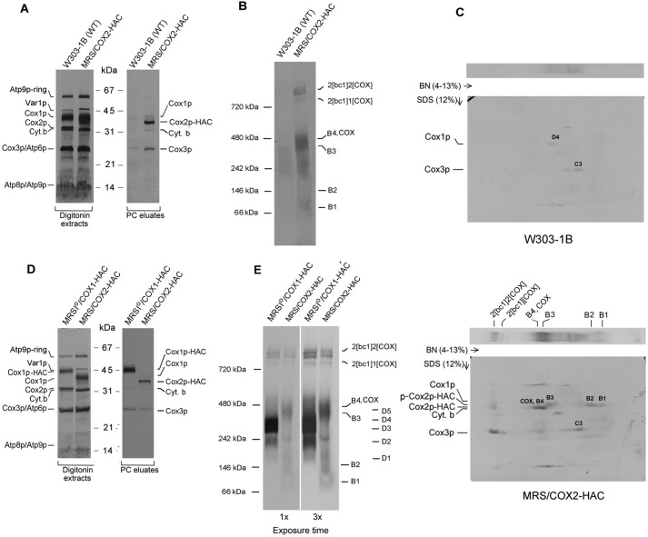
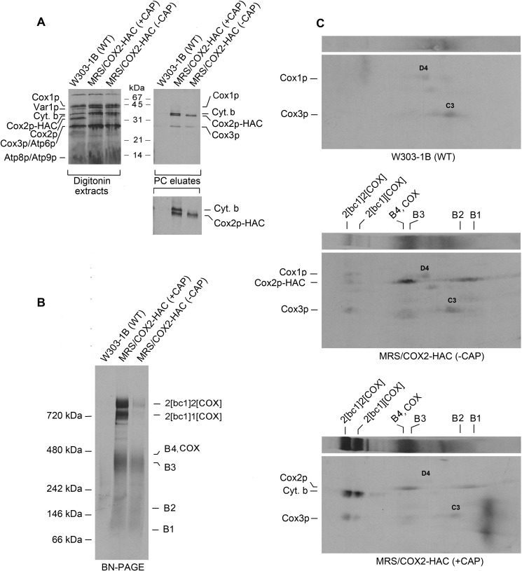
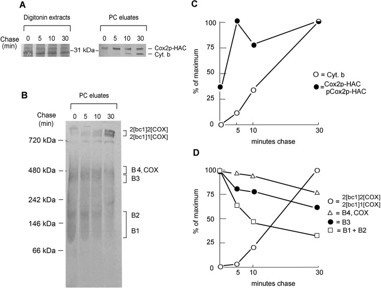
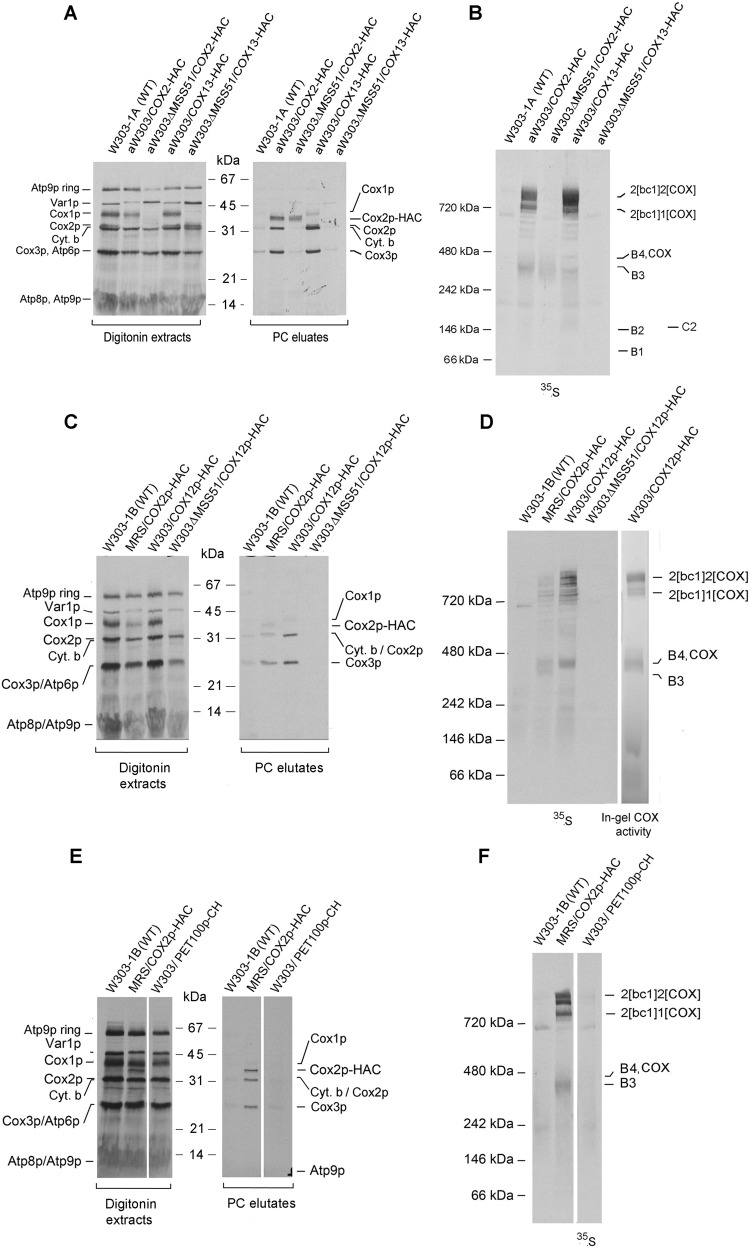
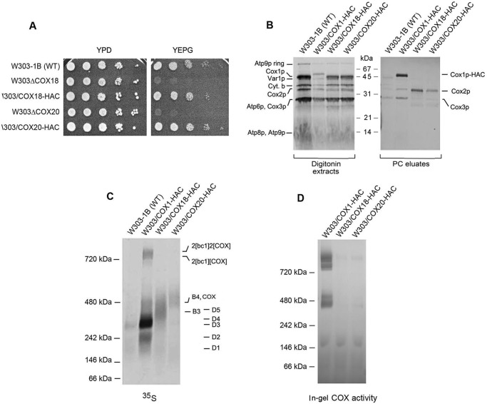
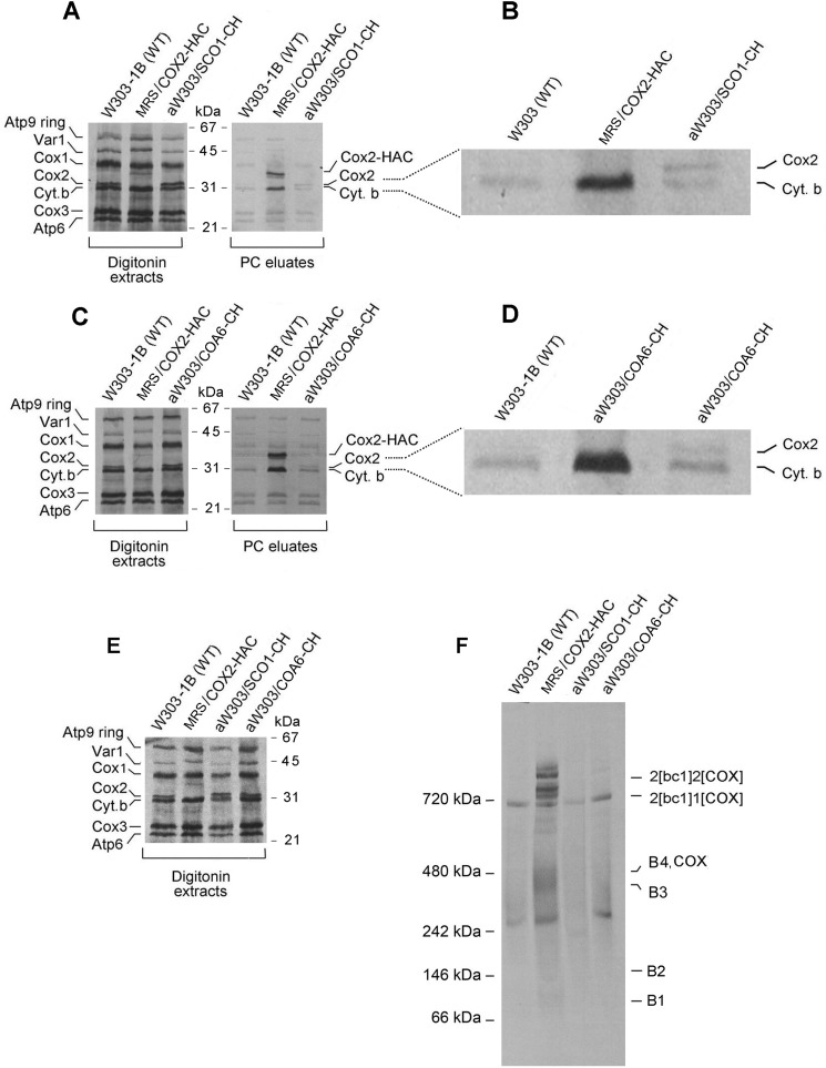
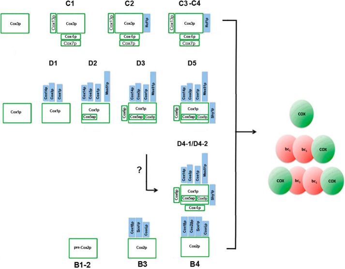
Cytochrome oxidase (COX) is a hetero-oligomeric complex of the mitochondrial inner membrane that reduces molecular oxygen to water, a reaction coupled to proton transfer from the mitochondrial matrix to the intermembrane space. In the yeast Saccharomyces cerevisiae, COX is composed of 11-13 different polypeptide subunits. Here, using pulse labeling of mitochondrial gene products in isolated yeast mitochondria, combined with purification of tagged COX subunits and ancillary factors, we studied the Cox2p assembly intermediates. Analysis of radiolabeled Cox2p obtained in pulldown assays by native gel electrophoresis revealed the existence of several assembly intermediates, the largest of which had an estimated mass of 450-550 kDa. None of the other known subunits of COX were present in these Cox2p intermediates. This was also true for the several ancillary factors having still undefined functions in COX assembly. In agreement with earlier evidence, Cox18p and Cox20p, previously shown to be involved in processing and in membrane insertion of the Cox2p precursor, were found to be associated with the two largest Cox2p intermediates. A small fraction of the Cox2p module contained Sco1p and Coa6p, which have been implicated in metalation of the binuclear copper site on this subunit. Our results indicate that following its insertion into the mitochondrial inner membrane, Cox2p assembles as a stand-alone protein with the compositionally more complex Cox1p and Cox3p modules.

Keywords: Cox2p module; Saccharomyces cerevisiae; biogenesis; cytochrome c oxidase (complex IV); cytochrome oxidase assembly; mitochondria; mitochondrial DNA (mtDNA); mitochondrial respiratory chain complex; yeast genetics.


Conflict of interest statement

The authors declare that they have no conflicts of interest with the contents of this article

Figures

Figure 1.
Figure 1.
Properties of yeast expressing HA- and HAC-tagged Cox2p. A, overnight cultures of the parental WT MRS-1A, aMRSΔCOX2, aMRS/COX2-HA, and aMRS/COX2-HAC were serially diluted and spotted on rich glucose (YPD) and rich glycerol/ethanol (YEPG) plates. The photograph was taken after 2 days' incubation at 30 °C. B, mitochondria were prepared from aMRS/COX2-HA and aMRS/COX2-HAC. The indicated amounts of mitochondria were applied to a 12% polyacrylamide gel and separated by SDS-PAGE. Western blots were incubated with polyclonal antibody against the protein C epitope followed by secondary peroxidase-coupled antibody against rabbit γ globulin. Proteins were visualized with SuperSignal chemiluminescent substrate kit (Pierce). C, protocol used to purify HAC-tagged cytochrome oxidase. D, two different concentrations of cytochrome oxidase purified as in C were depolymerized in Laemmli sample buffer (43) and separated by SDS-PAGE on a 12% polyacrylamide gel. Proteins were stained with silver. The subunits of cytochrome oxidase are identified in the right-hand margin. E, mitochondria from the indicated strains were extracted and separated by BN-PAGE. Proteins were transferred to a PVDF membrane, reacted with a polyclonal antibody against Cox2p and further processed as in B. F, mitochondria of the WT strain W303–1B and MRS/COX2-HAC expressing tagged Cox2p were assayed for oxidation of ferrocytochrome c at 550 nm. S. A. refers to specific activity expressed as micromoles of cytochrome c oxidized per minute per milligram protein.
Figure 2.
Figure 2.
In organello labeling of Cox2p intermediates. A, mitochondria were isolated from respiratory competent haploid strains W303–1B and MRS/COX2-HAC. Mitochondria (250 μg protein) were labeled in vitro with [35S]methionine plus [35S]cysteine and extracted with digitonin as described previously (10). The digitonin extract (5% of total) and the fraction purified on protein C antibody beads as in Fig. 1D (15% of total) were separated by SDS-PAGE on a 12% polyacrylamide gel, transferred to nitrocellulose and exposed to X-ray film. B, the fraction purified on the protein C beads was separated by BN-PAGE on a 4–13% polyacrylamide gel, transferred to a PVDF membrane, and exposed to X-ray. C, the purified PC eluates from WT and from MRS/COX2-HAC were separated by BN-PAGE on a 4–13% polyacrylamide gel in the first dimension and by SDS-PAGE in the second dimension. The background bands detected in the autoradiogram of the WT control (2D gel of top panel) correspond to the D3 and D4 intermediates of the Cox1p module and the C3 intermediate of Cox3p module. The radiolabeled bands pulled down by Cox2p are the B1, B2, and B3 intermediates. The Cox2p precursor (p-Cox2p), the B4 intermediate and COX with mature Cox2p and the nonspecific adsorbed C3 intermediate with Cox3p (2D gel of lower panel). The migration of Cox1p, Cox2p-HAC, and the Cox2p precursor are marked in the margins. D and E, same as A and B, except that MRSIO/COX1-HAC, a strain expressing Cox1p with an HAC tag was used for comparison. The Western blotting of the native gel in the right panel was exposed three times longer.
Figure 3.
Figure 3.
Effect of 2-h growth in the presence of chloramphenicol on translation of cytochrome b. A, mitochondria were isolated from the WT strain W303-1B and MRS/COX2-HAC, a strain expressing Cox2p-HAC, grown to early stationary phase in rich galactose (YPGal) medium without chloramphenicol (−CAP) and with an additional 2-h growth in YPGal containing 2 mg/ml chloramphenicol (+CAP). Mitochondria labeled for 20 min with [35S]methionine plus [35S]cysteine were extracted with digitonin and Cox2p-HAC purified on protein C antibody beads as described under “Materials and methods.” The digitonin extracts and purified fraction from the beads (PC eluates) were separated by SDS-PAGE (43) on a 17% polyacrylamide gel, transferred to nitrocellulose and exposed to an X-ray film. The radiolabeled mitochondrial gene products are identified in the margin. The migrations of Var1p relative to Cox1p and of Cox2p relative to cytochrome b are reversed at 17% compared with 12% acrylamide. The small inset panel of the PC eluates has been expanded to better show the separation of cytochrome b and Cox2p-HAC. B, the PC eluates of A were separated by BN-PAGE on a 4–13% polyacrylamide gel, transferred to a PVDF membrane and exposed to X-ray film. The bands corresponding to the supercomplexes, COX, and Cox2p intermediates are marked in the margin. C, the PC eluates of A were first separated by BN-PAGE as in B and by SDS-PAGE on a 12% polyacrylamide gel in the second dimension. The large area of radioactivity near the right edge of the lower gel is an artifact.
Figure 4.
Figure 4.
Pulse-chase analysis of mitochondria expressing Cox2p-HAC. A, mitochondria of MRS/COX2-HAC that were grown on YPGal with a subsequent incubation in the presence of chloramphenicol were labeled with [35S]methionine plus [35S]cysteine for 5 min. Translation was terminated by addition of puromycin and cold methionine and chased by further incubation for the indicated times. Mitochondria sampled at each time point were extracted with digitonin and Cox2p-HAC purified on protein C antibody beads (PC eluates). The digitonin extracts and PC eluates were separated by SDS-PAGE on a 12% polyacrylamide gel, transferred to nitrocellulose and exposed to X-ray. B, the PC eluates from A were separated by BN-PAGE on a 4–13% polyacrylamide gel, transferred to a PVDF membrane and exposed to X-ray film. C, radiolabeled Cox2p-HAC and cytochrome b on the Western blotting of A were quantified with a phosphorimager. D, the band of B corresponding to the radiolabeled supercomplexes, COX plus intermediates B4, B3, and B1 plus B2 were quantified with a phosphorimager.
Figure 5.
Figure 5.
Pulldown assays of radiolabeled mitochondrial gene products of strains expressing Cox12p-HAC, Cox13p-HAC, and Pet100p-CH. A and B, the parental WT and strains expressing Cox2p-HAC and Cox13p-HAC with and without an mss51 null mutation were grown in YPGal medium and incubated for an additional 2 h in fresh YPGal containing 2 mg/ml chloramphenicol. Mitochondria were labeled for 20 min and the tagged proteins purified on protein C beads as in Fig. 3A. A and B, the fractions purified on protein C antibody beads were separated on a 12% polyacrylamide gel by SDS-PAGE (A) and on a 4–13% polyacrylamide gel by BN-PAGE (B). Following transfer to nitrocellulose (A) or PVDF (B), the membranes were exposed to an X-ray film. The positions of the radiolabeled mitochondrial products and assembly intermediates are marked in the margins of the autoradiograms. C and D, yeast strains expressing Cox12p-HAC were analyzed as in A and B. E and F, yeast strains expressing Pet100p-CH were analyzed as in A and B. In D, the in-gel COX activity was measured as described previously (10).
Figure 6.
Figure 6.
Cox18p and Cox20p are components of the Cox2p module. A, the parental WT, the cox18 and cox20 null mutants, and the null mutants expressing HAC tagged Cox18p and Cox20p were serially diluted and spotted on rich glucose (YPD) and rich ethanol plus glycerol (YEPG). The plates were incubated for 2 days at 30 °C. B, mitochondria of the WT and the stains expressing HAC-tagged Cox1p (MRSIO/COX1-HAC), Cox18p (W303/COX18-HAC), and Cox20p (W303/COX20-HAC) were grown in YPGal without additional growth in medium containing chloramphenicol. Mitochondria were prepared and labeled as in Fig. 3A. The digitonin extracts and eluates from the protein C antibody beads (PC eluates) were separated by SDS-PAGE on a 12% polyacrylamide gel. C, the fractions of B eluted from the PC beads were separated by BN-PAGE on a 4–13% polyacrylamide gel, transferred to a PVDF membrane and exposed to X-ray. D, same as C except that the gel was stained for COX activity.
Figure 7.
Figure 7.
Association of Sco1p and Coa6p with Cox2p. A, the different strains were grown to early stationary phase in 2% YPGal with a final 2-h growth in the presence of 2 mg/ml chloramphenicol. Mitochondria were labeled for 20 min and fractionated as in Fig. 2A. The digitonin extracts and eluates from the protein C antibody beads were separated by SDS-PAGE on a 12% polyacrylamide gel containing 6 m urea and 10% by weight glycerol. B, an enlargement of the Cox2p and cytochrome b region of the gel shown in A. C, the different strains of yeast were grown and mitochondria were labeled and fractionated, and the digitonin extracts and eluates from the protein C antibody beads were electrophoresed as in A. D, an enlargement of the Cox2p and cytochrome b region of the gel shown in C. E, the indicated strains were grown and mitochondria were labeled and fractionated, and the digitonin extracts electrophoresed as in A. F, the digitonin extracts of E were purified on protein C antibody beads and separated by BN-PAGE on a 4–13% polyacrylamide gel.
Figure 8.
Figure 8.
The three cytochrome oxidase modules. The structural subunits and ancillary assembly factors present in each module are shown.

References

    1. Yoshikawa S., Muramoto K., Shinzawa-Itoh K., Aoyama H., Tsukihara T., Ogura T., Shimokata K., Katayama Y., and Shimada H. (2006) Reaction mechanism of bovine heart cytochrome c oxidase. Biochim. Biophys. Acta 1757, 395–400 10.1016/j.bbabio.2006.04.028 - DOI - PubMed
    1. Levchenko M., Wuttke J. M., Römpler K., Schmidt B., Neifer K., Juris L., Wissel M., Rehling P., and Deckers M. (2016) Cox26 is a novel stoichiometric subunit of the yeast cytochrome c oxidase. Biochim. Biophys. Acta. 1863, 1624–1632 10.1016/j.bbamcr.2016.04.007 - DOI - PubMed
    1. Strecker V., Kadeer Z., Heidler J., Cruciat C. M., Angerer H., Giese H., Pfeiffer K., Stuart R. A., and Wittig I. (2016) Supercomplex-associated Cox26 protein binds to cytochrome c oxidase. Biochim. Biophys. Acta 1863, 1643–1652 10.1016/j.bbamcr.2016.04.012 - DOI - PMC - PubMed
    1. Carlson C. G., Barrientos A., Tzagoloff A., and Glerum D. M. (2003) COX16 encodes a novel protein required for the assembly of cytochrome oxidase in Saccharomyces cerevisiae. J. Biol. Chem. 278, 3770–3775 10.1074/jbc.M209893200 - DOI - PubMed
    1. Su C. H., and Tzagoloff A. (2017) Cox16 protein is physically associated with Cox1p assembly intermediates and with cytochrome oxidase. J. Biol. Chem. 292, 16277–16283 10.1074/jbc.M117.801811 - DOI - PMC - PubMed

Publication types

MeSH terms

Substances