

A DNA nick at Ku-blocked double-strand break ends serves as an entry site for exonuclease 1 (Exo1) or Sgs1-Dna2 in long-range DNA end resection
- PMID: 30224356
- PMCID: PMC6222114
- DOI: 10.1074/jbc.RA118.004769
A DNA nick at Ku-blocked double-strand break ends serves as an entry site for exonuclease 1 (Exo1) or Sgs1-Dna2 in long-range DNA end resection
Abstract
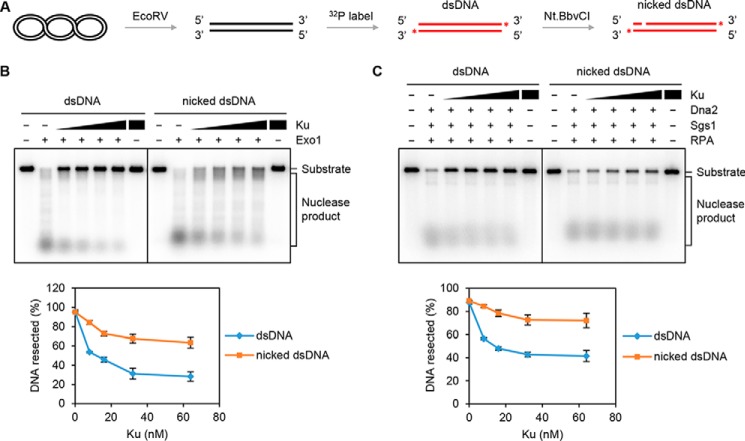
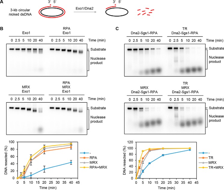
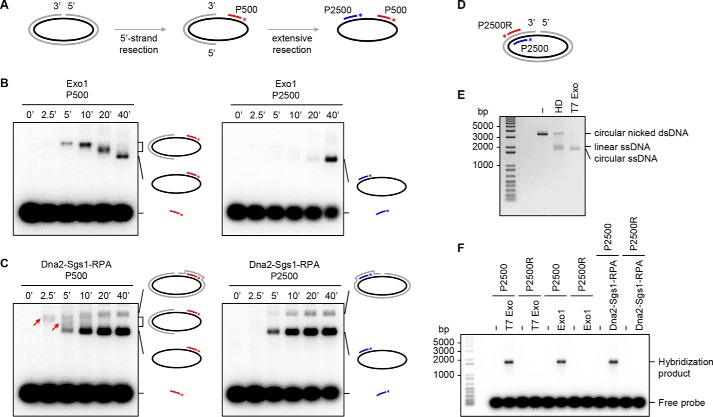
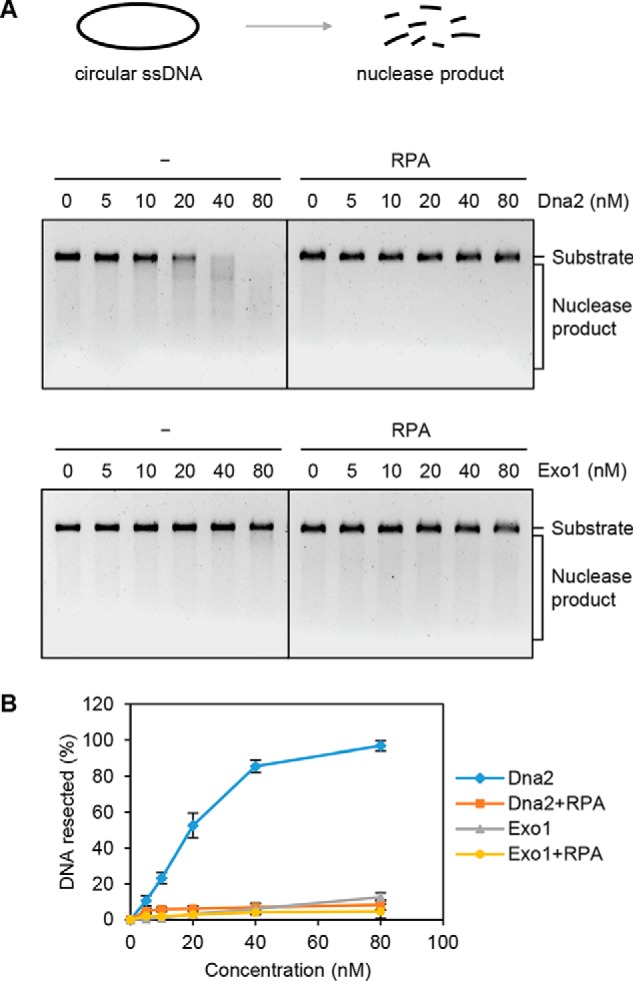
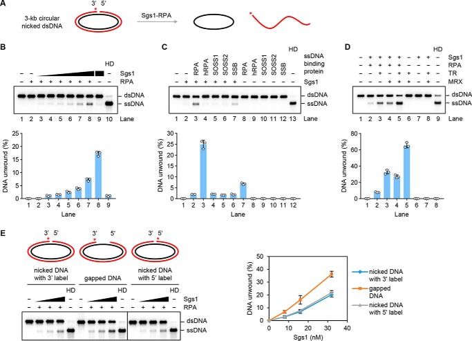
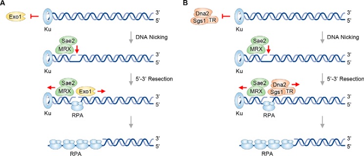
The repair of DNA double-strand breaks (DSBs) by homologous recombination (HR) is initiated by nucleolytic resection of the DNA break ends. The current model, being based primarily on genetic analyses in Saccharomyces cerevisiae and companion biochemical reconstitution studies, posits that end resection proceeds in two distinct stages. Specifically, the initiation of resection is mediated by the nuclease activity of the Mre11-Rad50-Xrs2 (MRX) complex in conjunction with its cofactor Sae2, and long-range resection is carried out by exonuclease 1 (Exo1) or the Sgs1-Top3-Rmi1-Dna2 ensemble. Using fully reconstituted systems, we show here that DNA with ends occluded by the DNA end-joining factor Ku70-Ku80 becomes a suitable substrate for long-range 5'-3' resection when a nick is introduced at a locale proximal to one of the Ku-bound DNA ends. We also show that Sgs1 can unwind duplex DNA harboring a nick, in a manner dependent on a species-specific interaction with the ssDNA-binding factor replication protein A (RPA). These biochemical systems and results will be valuable for guiding future endeavors directed at delineating the mechanistic intricacy of DNA end resection in eukaryotes.
Keywords: DNA binding protein; DNA damage; DNA end resection; DNA gap; DNA helicase; DNA nick; DNA repair; Exo1; Sgs1-Dna2; homologous recombination; nuclease.
© 2018 Wang et al.
Conflict of interest statement
The authors declare that they have no conflicts of interest with the contents of this article
Figures
Similar articles
-
DNA end resection by Dna2-Sgs1-RPA and its stimulation by Top3-Rmi1 and Mre11-Rad50-Xrs2.Nature. 2010 Sep 2;467(7311):112-6. doi: 10.1038/nature09355. Nature. 2010. PMID: 20811461 Free PMC article.
-
Relationship of DNA degradation by Saccharomyces cerevisiae exonuclease 1 and its stimulation by RPA and Mre11-Rad50-Xrs2 to DNA end resection.Proc Natl Acad Sci U S A. 2013 Apr 30;110(18):E1661-8. doi: 10.1073/pnas.1305166110. Epub 2013 Apr 15. Proc Natl Acad Sci U S A. 2013. PMID: 23589858 Free PMC article.
-
Sgs1 helicase and two nucleases Dna2 and Exo1 resect DNA double-strand break ends.Cell. 2008 Sep 19;134(6):981-94. doi: 10.1016/j.cell.2008.08.037. Cell. 2008. PMID: 18805091 Free PMC article.
-
DNA End Resection: Nucleases Team Up with the Right Partners to Initiate Homologous Recombination.J Biol Chem. 2015 Sep 18;290(38):22931-8. doi: 10.1074/jbc.R115.675942. Epub 2015 Jul 31. J Biol Chem. 2015. PMID: 26231213 Free PMC article. Review.
-
DNA end resection--unraveling the tail.DNA Repair (Amst). 2011 Mar 7;10(3):344-8. doi: 10.1016/j.dnarep.2010.12.004. Epub 2011 Jan 11. DNA Repair (Amst). 2011. PMID: 21227759 Free PMC article. Review.
Cited by
-
Mechanisms underlying genome instability mediated by formation of foldback inversions in Saccharomyces cerevisiae.Elife. 2020 Aug 7;9:e58223. doi: 10.7554/eLife.58223. Elife. 2020. PMID: 32762846 Free PMC article.
-
Human RPA activates BLM's bidirectional DNA unwinding from a nick.Elife. 2020 Feb 26;9:e54098. doi: 10.7554/eLife.54098. Elife. 2020. PMID: 32101168 Free PMC article.
-
Perturbation of base excision repair sensitizes breast cancer cells to APOBEC3 deaminase-mediated mutations.Elife. 2020 Jan 6;9:e51605. doi: 10.7554/eLife.51605. Elife. 2020. PMID: 31904337 Free PMC article.
-
Active Replication Checkpoint Drives Genome Instability in Fission Yeast mcm4 Mutant.Mol Cell Biol. 2020 Jun 29;40(14):e00033-20. doi: 10.1128/MCB.00033-20. Print 2020 Jun 29. Mol Cell Biol. 2020. PMID: 32341083 Free PMC article.
-
Mouse MRE11-RAD50-NBS1 is needed to start and extend meiotic DNA end resection.Nat Commun. 2025 Apr 16;16(1):3613. doi: 10.1038/s41467-025-57928-x. Nat Commun. 2025. PMID: 40240347 Free PMC article.
References
Publication types
MeSH terms
Substances
Grants and funding
LinkOut - more resources
Full Text Sources
Other Literature Sources
Molecular Biology Databases
Research Materials
Miscellaneous
