Review
. 2018 Oct;10(10):e8259.
doi: 10.15252/emmm.201708259.

Modulation of three key innate immune pathways for the most common retinal degenerative diseases

Affiliations
Review

Modulation of three key innate immune pathways for the most common retinal degenerative diseases

Isha Akhtar-Schäfer et al. EMBO Mol Med. 2018 Oct.

Abstract

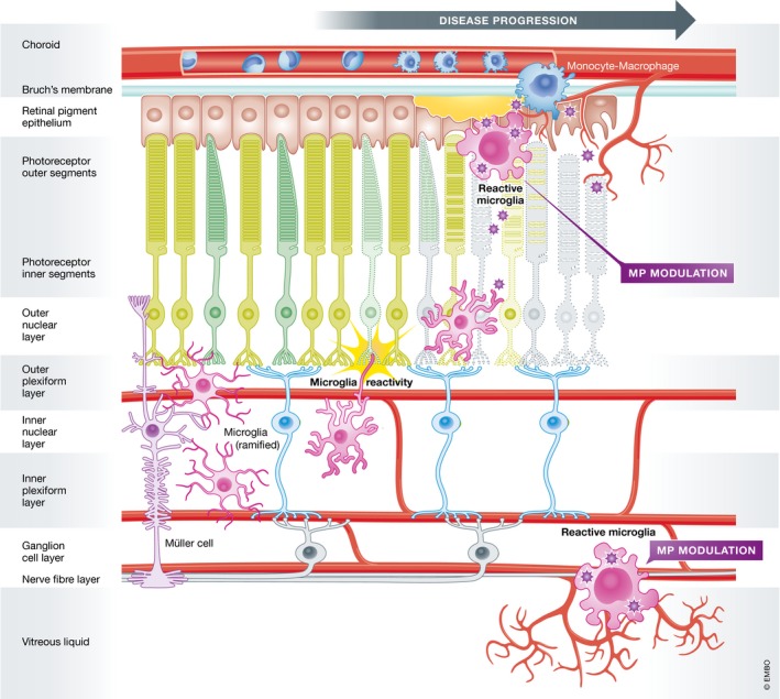
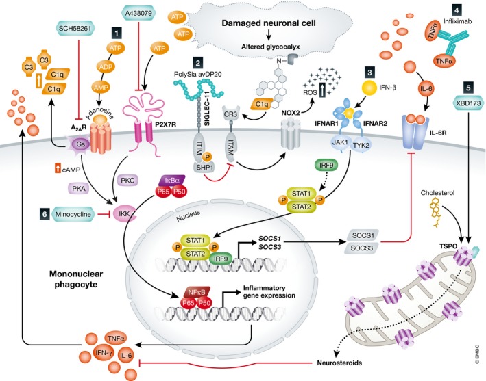
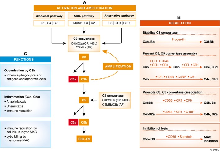
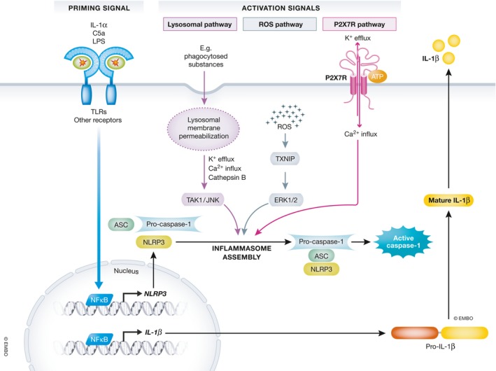
This review highlights the role of three key immune pathways in the pathophysiology of major retinal degenerative diseases including diabetic retinopathy, age-related macular degeneration, and rare retinal dystrophies. We first discuss the mechanisms how loss of retinal homeostasis evokes an unbalanced retinal immune reaction involving responses of local microglia and recruited macrophages, activity of the alternative complement system, and inflammasome assembly in the retinal pigment epithelium. Presenting these key mechanisms as complementary targets, we specifically emphasize the concept of immunomodulation as potential treatment strategy to prevent or delay vision loss. Promising molecules are ligands for phagocyte receptors, specific inhibitors of complement activation products, and inflammasome inhibitors. We comprehensively summarize the scientific evidence for this strategy from preclinical animal models, human ocular tissue analyses, and clinical trials evolving in the last few years.

Keywords: complement; inflammasome; microglia; mononuclear phagocytes; retina.


Figures

Figure 1
Figure 1. Schematic representation of microglial activity in the retina
Under homeostatic conditions, resident microglia mainly populate the plexiform layers. With their long protrusions, they continuously scan their environment and phagocytose cell debris. Different insults leading to abnormal cell functions or degeneration in the RPE, the photoreceptor layer, and the ganglion cell layer rapidly alert microglia. Resident microglia migrate to the lesion sites, where they transform into amoeboid full‐blown phagocytes and recruit macrophages from the periphery. Modified from Karlstetter et al (2010).
Figure 2
Figure 2. Activation pathways and therapy targets for mononuclear phagocytes
(1) The purines ATP and adenosine ligate to their receptors P2X7 and A2AR, respectively, which act through PKA/PKC signaling and thereby activate IKK. IKK aids to cleave and translocate NF‐κB into the nucleus and induce inflammatory gene expression and cytokine release. A2AR additionally potentiates the expression of the complement component C3 and the opsonin C1q. Neuroprotective effects were observed when A2AR and P2X7R were blocked using the selective inhibitors SCH58261 and A438079, respectively. (2) Intravitreal administration of polySia avDP20 compensates for desialylated neurons by binding to its receptor SIGLEC‐11 and inhibiting neurotoxic inflammation through its ITIM domain. During inflammation, desialylated neurons activate CR3 associated ITAM and thereby trigger excessive release of ROS via NOX2. (3) The IFN‐β pathway involves STAT1‐ and STAT2‐induced SOCS1/SOCS3 release which reduces cytokine expression through a negative feedback and inhibits IL‐6 signaling. (4) Neutralizing antibodies such as infliximab aid to neutralize cytosolic TNF‐α. (5) TSPO ligands such as XBD173 stimulate the production of neurosteroids which limit the release of pro‐inflammatory cytokines. (6) Minocycline is a potent inhibitor of NF‐κB signaling.
Figure 3
Figure 3. Complement activation, regulation, and immune functions
(A) The complement system can be activated by three pathways: the classical pathway (CP), mannose‐binding lectin (MBL) pathway, and the alternative pathway (AP); all lead to the cleavage of C3 and C5 and the formation of C5b‐C9. In the CP and MBL pathways, the C3 and C5 convertases are C4b2a and C4b2a3b, whereas in the AP, they are C3bBb and C3bBbC3b, respectively. Once C3 is cleaved into C3a and C3b, the C3b fragment can form C3bBb to amplify the complement activation cascade. Therefore, even if the initial activation is mediated by CP or MBL pathway, the cascade is ultimately amplified by the AP. (B) The complement activation cascades are regulated at multiple levels. Properdin is the only factor that stabilizes C3bBb and enhances complement activation. CFI, CD46, CFH, CR1, and C4BP prevent the assembly of C3 and C5 convertases by further breaking down C3b and C4b, whereas CD55, CR1, CFH, and C4BP can dissociate C3 and C5 convertases. CD59 and S protein can prevent the assembly of C5b‐C9. (C) Activation of the complement system generates C3a, C3b, and C5a fragments that are actively involved in immune responses. The soluble form of and sublytic levels of C5b‐C9 can regulate immune cell functions, whereas the membrane MACs directly kill pathogens and cells.
Figure 4
Figure 4. Molecular mechanisms of NLRP3 inflammasome priming and activation
Schematic representation of the NLRP3 inflammasome pathway which requires two signals: (i) a priming signal which activates NF‐kB, subsequently promoting the transcription of NLRP3 and pro‐IL‐1β, and (ii) an activation signal which facilitates the oligomerization of NLRP3, ASC, and procaspase‐1, resulting in the activation of NLRP3 inflammasome and secretion of mature IL‐1β and IL‐18.

Similar articles

Cited by

References

    1. Abdelsalam A, Del Priore L, Zarbin MA (1999) Drusen in age‐related macular degeneration: pathogenesis, natural course, and laser photocoagulation‐induced regression. Surv Ophthalmol 44: 1–29 - PubMed
    1. Abri Aghdam K, Pielen A, Framme C, Junker B (2015) Correlation between hyperreflective foci and clinical outcomes in neovascular age‐related macular degeneration after switching to aflibercept. Invest Ophthalmol Vis Sci 56: 6448–6455 - PubMed
    1. Adamis AP (2002) Is diabetic retinopathy an inflammatory disease? Br J Ophthalmol 86: 363–365 - PMC - PubMed
    1. Al‐Gayyar MM, Elsherbiny NM (2013) Contribution of TNF‐alpha to the development of retinal neurodegenerative disorders. Eur Cytokine Netw 24: 27–36 - PubMed
    1. Altay L, Scholz P, Schick T, Felsch M, Hoyng CB, den Hollander AI, Langmann T, Fauser S (2016) Association of hyperreflective foci present in early forms of age‐related macular degeneration with known age‐related macular degeneration risk polymorphisms. Invest Ophthalmol Vis Sci 57: 4315–4320 - PubMed

Publication types

MeSH terms