. 2018 Nov;178(3):1207-1221.
doi: 10.1104/pp.18.00978. Epub 2018 Sep 17.

A Trihelix Family Transcription Factor Is Associated with Key Genes in Mixed-Linkage Glucan Accumulation

Affiliations

A Trihelix Family Transcription Factor Is Associated with Key Genes in Mixed-Linkage Glucan Accumulation

Mingzhu Fan et al. Plant Physiol. 2018 Nov.

Abstract

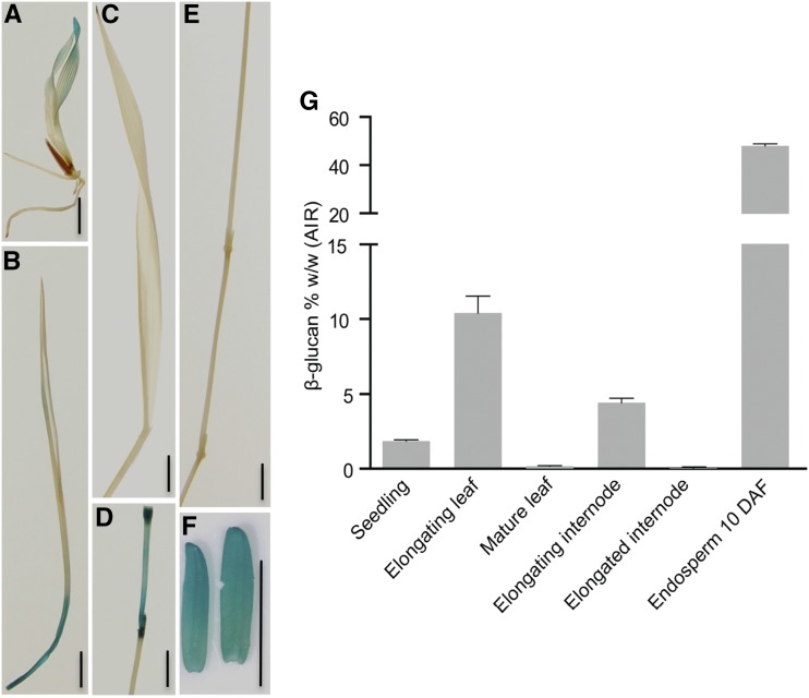
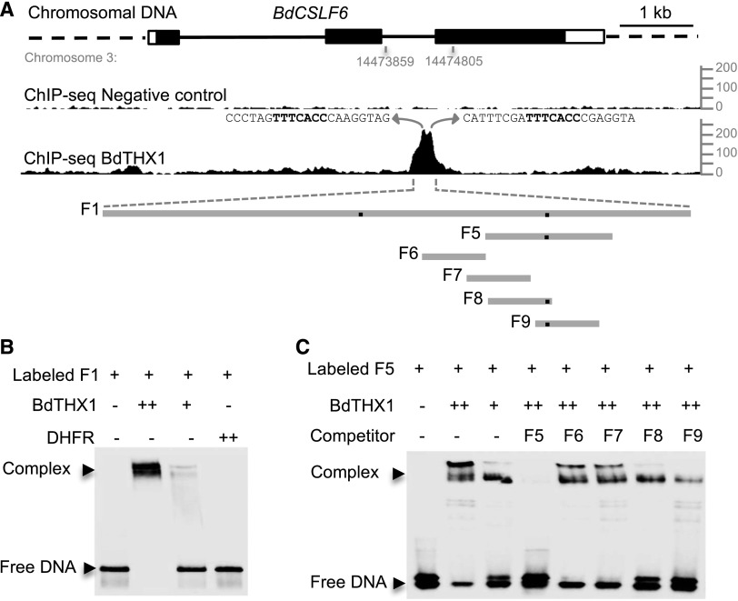
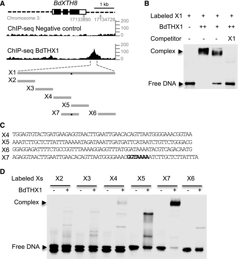
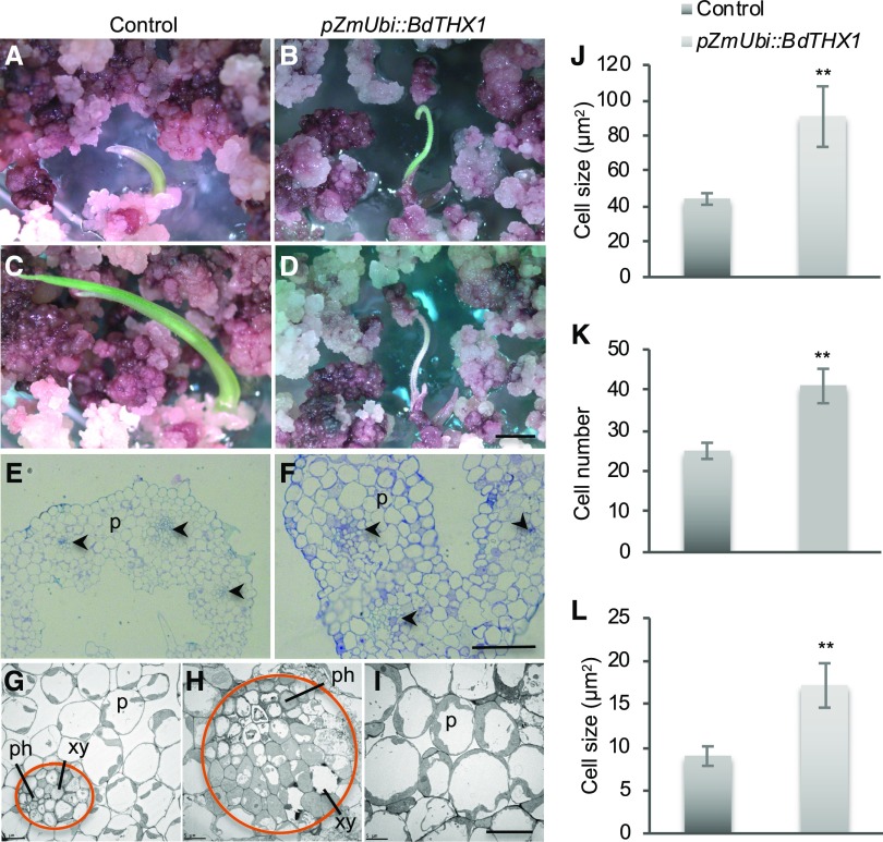
Mixed-linkage glucan (MLG) is a polysaccharide that is highly abundant in grass endosperm cell walls and present at lower amounts in other tissues. Cellulose synthase-like F (CSLF) and cellulose synthase-like H genes synthesize MLG, but it is unknown if other genes participate in the production and restructuring of MLG. Using Brachypodium distachyon transcriptional profiling data, we identified a B distachyon trihelix family transcription factor (BdTHX1) that is highly coexpressed with the B distachyon CSLF6 gene (BdCSLF6), which suggests that BdTHX1 is involved in the regulation of MLG biosynthesis. To determine the genes regulated by this transcription factor, we conducted chromatin immunoprecipitation sequencing (ChIP-seq) experiments using immature B distachyon seeds and an anti-BdTHX1 polyclonal antibody. The ChIP-seq experiment identified the second intron of BdCSLF6 as one of the most enriched sequences. The binding of BdTHX1 to the BdCSLF6 intron sequence was confirmed using electrophoretic mobility shift assays (EMSA). ChIP-seq also showed that a gene encoding a grass-specific glycoside hydrolase family 16 endotransglucosylase/hydrolase (BdXTH8) is bound by BdTHX1, and the binding was confirmed by EMSA. Radiochemical transglucanase assays showed that BdXTH8 exhibits predominantly MLG:xyloglucan endotransglucosylase activity, a hetero-transglycosylation reaction, and can thus produce MLG-xyloglucan covalent bonds; it also has a lower xyloglucan:xyloglucan endotransglucosylase activity. B distachyon shoots regenerated from transformed calli overexpressing BdTHX1 showed an abnormal arrangement of vascular tissue and seedling-lethal phenotypes. These results indicate that the transcription factor BdTHX1 likely plays an important role in MLG biosynthesis and restructuring by regulating the expression of BdCSLF6 and BdXTH8.


Figures

Figure 1.
Figure 1.
Gus expression driven by the 3.5-kb upstream region of BdTHX1 and MLG content in different B. distachyon tissues. A, GUS staining of 5-d-old pBdTHX1::GUS T1 seedling. B to F, GUS staining of elongating leaf (B), mature leaf (C), elongating internode (D), elongated internode (E), and 10 d-after-fertilization (DAF) endosperm (F) of pBdTHX1::GUS T1 plant. Scale bars, 5 mm. G, MLG amounts in various B. distachyon tissues are shown as means ± sd from three biological replicates. Alcohol-insoluble residue (AIR) extracted from seedlings and different tissues of Bd21-3 plant were subjected to MLG assay.
Figure 2.
Figure 2.
Immunolabeling and expression of BdCSLF6 and BdTHX1 in leaf. A, Immunolabeling of elongating leaf and leaf sheath with MLG monoclonal antibody. B and C, RNA in situ hybridization of BdCSLF6 (B) and BdTHX1 (C) in elongating leaf and leaf sheath. Sections hybridized with sense probes are shown as controls. The arrowheads indicate the vascular bundle. P, Parenchyma cells; ls, leaf sheath; el, elongating leaf. Scale bars, 100 μm.
Figure 3.
Figure 3.
Binding of BdTHX1 to BdCSLF6 intron sequences. A, ChIP-seq profile for BdCSLF6 using BdTHX1 antibody and schematic diagram of the probes used in electrophoretic mobility-shift assays. The ChIP-seq reads were mapped to B. distachyon Bd21 v1.0 chromosome 3: 14473859 to 14474805, corresponding to the second intron of BdCSLF6. y axis shows normalized read counts. The exons and introns of BdCSLF6 were shown as rectangles (filled, translated regions; open, untranslated regions) and black lines, respectively. Preimmune serum was used as a negative control. The sequences that correspond to the two peaks are shown, and GT motifs are highlighted in bold. F1 is a 272-bp-long probe located in the second intron of BdCSLF6. Probe F5 is 60 bp long. Probes F6, F7, F8, and F9 are 30 bp long. The black dots on the gray lines represent the binding site. B and C, EMSA was performed with biotin-labeled probes and wheat germ cell-free synthesized BdTHX1. Two different amounts of BdTHX1 were used to test if the binding is BdTHX1 amount dependent. ++ represents that 4-fold excess amount of protein was added in the reaction. Free DNA and protein-DNA complex were separated by 5% TBE polyacrylamide gels. B, The binding of BdTHX1 to probe F1. Wheat germ cell-free synthesized protein DHFR was used as negative control. C, The specific binding of BdTHX1 to probe F5 was tested by using a competition assay. Excess amounts of unlabeled DNA fragments (200-fold molar excess over labeled DNA) were included in the assay as competitors.
Figure 4.
Figure 4.
Analysis of BdTHX1 binding site. A and B, BdTHX1 binding site was analyzed by using mutant competitors in EMSA. The sequences of mutant competitors used in EMSA (B) are shown in A. A, The binding motif is highlighted in bold, and the mutated nucleotide of each competitor is lowercased. B, EMSA was performed with biotin-labeled probe F5 and wheat germ cell-free synthesized BdTHX1. Free DNA and protein-DNA complex were separated by 5% TBE polyacrylamide gels. Excess amounts of unlabeled probes F5, M1, M2, M3, M4, M5, M6, M7, M8, and M9 (200-fold molar excess over labeled DNA) were included as competitors. C, The motif created using program MEME motif analysis of the top 100 peaks by IDR score of ChIP-seq data. The height the label of each residues represents its frequency at that position. The GT-motif nucleotides are underlined.
Figure 5.
Figure 5.
Binding of BdTHX1 to the 3′ proximal region of BdXTH8. A, ChIP-seq profile for BdXTH8 using BdTHX1 antibody and schematic diagram of the probes used in EMSA. The ChIP-seq reads were mapped to B. distachyon Bd21 v1.0 chromosome 3: 17133890 to 17134728, corresponding to the 3′ proximal region of BdXTH8. y axis shows normalized read counts. The exons and introns of BdXTH8 are shown as rectangles (filled, translated regions; open, untranslated regions) and black lines, respectively. Dashed lines represents upstream and downstream sequences. X1 is a 328-bp sequence located downstream of BdXTH8. X2 to X7 vary between 57 and 60 bp long. The black dots on the gray lines represent the binding site. B, EMSA was performed with biotin-labeled probe X1 and wheat germ cell-free synthesized BdTHX1. Free DNA and protein-DNA complex were separated by 5% TBE polyacrylamide gels. Excess amounts of unlabeled X1 (200-fold molar excess over labeled X1) were included as specific competitor. Two different amounts of BdTHX1 were used. ++ represents that 4-fold excess amount of protein was added in the reaction. C, Probe sequence for X4, X5, X6, and X7. Both X4 and X5 contain part of GT motif. The binding motif in X7 is highlight in bold. D, Verification of binding motif using labeled probes from different regions of X1. BdTHX1 binds to X7, which probe contains a typical GT motif.
Figure 6.
Figure 6.
Activities of Pichia expressed BdXTH8. A, XET, MXE, and CXE (WSCA) activities were measured using soluble donor substrates (xyloglucan, MLG, or water-soluble cellulose acetate; WSCA); CXE (AP) activity was assayed using insoluble alkali-treated filter paper (AP) as a donor. [3H]XXXGol served as acceptor. Statistically significant differences (P < 0.01, ANOVA + Tukey’s post-hoc test) among activities after 24 h or 36 h incubation are indicated by lower- or uppercase letters, respectively. B, pH dependence of XET and MXE activity expressed as % of pH optima. Activities are shown as means ± sd from four (A) or three (B) independent experiments.
Figure 7.
Figure 7.
Phenotype of pZmUbi::BdTHX1 transgenic shoots. A to D, Transgenic shoots of control (A) and pZmUbi::BdTHX1 (B) regenerated on shoot-regeneration medium. After 4 days (control) or 7 days (pZmUbi::BdTHX1), shoots of control plants continued to grow (C), but the transgenic shoots of pZmUbi::THX1 stopped growing and died (D). Scale bar, 2 mm. E and F, Cross sections of control (E) and pZmUbi::BdTHX1 (F) shoots stained with toluidine blue. The arrowheads indicate the vascular bundle. P, parenchyma cells. Scale bar, 50 μm. G to I, Transmission electron microscopy of control (G) and pZmUbi::BdTHX1 shoots (H and I). The circle in orange indicates the area of vascular bundle. xy, Xylem; ph, phloem; P, parenchyma cells. Scale bar, 10 μm. J, Average cell size of control and pZmUbi::BdTHX1 were shown as means ± sd from different transgenic shoots (n = 3). **P < 0.01 compared with control (unpaired t test). K and L, Cell number (K) and average cell size (L) of cells in vascular bundle of control and pZmUbi::BdTHX1 transgenic shoots were shown as means ± sd (n = 3). **P < 0.01 compared with control (unpaired t test).

Similar articles

Cited by

References

    1. Bailey TL, Elkan C (1994) Fitting a Mixture Model by Expectation Maximization to Discover Motifs in Biopolymers. Proc Second Int Conf Intell Syst Mol Biol. 28–36 - PubMed
    1. Bianchi M, Crinelli R, Giacomini E, Carloni E, Magnani M (2009) A potent enhancer element in the 5′-UTR intron is crucial for transcriptional regulation of the human ubiquitin C gene. Gene 448: 88–101 - PubMed
    1. Brown DM, Zeef LAH, Ellis J, Goodacre R, Turner SR (2005) Identification of novel genes in Arabidopsis involved in secondary cell wall formation using expression profiling and reverse genetics. Plant Cell 17: 2281–2295 - PMC - PubMed
    1. Burton RA, Fincher GB (2012) Current challenges in cell wall biology in the cereals and grasses. Front Plant Sci 3: 130. - PMC - PubMed
    1. Burton RA, Fincher GB (2014) Evolution and development of cell walls in cereal grains. Front Plant Sci 5: 456. - PMC - PubMed

Publication types

MeSH terms

LinkOut - more resources