. 2018 Oct 2;115(40):10100-10105.
doi: 10.1073/pnas.1812622115. Epub 2018 Sep 17.

Fibroblasts lacking nuclear lamins do not have nuclear blebs or protrusions but nevertheless have frequent nuclear membrane ruptures

Affiliations

Fibroblasts lacking nuclear lamins do not have nuclear blebs or protrusions but nevertheless have frequent nuclear membrane ruptures

Natalie Y Chen et al. Proc Natl Acad Sci U S A. .

Abstract

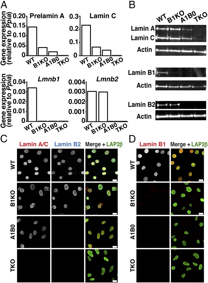
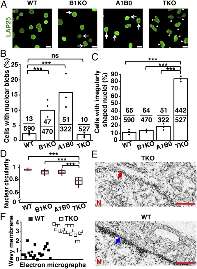
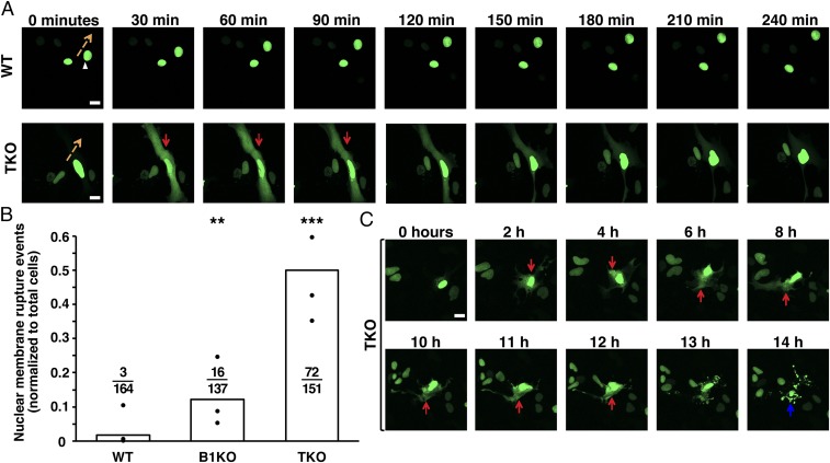
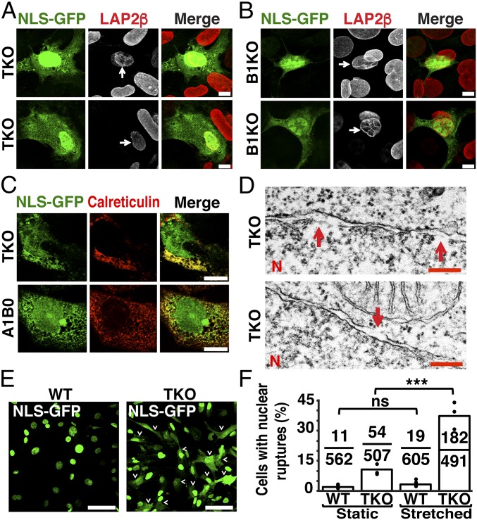
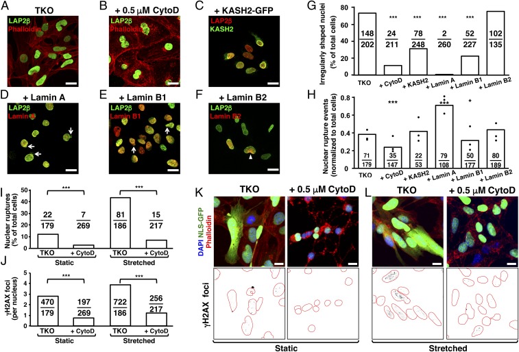
The nuclear lamina, an intermediate filament meshwork lining the inner nuclear membrane, is formed by the nuclear lamins (lamins A, C, B1, and B2). Defects or deficiencies in individual nuclear lamin proteins have been reported to elicit nuclear blebs (protrusions or outpouchings of the nuclear envelope) and increase susceptibility for nuclear membrane ruptures. It is unclear, however, how a complete absence of nuclear lamins would affect nuclear envelope morphology and nuclear membrane integrity (i.e., whether nuclear membrane blebs or protrusions would occur and, if not, whether cells would be susceptible to nuclear membrane ruptures). To address these issues, we generated mouse embryonic fibroblasts (MEFs) lacking all nuclear lamins. The nuclear lamin-deficient MEFs had irregular nuclear shapes but no nuclear blebs or protrusions. Despite a virtual absence of nuclear blebs, MEFs lacking nuclear lamins had frequent, prolonged, and occasionally nonhealing nuclear membrane ruptures. By transmission electron microscopy, the inner nuclear membrane in nuclear lamin-deficient MEFs have a "wavy" appearance, and there were discrete discontinuities in the inner and outer nuclear membranes. Nuclear membrane ruptures were accompanied by a large increase in DNA damage, as judged by γ-H2AX foci. Mechanical stress increased both nuclear membrane ruptures and DNA damage, whereas minimizing transmission of cytoskeletal forces to the nucleus had the opposite effects.

Keywords: nuclear envelope; nuclear lamina; nuclear membrane rupture.


Conflict of interest statement

The authors declare no conflict of interest.

Figures

Fig. 1.
Fig. 1.
Mouse embryonic fibroblasts (MEFs) lacking nuclear lamins. (A) Transcript levels for prelamin A, lamin C, lamin B1, and lamin B2 in Lmna+/+Lmnb1+/+Lmnb2+/+ [wild-type (WT)], Lmna+/+Lmnb1−/−Lmnb2+/+ [lamin B1 knockout (B1KO)], Lmna+/−Lmnb1−/−Lmnb2−/− [expressing lamin A/C from one allele but no B-type lamins (A1B0)], and Lmna−/−Lmnb1−/−Lmnb2−/− [triple-knockout (TKO)] MEFs. Expression was normalized to Ppia; mean of two independent experiments. (B) Western blots showing nuclear lamin expression in MEFs. Actin was used as a loading control. (C) Immunofluorescence microscopy of MEFs with antibodies against lamin A/C (red), lamin B2 (blue), and LAP2β (green). (Scale bars, 20 μm.) (D) Immunofluorescence microscopy of MEFs with antibodies against lamin B1 (red) and LAP2β (green). (Scale bars, 20 μm.)
Fig. 2.
Fig. 2.
Morphological abnormalities in WT, B1KO, A1B0, and TKO MEFs. (A) Immunofluorescence microscopy of MEFs stained with an antibody against LAP2β (green). The arrows point to nuclear blebs; the arrowheads point to irregularly shaped nuclei. (Scale bars, 20 μm.) (B) Percentages of cells with nuclear blebs. The black circles show the averages for three independent experiments; fractions show numbers of cells with blebs divided by the total number of cells examined. ***P < 0.0001 by χ2 test; ns, nonsignificant, P > 0.05. (C) Percentages of cells with irregularly shaped nuclei. The black circles indicate the averages for three independent experiments; fractions show the numbers of cells with irregularly shaped nuclei divided by the total number of cells examined. ***P < 0.0001 by χ2 test. (D) Box plots showing reduced circularity of the nucleus in TKO MEFs. The red line denotes the population median; boxes show 25th and 75th percentiles; and vertical lines show the 10th and 90th percentiles. ***P < 0.0001 by unpaired Student’s t test. (E) Electron micrographs showing that the inner nuclear membrane in TKO MEFs is wavy (red arrow), whereas it is straighter in WT MEFs (blue arrow). N, nuclei. (Scale bar, 500 nm.) (F) Scatter plot showing the average “wavy membrane score” for 38 electron micrographs (19 WT and 19 TKO MEFs) by 10 observers blinded to genotype. Each square represents the average score for an image (0 representing the “least wavy” and 4 representing the “most wavy”). WT MEFs (black squares); TKO MEF electron micrographs (white squares).
Fig. 3.
Fig. 3.
Nuclear membrane ruptures in TKO MEFs. (A) Sequential images of WT and TKO MEFs expressing a green fluorescent protein fused to a nuclear localization signal (NLS-GFP) (green) and imaged by live-cell fluorescence microscopy for 240 min. Images at 30-min intervals are shown. The orange arrows indicate the direction of nuclear movement, and the red arrows point to a nuclear membrane rupture event in a TKO MEF. (B) Bar graph showing the number of nuclear membrane ruptures as a percentage of the total number of cells evaluated. The black circles show the averages for three independent experiments. Ratios above each genotype show the total number of nuclear membrane rupture events divided by the total number of cells evaluated. Nuclear membrane ruptures were more frequent in TKO and B1KO MEFs than in WT MEFs. **P < 0.001; ***P < 0.0001 by χ2 test. (C) Sequential images showing a nonhealing nuclear membrane rupture in a TKO MEF (NLS-GFP remains in the cytoplasm) and cell death after 14 h. (Scale bars: A and C, 20 μm.)
Fig. 4.
Fig. 4.
Increased nuclear membrane ruptures in MEFs lacking nuclear lamins. (A and B) Immunofluorescence microscopy showing abnormal LAP2β distribution (red) in TKO (A) and B1KO (B) cells harboring nuclear membrane ruptures (arrows). (Scale bars, 10 μm.) (C) Fluorescence microscopy of MEFs with nuclear membrane ruptures (i.e., NLS-GFP in the cytoplasm) and stained with an antibody against calreticulin (ER marker; red). Cytoplasmic NLS-GFP was detected within the ER (yellow) and outside the ER (green). (Scale bars, 10 μm.) (D) Electron micrographs showing breaks (red arrows) in the inner (Top) and the outer nuclear membrane (Bottom) of TKO MEFs. N, nuclei. (Scale bars, 200 nm.) (E) Fluorescence microscopy showing larger numbers of nuclear membrane ruptures in TKO MEFs subjected to biaxial stretching. WT and TKO MEFs were subjected to biaxial stretching for 24 h. Nuclear membrane ruptures were frequent, as judged by NLS-GFP (green) in the cytoplasm (arrowheads). (Scale bars, 50 μm.) (F) Bar graph showing effects of stretching on nuclear membrane ruptures in WT and TKO MEFs. The black circles indicate frequencies in three independent experiments. Ratios above each genotype show the number of cells with NLS-GFP in the cytoplasm divided by the number of cells scored. ***P < 0.0005; ns, nonsignificant, P > 0.05 by χ2 test.
Fig. 5.
Fig. 5.
Impact of actin depolymerization, disrupting the LINC complex, and nuclear lamin expression on nuclear morphology, nuclear membrane ruptures, and DNA damage in TKO MEFs. (AF) Fluorescence microscopy images of TKO MEFs treated with 0.5 μM cytochalasin D (to disrupt the cytoskeleton); TKO MEFs expressing the KASH domain of nesprin 2 (KASH2-EGFP; to disrupt the LINC complex); and expressing human prelamin A (pTRIPZ-hu-prelamin A), human lamin B1 (pTRIPZ-LMNB1), or human lamin B2 (pTRIPZ-LMNB2). Cells were stained to visualize LAP2β, actin (with phalloidin), KASH2, lamin A, lamin B1, or lamin B2. Expression of lamin A and lamin B1 elicited nuclear blebs (arrows). In most cells, lamin B2 was not expressed uniformly in the nucleus (arrowhead). (Scale bars, 20 μm.) (G) Percentage of TKO MEFs with irregularly shaped nuclei. The ratios show the number of cells with irregularly shaped nuclei divided by the total number of cells examined. Treatment groups vs. control: ***P < 0.0001 by χ2 test. (H) Number of nuclear membrane ruptures in TKO MEFs. The results were analyzed as described in G. Treatment groups vs. control: *P < 0.01, ***P < 0.0001 by χ2 test. (I) Bar graph showing that cytochalasin D reduces the frequency of nuclear membrane ruptures in static and stretched TKO MEFs. The results were analyzed as described in G. ***P < 0.0001 by χ2 test. (J) Bar graph showing that cytochalasin D reduces γ-H2AX foci in static and stretched TKO MEFs. The results were analyzed as described in G. ***P < 0.0005 by Student’s t test. (K and L) Fluorescence microscopy images showing that cytochalasin D reduces γ-H2AX foci in static (K) and stretched (L) TKO MEFs. Panels below images show γ-H2AX foci (black) inside nuclei (outlined in red). (Scale bars: K and L, 10 μm.)

References

    1. Worman HJ, Fong LG, Muchir A, Young SG. Laminopathies and the long strange trip from basic cell biology to therapy. J Clin Invest. 2009;119:1825–1836. - PMC - PubMed
    1. Burke B, Stewart CL. The nuclear lamins: Flexibility in function. Nat Rev Mol Cell Biol. 2013;14:13–24. - PubMed
    1. Moir RD, Montag-Lowy M, Goldman RD. Dynamic properties of nuclear lamins: Lamin B is associated with sites of DNA replication. J Cell Biol. 1994;125:1201–1212. - PMC - PubMed
    1. Moir RD, et al. Review: The dynamics of the nuclear lamins during the cell cycle—relationship between structure and function. J Struct Biol. 2000;129:324–334. - PubMed
    1. Ellis DJ, Jenkins H, Whitfield WG, Hutchison CJ. GST-lamin fusion proteins act as dominant negative mutants in Xenopus egg extract and reveal the function of the lamina in DNA replication. J Cell Sci. 1997;110:2507–2518. - PubMed

Publication types