. 2018 Oct 2;115(40):E9298-E9307.
doi: 10.1073/pnas.1807704115. Epub 2018 Sep 17.

Control of CCND1 ubiquitylation by the catalytic SAGA subunit USP22 is essential for cell cycle progression through G1 in cancer cells

Affiliations

Control of CCND1 ubiquitylation by the catalytic SAGA subunit USP22 is essential for cell cycle progression through G1 in cancer cells

Victoria J Gennaro et al. Proc Natl Acad Sci U S A. .

Abstract

Overexpression of the deubiquitylase ubiquitin-specific peptidase 22 (USP22) is a marker of aggressive cancer phenotypes like metastasis, therapy resistance, and poor survival. Functionally, this overexpression of USP22 actively contributes to tumorigenesis, as USP22 depletion blocks cancer cell cycle progression in vitro, and inhibits tumor progression in animal models of lung, breast, bladder, ovarian, and liver cancer, among others. Current models suggest that USP22 mediates these biological effects via its role in epigenetic regulation as a subunit of the Spt-Ada-Gcn5-acetyltransferase (SAGA) transcriptional cofactor complex. Challenging the dogma, we report here a nontranscriptional role for USP22 via a direct effect on the core cell cycle machinery: that is, the deubiquitylation of the G1 cyclin D1 (CCND1). Deubiquitylation by USP22 protects CCND1 from proteasome-mediated degradation and occurs separately from the canonical phosphorylation/ubiquitylation mechanism previously shown to regulate CCND1 stability. We demonstrate that control of CCND1 is a key mechanism by which USP22 mediates its known role in cell cycle progression. Finally, USP22 and CCND1 levels correlate in patient lung and colorectal cancer samples and our preclinical studies indicate that targeting USP22 in combination with CDK inhibitors may offer an approach for treating cancer patients whose tumors exhibit elevated CCND1.

Keywords: CCND1; SAGA; USP22; cell cycle; deubiquitylation.


Conflict of interest statement

Conflict of interest statement: The authors declare no conflict of interest.

Figures
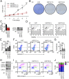
Fig. 1.
Fig. 1.
Loss of USP22 in cancer cells results in G1-phase cell cycle arrest. H1299 human lung cancer cells depleted of USP22 via infection with an shRNA-encoding lentivirus (or a luciferase shRNA as a control). After selection, cells counted and plated at 80,000 cells/mL on day 2 postinfection. (A) Cell number determined by direct counting of triplicate wells in a six-well plate via hemocytometer at the indicated time points. (B) Colony growth assessed via fixation and methylene blue staining of foci at day 7 postinfection. (C) Efficient knockdown of USP22 confirmed by both qRT-PCR and immunoblot (IB). (D) Cell viability quantified via flow cytometry. (E) Quantification of three experimental replicates representing average and SD of percent cell death, based on permeability. (F) Apoptosis measured by Annexin V-PE and 7AAD DNA staining, quantified by flow cytometry. (G) Quantification of three experimental replicates representing average and SD of percent apoptosis based on population of Annexin V-PE+ cells. NS, not significant. (H) PARP and CASPASE-3 (CAS-3) cleavage demonstrated by IB. The black arrowhead indicates cleaved species. (I) Progression through the cell cycle determined by EdU incorporation and PI staining followed by flow cytometry; the percent of cells in G1 phase is represented in blue. (J) Quantification of three experimental replicates of cell cycle phase distribution. Error bars indicate SD based on three independent experiments. *P < 0.05, **P < 0.02, ***P < 0.005.
Fig. 2.
Fig. 2.
Proteomic analysis (UbiScan) identifies CCND1 as a candidate substrate of USP22. (A) USP22-dependent regulation of CCND1 ubiquitylation status identified by UbiScan. This unbiased proteomic analysis was performed in HCT116 cells following USP22 depletion and proteasome inhibition. Ubiquitylated lysine residues and their reported functional roles in CCND1 are listed, along with potential links to cancer mutations. (B) Efficient knockdown of USP22 shown by IB for samples subjected to UbiScan analysis. (C) Schematic representation of CCND1 indicating known posttranslational modifications and ubiquitylated residues identified by UbiScan. (D) Levels of CCND1 protein following USP22 depletion determined by IB in HCT116, H1299, MCF7, and PC3 cells. (E) CCND1 protein levels following loss of USP22 evaluated in nonmalignant, human fibroblast 2091 cells. (F) FLAG-tagged USP22 was ectopically expressed in HCT116 cells via a stably integrated tetracycline-inducible vector. CCND1 and CDK4/6 protein levels assessed following USP22 induction for the indicated time points. (G) Protein levels of phosphorylated pRB, RB, CCND1, CCND2, CCND3 CDK4, and CDK6 assessed by IB in H1299 cells. (H) CCND1 mRNA levels measured by qRT-PCR following USP22 depletion in H1299, MCF7, and PC3 cells.
Fig. 3.
Fig. 3.
USP22 protects CCND1 protein from proteasome-mediated degradation. (A) CCND1 protein levels assessed in the presence of the proteasome inhibitors MG132 or EPX or the Calpain I inhibitor ALLN in H1299 cells. (B) USP22 depletion in H1299, MCF7, and PC3 cells accomplished using two distinct shRNA constructs. CCND1 protein levels assessed in the presence or absence of the proteasome inhibitor EPX. (C) The effect of USP22 depletion on the levels of ectopically expressed CCND1 assessed in H1299 cells. The impact of proteasome inhibition examined following MG132 treatment. (D) CCND1 protein half-life evaluated by treatment of cells with CHX for the indicated times. (E) CCND1 protein levels quantified for three CHX time-course experiments, followed by normalization to actin. Initial CCND1 levels set to 100%. (F) Bar graph representing the average and SD of CCND1 protein half-life for the experiments plotted in E. Error bars indicate SD based on three independent experiments. **P < 0.02.
Fig. 4.
Fig. 4.
USP22 directly deubiquitylates CCND1 protein independent of phosphorylation at T286. (A) H1299 cell lysates subjected to an endogenous CCND1 IP under nondenaturing conditions using A/G beads. Precipitates probed for CCND1 to detect high molecular-weight species, presumably ubiquitylated CCND1. (B) Sf9 cells were infected with baculovirus-expressing Flag-Fbxo4, Flag-cyclin D1, CDK4, HA-α-B-crystallin, HA-Cul1, HA-Skp1, and HA-Rbx1. SCFFbxo4 complex and CCND1/CDK4 complex then purified using anti-FLAG M2 and combined with E1/E2 ligases, ATP, and ubiquitin for 30 min at 37 °C to ubiquitylate CCND1 protein. Postubiquitylation assay, the CCND1/CDK4 complex was isolated and incubated with or without USP22 at 37 °C for indicated times in deubiquitylation buffer. Protein levels detected by IB; high molecular-weight CCND1 species indicative of polyubiquitylated CCND1. (C) HCT116 cell lysates subjected to in vitro analysis of CCND1 ubiquitylation status following USP22 depletion using UbiTest, as described in the Materials and Methods. Cells treated with MG132 before harvest and lysates generated using buffer containing protease mixture inhibitor, pan DUB inhibitor PR619, and the conventional chelator o-phenanthroline. Eluates then either left undigested (lanes 1 and 2) or subsequently digested with a global-DUB to strip polyubiquitin (lanes 3 and 4). Protein levels were detected by IB. The black arrowheads indicate unit length CCND1 (nonubiquitylated) and the green arrowheads indicate high molecular-weight species presumably representing ubiquitylated CCND1. (D) Schematic representing UbiTest experimental design from C. (E) H1299 cells made to express HA-tagged CCND1 WT or mutant with lysines from K33-K123. USP22 depleted in cells with shRNA-mediated lentivirus and CCND1 protein levels were assessed via anti-HA by IB. (F) HA-tagged CCND1 was expressed in H1299 cells as either WT or with phosphorylation site mutation T286A. Following USP22 depletion with two distinct shRNA constructs, cells were lysed and CCND1 protein levels were assessed by IB. (G) H1299 cells expressing the WT or T286A isoforms of HA-tagged CCND1 were subjected to USP22 depletion and subsequent analysis by IB in the presence or absence of proteasome inhibition (EPX). (H) Phosphorylation-dependence of USP22-depletion induced CCND1 degradation was assessed by inhibition of the T286 kinase GSK3β via treatment with TSW-119. Following depletion of USP22, CCND1 protein levels were assessed by IB. The established GSK3β pathway substrate β-catenin was included as control for TSW119 efficacy.
Fig. 5.
Fig. 5.
Ectopic expression of CCND1 provides a partial genetic rescue of the cell cycle phenotype observed with USP22 depletion. H1299 cells were transfected with a vector encoding 3XFLAG-tagged CCND1, followed by USP22 depletion. (A) Immunoblot demonstrating efficient knockdown of USP22, exogenous 3XFLAG-CCND1, and endogenous CCND1 expression. (B) Cell number was determined by manual counting of experimental triplicates at each of the postdepletion time points indicated. Error bars indicate SD based on three independent experiments. (C) Methylene blue-stained foci of representative plates from B. (D) At day 7 following USP22 depletion, cells were harvested and cell cycle profile was determined by PI staining and flow cytometry. G1-phase cells were gated (in blue) as percent of the total population. **P < 0.02.
Fig. 6.
Fig. 6.
CCND1 and USP22 protein levels correlate in patient samples from lung and colon adenocarcinoma and CDK4/6i treatment rescues the G1 phenotype associated with USP22 overexpression. (A) Representative images of serial sections of colon adenocarcinoma or lung adenocarcinoma BioMax tissue microarrays stained with DAPI and either USP22 or CCND1 antibodies. (B) Graphical representation of USP22(log) expression against CCND1(log) expression in each of the 120 cases with representative cases is indicated on the graph. [Pearson’s correlation coefficient (PCC) of 0.338 for colon adenocarcinoma or 0.357 for lung adenocarcinoma.] (C) FLAG-tagged USP22 was ectopically expressed in HCT116 cells via a stably integrated tetracycline-inducible vector. Following USP22 induction and treatment with selective CDK4/6 inhibitor PD-0332991 (PD), cells were harvested and cell cycle profile was determined by assessment of DNA content with PI staining and flow cytometry. G1-phase cells were gated (in blue) as percent of the total population. (D) Quantification of cell cycle phase distribution. Error bars indicate SD based on three independent experiments. (E) Induction of ectopic USP22 and increased CCND1 protein levels were assessed by IB. **P < 0.02, ***P < 0.005. (F) A schematic representing the proposed model of CCND1 stabilization by deubiquitylase USP22. CCND1–CDK4/6 complex advances cell cycling via hyperphosphorylating RB, which in turn releases E2F transcription factor from an inhibitory constraint and enables the expression of genes required for G1–S phase transition. To regulate rapid turnover, CCND1–CDK4/6 can incur phosphorylation at Thr286 by GSK3β, a precursor to nuclear export and subsequent polyubiquitylation by distinct E3 ligases (e.g., FBX4, PARK2, SKP2, FBXW8…) (–71). Polyubiquitylated CCND1–CDK4/6 is then targeted for proteasomal degradation. USP22 promotes CCND1 stabilization via removing CCND1 ubiquitin within the nucleus and/or cytoplasm and blocking degradation by the proteasome.

References

    1. Zhang XY, et al. The putative cancer stem cell marker USP22 is a subunit of the human SAGA complex required for activated transcription and cell-cycle progression. Mol Cell. 2008;29:102–111. - PMC - PubMed
    1. Zhao Y, et al. A TFTC/STAGA module mediates histone H2A and H2B deubiquitination, coactivates nuclear receptors, and counteracts heterochromatin silencing. Mol Cell. 2008;29:92–101. - PubMed
    1. Weake VM, et al. SAGA-mediated H2B deubiquitination controls the development of neuronal connectivity in the Drosophila visual system. EMBO J. 2008;27:394–405. - PMC - PubMed
    1. Zhang XY, Pfeiffer HK, Thorne AW, McMahon SB. USP22, an hSAGA subunit and potential cancer stem cell marker, reverses the polycomb-catalyzed ubiquitylation of histone H2A. Cell Cycle. 2008;7:1522–1524. - PMC - PubMed
    1. Glinsky GV. Death-from-cancer signatures and stem cell contribution to metastatic cancer. Cell Cycle. 2005;4:1171–1175. - PubMed

Publication types