. 2018 Oct 2;115(40):10112-10117.
doi: 10.1073/pnas.1810927115. Epub 2018 Sep 17.

Influenza hemagglutinin membrane anchor

Affiliations

Influenza hemagglutinin membrane anchor

Donald J Benton et al. Proc Natl Acad Sci U S A. .

Abstract

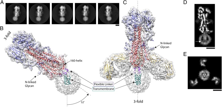
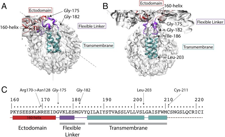
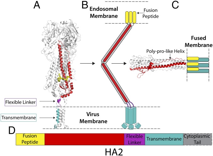
Viruses with membranes fuse them with cellular membranes, to transfer their genomes into cells at the beginning of infection. For Influenza virus, the membrane glycoprotein involved in fusion is the hemagglutinin (HA), the 3D structure of which is known from X-ray crystallographic studies. The soluble ectodomain fragments used in these studies lacked the "membrane anchor" portion of the molecule. Since this region has a role in membrane fusion, we have determined its structure by analyzing the intact, full-length molecule in a detergent micelle, using cryo-EM. We have also compared the structures of full-length HA-detergent micelles with full-length HA-Fab complex detergent micelles, to describe an infectivity-neutralizing monoclonal Fab that binds near the ectodomain membrane anchor junction. We determine a high-resolution HA structure which compares favorably in detail with the structure of the ectodomain seen by X-ray crystallography; we detect, clearly, all five carbohydrate side chains of HA; and we find that the ectodomain is joined to the membrane anchor by flexible, eight-residue-long, linkers. The linkers extend into the detergent micelle to join a central triple-helical structure that is a major component of the membrane anchor.

Keywords: cryo-EM; hemagglutinin; influenza; membrane fusion; membrane protein.


Conflict of interest statement

Conflict of interest statement: A.L. is the scientific founder of Humabs BioMed SA. A.L. holds shares in Humabs BioMed. D.C. is an employee of Humabs Biomed. N.L.K. was employed by MedImmune, LLC when work was executed and may currently hold AstraZeneca stock or stock options.

Figures

Fig. 1.
Fig. 1.
The structure of full-length hemagglutinin from A/duck/Alberta/35/76 H1N1 determined by cryo-EM. (A) Image classes of similarly oriented HAs illustrating the flexibility of the detergent micelle containing the transmembrane domain. The threefold axis of symmetry of the ectodomain trimer is marked in each frame by a dashed line. (B and C) Two structures of HA with density suitable for model building of the transmembrane domain: (B) the transmembrane domain in a detergent micelle exhibits a tilt of 52° from the threefold axis of the ectodomain, and (C) a complex of HA with the Fab of the human monoclonal antibody FISW84 where classification led to identification of a subset of molecules in which the transmembrane region follows the threefold symmetry axis of the ectodomain. In both B and C, examples of well-defined density for N-linked glycans are indicated. Both structures indicate a linker region (purple) between the base of the ectodomain (HA1 shown in blue, HA2 shown in red), defined by the 160 helix, and the transmembrane region (cyan) formed of a bundle of three α-helices. (D and E) Slices of map of HA−Fab complex shown in C. At the C termini of the transmembrane α-helices, they begin to splay apart, and extend toward the surface of the micelle, indicated by arrow in D. Also, in E, the micelle is seen to adopt the trimeric shape of the α-helical bundle, with Fab fragments visible in the periphery. (Scale bars: 5 nm.)
Fig. 2.
Fig. 2.
The structure of the membrane-associated region. Detailed views of (A) tilted and (B) straight micelles, as shown in Fig. 1 B and C respectively. The flexible linker region (purple) runs between Gly-175, at the C terminus of the 160 helix, and Gly-182 and extends to the N termini of the α-helices of a trimeric α-helical bundle, residues 186 to 203 (cyan). (C) The amino acid sequence of the transmembrane domain. The sequence shown begins at the 160 helix to the C terminus of HA2. Color-coded block diagrams indicate the positions of these structural elements in the sequence.
Fig. 3.
Fig. 3.
A comparison of the overall structures of tilted and straight transmembrane domains. Schematic diagrams of (A) 52° tilted and (B) straight transmembrane domains to indicate that the linker regions undergo extensive rearrangements in the tilted domain by comparison with the straight, and that each linker adopts a different structure. The individual subunits of the trimer are colored differently as blue, yellow, and silver, while the 160 helices are in red. The α-helices of the α-helical bundle in the tilted domain are also extensively rearranged. (C and D) Details of the map (gray) used to build the structure of the linker region of the straight transmembrane domain. C shows the short α-helix of the linker region (purple) residues 176 to 180. (D) The N-terminal region of the bundled α-helices (cyan) and its junction with the linker region (purple) and conserved residues Gln-185 and Tyr-184 at the linker−α-helix junction. (E) The stabilizing interaction of the 160-helix between Asn-128 and Arg-170.
Fig. 4.
Fig. 4.
HA reconstituted in liposomes. (A) Cryotomogram section showing cross-section of a liposome with examples of HAs tilted with respect to the lipid bilayer (white boxes). (Scale bar: 20 nm.) (B) Gallery of subtomograms of tilted HA in liposomes. Images in the second row are identical to those above but indicate HAs (blue) and liposome bilayer (red lines). (Scale bar: 10 nm.)
Fig. 5.
Fig. 5.
Structural snapshots toward membrane fusion. (A) The structure of straight full-length HA from cryo-EM (this study). (B) Schematic representation of possible fusion pH intermediate (46). (C) The structure of fusion pH HA2 (18), with modeled membrane anchor and fusion peptide colocalized in a fused membrane. (D) A color-coded diagram showing the location of the different regions of HA2 in its primary structure.

References

    1. Skehel JJ, Wiley DC. Receptor binding and membrane fusion in virus entry: The influenza hemagglutinin. Annu Rev Biochem. 2000;69:531–569. - PubMed
    1. Cossart P, Helenius A. Endocytosis of viruses and bacteria. Cold Spring Harb Perspect Biol. 2014;6:a016972. - PMC - PubMed
    1. Skehel JJ, et al. Changes in the conformation of influenza virus hemagglutinin at the pH optimum of virus-mediated membrane fusion. Proc Natl Acad Sci USA. 1982;79:968–972. - PMC - PubMed
    1. Bullough PA, Hughson FM, Skehel JJ, Wiley DC. Structure of influenza haemagglutinin at the pH of membrane fusion. Nature. 1994;371:37–43. - PubMed
    1. Gerhard W, Yewdell J, Frankel ME, Webster R. Antigenic structure of influenza virus haemagglutinin defined by hybridoma antibodies. Nature. 1981;290:713–717. - PubMed

Publication types

MeSH terms

Substances