. 2018 Nov 13;38(23):e00385-18.
doi: 10.1128/MCB.00385-18. Print 2018 Dec 1.

Deficiency of the Endocytic Protein Hip1 Leads to Decreased Gdpd3 Expression, Low Phosphocholine, and Kypholordosis

Affiliations

Deficiency of the Endocytic Protein Hip1 Leads to Decreased Gdpd3 Expression, Low Phosphocholine, and Kypholordosis

Ranjula Wijayatunge et al. Mol Cell Biol. .

Abstract

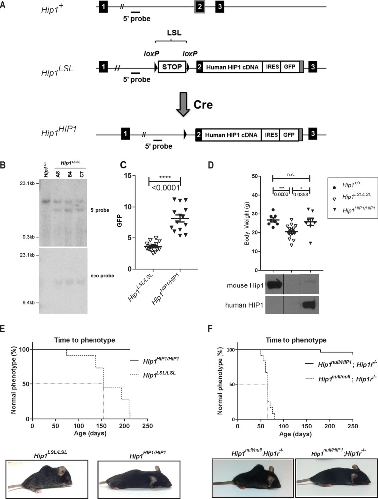
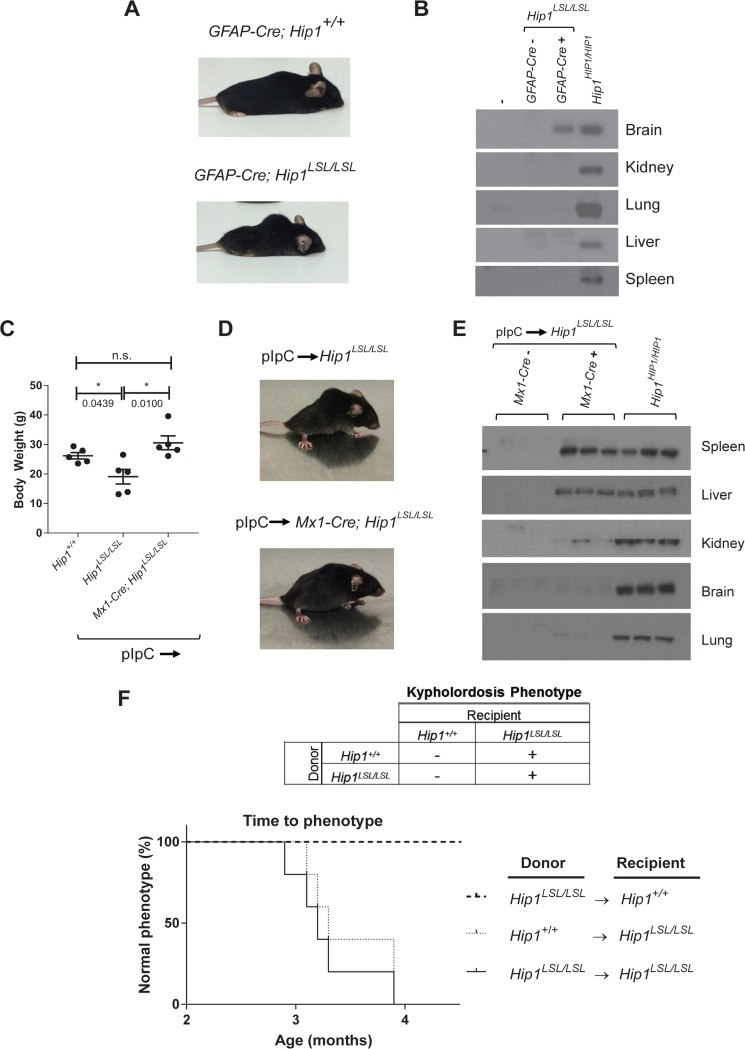
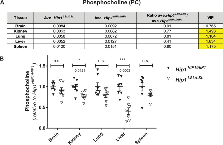
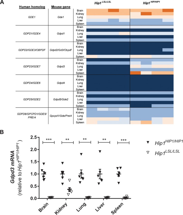
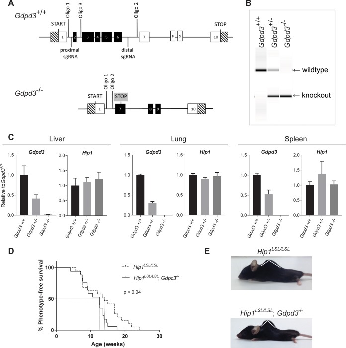
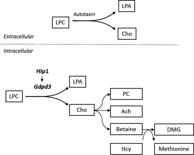
Deficiency of huntingtin-interacting protein 1 (Hip1) results in degenerative phenotypes. Here we generated a Hip1 deficiency allele where a floxed transcriptional stop cassette and a human HIP1 cDNA were knocked into intron 1 of the mouse Hip1 locus. CMV-Cre-mediated germ line excision of the stop cassette resulted in expression of HIP1 and rescue of the Hip1 knockout phenotype. Mx1-Cre-mediated excision led to HIP1 expression in spleen, kidney and liver, and also rescued the phenotype. In contrast, hGFAP-Cre-mediated, brain-specific HIP1 expression did not rescue the phenotype. Metabolomics and microarrays of several Hip1 knockout tissues identified low phosphocholine (PC) levels and low glycerophosphodiester phosphodiesterase domain containing 3 (Gdpd3) gene expression. Since Gdpd3 has lysophospholipase D activity that results in the formation of choline, a precursor of PC, Gdpd3 downregulation could lead to the low PC levels. To test whether Gdpd3 contributes to the Hip1 deficiency phenotype, we generated Gdpd3 knockout mice. Double knockout of Gdpd3 and Hip1 worsened the Hip1 phenotype. This suggests that Gdpd3 compensates for Hip1 loss. More-detailed knowledge of how Hip1 deficiency leads to low PC will improve our understanding of HIP1 in choline metabolism in normal and disease states.

Keywords: Cre recombinase; GDE7; GDPD3; HIP1; endocytosis; kypholordosis; phosphocholine.


Figures

FIG 1
FIG 1
Generation of Hip1-deficient and conditionally HIP1-humanized mice. (A) Schematic of the first 3 exons of the 32-exon murine Hip1 locus (Hip1+), the targeted knock-in allele with a floxed stop cassette (LSL), and a human HIP1 cDNA inserted into the murine Hip1 locus by homologous recombination (Hip1LSL) and the Cre-mediated recombined allele with the excised stop cassette (Hip1HIP1). The following features are indicated: stop cassette bracketed by loxP recombination sequences (LSL), partial mouse exon 2 fused to a human HIP1 cDNA-IRES-eGFP poly(A) tail (gray box), and 5′ genomic hybridization probe. (B) Southern blot confirmation of successfully targeted ES cell lines. Shown are ES cell lines carrying wild-type (Hip1+/+) or Hip1LSL targeted alleles (A8, B4, and C7). Genomic DNA was digested with EcoRI or HindIII and Southern blotted either with the 5′ probe to yield a 15.7-kb band corresponding to the wild-type allele and a 13.4-kb band corresponding to the recombined allele or with the 3′ neomycin probe to yield a 15.5-kb band corresponding to the recombined allele. The neomycin expression cassette was then excised by transfection of the cells with an Flp-expressing plasmid. (C) A CMV-Cre transgene was used to generate a germ line Cre-mediated recombined allele, Hip1HIP1, as delineated in panel A. Here GFP expression in peripheral blood white blood cells (WBCs) was quantitated to confirm that excision of the stop cassette leads to GFP expression in Hip1HIP1/HIP1 mouse tissue. Data represent mean ± standard error of the mean (SEM) (n = 14 per group). (D) Top, expression of human HIP1 from the Hip1 locus prevents the weight loss of Hip1-deficient mice. Data represent mean ± SEM (n = 8 to 16). ***, P < 0.005; *, P < 0.05; n.s., not significant. Ages ranged between 5 and 6.5 months. Bottom, Western blot analysis confirmed that expression of human HIP1 protein replaces mouse Hip1 protein in lung tissue from Hip1HIP1/HIP1 mice. The top panel represents Western blot with a mouse-specific monoclonal antibody (UM1B11) and the bottom panel a human-specific polyclonal antibody (UM323). (E) Expression of human HIP1 from the Hip1 locus prevents kypholordotic and weight loss phenotypes in all mice. Top, the Kaplan-Meier curves depict the percentage of mice with a normal phenotype as a function of time (in days) since birth. Hip1-deficient mice (Hip1LSL/LSL) are represented by the dotted line, and the fully humanized HIP1 mice (Hip1HIP1/HIP1) are represented by the solid line (n = 11 per genotype). Bottom, representative mice at 6 months of age with and without the Hip1 deficiency-associated kypholordosis and diminished weight. Note that the HIP1-humanized mouse (Hip1HIP1/HIP1) displays a slight kyphosis that is also observed in wild-type mice. Without the lordosis displayed by the Hip1LSL/LSL mouse, this slight kyphosis is normal. (F) The kypholordotic phenotype of Hip1 and Hip1r double knockout mice is rescued by a single copy of human HIP1. Top, the Kaplan-Meier curves depict the percentage of mice with a normal phenotype as a function of time (in days) since birth. Rescued Hip1null/HIP1; Hip1r−/− mice are represented by the solid line and double-knockout Hip1null/null; Hip1r−/− mice by the dotted line. Bottom, representative photographs of 2-month-old Hip1null/null; Hip1r−/− mouse with kypholordosis and an age- and gender-matched Hip1null/HIP1; Hip1r−/− rescue mouse with no phenotype.
FIG 2
FIG 2
Tissue-specific rescue of Hip1 deficiency with human HIP1. (A) The Hip1 deficiency phenotype was not rescued by expression of human HIP1 in the nervous system with the hGFAP-Cre transgene. Representative photographs of 6-month-old hGFAP-Cre; Hip1LSL/LSL and hGFAP-Cre; Hip1+/+ mice are shown. (B) Western blotting for tissue-specific expression analysis of human HIP1 in brain from hGFAP-Cre; Hip1LSL/LSL mice. Tissue extracts from representatives of each genotype were analyzed. The human-specific HIP1 antibody UM323 was used to detect HIP1 in the brain and showed no detectable HIP1 in spleen, liver, kidney, and lung tissues. (C) At 6 weeks of age, Hip1LSL/LSL and Mx1-cre; Hip1LSL/LSL mice were treated with pIpC to induce Mx1-Cre-mediated expression of human HIP1 in the hematopoietic system, kidney, and liver. Expression of Mx1-Cre prevented the weight loss observed in Hip1-deficient mice (Hip1LSL/LSL). All mice were between 5.5 and 6 months of age, as this is when the weight loss was most apparent. Data represent mean ± SEM (n = 5). n.s., not significant; *, P < 0.05. (D) The kypholordotic spinal curvature in Hip1-deficient mice is rescued by Mx1-Cre-mediated expression of human HIP1. Representative photographs of 4-month-old pIpC-treated Hip1LSL/LSL and Mx1-cre; Hip1LSL/LSL mice are shown. (E) Western blotting for tissue-specific expression analysis of human HIP1 in spleen, liver, and kidney tissues. Tissue extracts from three mice of each genotype were analyzed. The human-specific HIP1 antibody UM323 was used to detect HIP1 in the spleen, liver, and kidney, with no detectable HIP1 in brain and lung tissues. (F) Kaplan-Meier curves depicting the lack of or the progression of the development of the kypholordotic phenotype in bone marrow-transplanted mice. Transplantation of Hip1-deficient (Hip1LSL/LSL) bone marrow into irradiated wild-type (Hip1+/+) mice does not lead to the development of the kypholordotic phenotype. Transplantation of wild-type (Hip1+/+) bone marrow into irradiated Hip1-deficient (Hip1LSL/LSL) mice does not prevent the development of the kypholordotic phenotype (n = 5 per group).
FIG 3
FIG 3
Hip1-deficient mice have low phosphocholine levels. (A) Of the 120 metabolites that were measured with LC-MS/MS, a robust and consistent metabolic change was decreased phosphocholine (PC) levels. All tissues except brain from Hip1-deficient mice (Hip1LSL/LSL) showed significantly decreased PC levels compared to those in HIP1-rescued mice (Hip1HIP1/HIP1). The units are peak areas normalized to total ion current (a marker of total metabolite abundance).The VIP (variable importance in projection) number is a measure of statistical significance. When it is above 1.0, the metabolite change is considered statistically significant. (B) The same data in panel A are shown as dot plots to visualize variability between samples. Data are expressed as relative to the average of levels in HIP1-rescued tissues (Hip1HIP1/HIP1). Data represent mean ± SEM (n = 7 per group). n.s., not significant; *, P < 0.05; ***, P < 0.0001.
FIG 4
FIG 4
Decreased Gdpd3 expression in Hip1-deficient mice. (A) Heat map of RNA expression of the seven members of the GDPD family in Hip1-deficient and HIP1-rescued mice. Dark blue is the lowest and orange is the highest expression. Only Gdpd3 (Gde7) expression is lost in Hip1-deficient mouse tissues (asterisk). (B) Real-time PCR analysis of Gdpd3 expression. Data were normalized to Gapdh and expressed as relative to the average of Gdpd3 levels in HIP1-rescued tissues (Hip1HIP1/HIP1). Data represent mean ± SEM (n = 6). **, P < 0.005; ***, P < 0.0001.
FIG 5
FIG 5
Acceleration of kypholordosis onset in Hip1 and Gdpd3 DKO mice compared to Hip1 single-knockout mice. (A) Schematic of the targeting strategy used to generate the Gdpd3 knockout allele. (B) PCR analysis of genomic DNA isolated from tail biopsy specimens of Gdpd3 wild-type (Gdpd3+/+), heterozygous (Gdpd3+/−), and homozygous (Gdpd3−/−) mice. The wild-type Gdpd3 allele generates a 501-bp band and the knockout allele a 290-bp band with the use of oligonucleotides 1 and 3 and oligonucleotides 1 and 2, respectively. (C) qPCR analysis of Gdpd3 mRNA expression levels in liver, lung, and spleen tissue of Gdpd3+/+, Gdpd3+/−, and Gdpd3−/− mice. Data were normalized to Gapdh and expressed as relative to the average of Gdpd3 or Hip1 levels in Gdpd3+/+ tissue (n = 3 per genotype). (D) Kaplan-Meier curves of phenotype onset in Hip1 knockout mice (dotted line) and Hip1 and Gdpd3 DKO mice (solid line). The log-rank test was used to calculate significance. Mice were phenotyped for kypholordosis as described in Materials and Methods without prior knowledge of mouse genotypes. (E) Representative 10-month-old male Hip1LSL/LSL single-knockout and Hip1LSL/LSL; Gdpd3−/− DKO mice.
FIG 6
FIG 6
Schematic depicting a potential link between reduced Gdpd3 and alterations in choline-related metabolites in Hip1-deficient mice. Extracellularly, LPC is metabolized to LPA and choline by the lysoPLD autotaxin. Hip1 deficiency results in decreased Gdpd3. LysoPLD activity of Gdpd3 predicts that alterations seen in choline-related metabolites with Hip1 deficiency are due to reduced Gdpd3 levels. Changes in choline-related metabolites in liver of Hip1-deficient mice are depicted. LPC, lysophosphatidylcholine; LPA, lysophosphatidic acid; Cho, choline; PC, phosphocholine; Ach, acetylcholine; DMG, dimethylglycine; Hcy, homocysteine.

Similar articles

Cited by

References

    1. Kalchman MA, Koide HB, McCutcheon K, Graham RK, Nichol K, Nishiyama K, Kazemi-Esfarjani P, Lynn FC, Wellington C, Metzler M, Goldberg YP, Kanazawa I, Gietz RD, Hayden MR. 1997. HIP1, a human homologue of S. cerevisiae Sla2p, interacts with membrane-associated huntingtin in the brain. Nat Genet 16:44–53. doi:10.1038/ng0597-44. - DOI - PubMed
    1. Wanker EE, Rovira C, Scherzinger E, Hasenbank R, Walter S, Tait D, Colicelli J, Lehrach H. 1997. HIP-I: a huntingtin interacting protein isolated by the yeast two-hybrid system. Hum Mol Genet 6:487–495. doi:10.1093/hmg/6.3.487. - DOI - PubMed
    1. Metzler M, Legendre-Guillemin V, Gan L, Chopra V, Kwok A, McPherson PS, Hayden MR. 2001. HIP1 functions in clathrin-mediated endocytosis through binding to clathrin and adaptor protein 2. J Biol Chem 276:39271–39276. doi:10.1074/jbc.C100401200. - DOI - PubMed
    1. Mishra SK, Agostinelli NR, Brett TJ, Mizukami I, Ross TS, Traub LM. 2001. Clathrin- and AP-2-binding sites in HIP1 uncover a general assembly role for endocytic accessory proteins. J Biol Chem 276:46230–46236. doi:10.1074/jbc.M108177200. - DOI - PubMed
    1. Rao DS, Chang JC, Kumar PD, Mizukami I, Smithson GM, Bradley SV, Parlow AF, Ross TS. 2001. Huntingtin interacting protein 1 is a clathrin coat binding protein required for differentiation of late spermatogenic progenitors. Mol Cell Biol 21:7796–7806. doi:10.1128/MCB.21.22.7796-7806.2001. - DOI - PMC - PubMed

Publication types