Preexisting Simian Immunodeficiency Virus Infection Increases Susceptibility to Tuberculosis in Mauritian Cynomolgus Macaques
- PMID: 30224552
- PMCID: PMC6246917
- DOI: 10.1128/IAI.00565-18
Preexisting Simian Immunodeficiency Virus Infection Increases Susceptibility to Tuberculosis in Mauritian Cynomolgus Macaques
Abstract
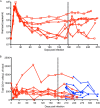
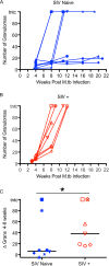
Tuberculosis (TB), caused by Mycobacterium tuberculosis, is the leading cause of death among human immunodeficiency virus (HIV)-positive patients. The precise mechanisms by which HIV impairs host resistance to a subsequent M. tuberculosis infection are unknown. We modeled this coinfection in Mauritian cynomolgus macaques (MCM) using simian immunodeficiency virus (SIV) as an HIV surrogate. We infected seven MCM with SIVmac239 intrarectally and 6 months later coinfected them via bronchoscope with ∼10 CFU of M. tuberculosis Another eight MCM were infected with M. tuberculosis alone. TB progression was monitored by clinical parameters, by culturing bacilli in gastric and bronchoalveolar lavages, and by serial [18F]fluorodeoxyglucose (FDG) positron emission tomography/computed tomography (PET/CT) imaging. The eight MCM infected with M. tuberculosis alone displayed dichotomous susceptibility to TB, with four animals reaching humane endpoint within 13 weeks and four animals surviving >19 weeks after M. tuberculosis infection. In stark contrast, all seven SIV+ animals exhibited rapidly progressive TB following coinfection and all reached humane endpoint by 13 weeks. Serial PET/CT imaging confirmed dichotomous outcomes in MCM infected with M. tuberculosis alone and marked susceptibility to TB in all SIV+ MCM. Notably, imaging revealed a significant increase in TB granulomas between 4 and 8 weeks after M. tuberculosis infection in SIV+ but not in SIV-naive MCM and implies that SIV impairs the ability of animals to contain M. tuberculosis dissemination. At necropsy, animals with preexisting SIV infection had more overall pathology, increased bacterial loads, and a trend towards more extrapulmonary disease than animals infected with M. tuberculosis alone. We thus developed a tractable MCM model in which to study SIV-M. tuberculosis coinfection and demonstrate that preexisting SIV dramatically diminishes the ability to control M. tuberculosis coinfection.
Keywords: Mauritian cynomolgus macaque; Mycobacterium tuberculosis; PET/CT imaging; bacterial burden; disease pathology; necropsy; simian immunodeficiency virus.
Copyright © 2018 American Society for Microbiology.
Figures


Similar articles
-
Spontaneous Control of SIV Replication Does Not Prevent T Cell Dysregulation and Bacterial Dissemination in Animals Co-Infected with M. tuberculosis.Microbiol Spectr. 2022 Jun 29;10(3):e0172421. doi: 10.1128/spectrum.01724-21. Epub 2022 Apr 25. Microbiol Spectr. 2022. PMID: 35467372 Free PMC article.
-
Pre-existing Simian Immunodeficiency Virus Infection Increases Expression of T Cell Markers Associated with Activation during Early Mycobacterium tuberculosis Coinfection and Impairs TNF Responses in Granulomas.J Immunol. 2021 Jul 1;207(1):175-188. doi: 10.4049/jimmunol.2100073. Epub 2021 Jun 18. J Immunol. 2021. PMID: 34145063 Free PMC article.
-
Transiently boosting Vγ9+Vδ2+ γδ T cells early in Mtb coinfection of SIV-infected juvenile macaques does not improve Mtb host resistance.Infect Immun. 2024 Dec 10;92(12):e0031324. doi: 10.1128/iai.00313-24. Epub 2024 Oct 30. Infect Immun. 2024. PMID: 39475292 Free PMC article.
-
Monkeying around with MAIT Cells: Studying the Role of MAIT Cells in SIV and Mtb Co-Infection.Viruses. 2021 May 8;13(5):863. doi: 10.3390/v13050863. Viruses. 2021. PMID: 34066765 Free PMC article. Review.
-
Chronic Immune Activation in TB/HIV Co-infection.Trends Microbiol. 2020 Aug;28(8):619-632. doi: 10.1016/j.tim.2020.03.015. Epub 2020 Apr 22. Trends Microbiol. 2020. PMID: 32417227 Free PMC article. Review.
Cited by
-
The Mtb-HIV syndemic interaction: why treating M. tuberculosis infection may be crucial for HIV-1 eradication.Future Virol. 2020 Feb;15(2):101-125. doi: 10.2217/fvl-2019-0069. Future Virol. 2020. PMID: 32273900 Free PMC article. Review.
-
Transiently boosting Vγ9+Vδ2+ γδ T cells early in Mtb coinfection of SIV-infected juvenile macaques does not improve Mtb host resistance.bioRxiv [Preprint]. 2024 Jul 23:2024.07.22.604654. doi: 10.1101/2024.07.22.604654. bioRxiv. 2024. Update in: Infect Immun. 2024 Dec 10;92(12):e0031324. doi: 10.1128/iai.00313-24. PMID: 39091843 Free PMC article. Updated. Preprint.
-
HIV-MTB Co-Infection Reduces CD4+ T Cells and Affects Granuloma Integrity.Viruses. 2024 Aug 21;16(8):1335. doi: 10.3390/v16081335. Viruses. 2024. PMID: 39205309 Free PMC article.
-
Antibody glycosylation correlates with disease progression in SIV-Mycobacterium tuberculosis coinfected cynomolgus macaques.Clin Transl Immunology. 2023 Nov 20;12(11):e1474. doi: 10.1002/cti2.1474. eCollection 2023. Clin Transl Immunology. 2023. PMID: 38020728 Free PMC article.
-
MAIT cells are functionally impaired in a Mauritian cynomolgus macaque model of SIV and Mtb co-infection.PLoS Pathog. 2020 May 20;16(5):e1008585. doi: 10.1371/journal.ppat.1008585. eCollection 2020 May. PLoS Pathog. 2020. PMID: 32433713 Free PMC article.
References
-
- WHO. 2017. Global tuberculosis report 2017. World Health Organization, Geneva, Switzerland.
-
- UNAIDS. 2017. UNAIDS data 2017. UNAIDS, Geneva, Switzerland.
-
- Houben RM, Crampin AC, Ndhlovu R, Sonnenberg P, Godfrey-Faussett P, Haas WH, Engelmann G, Lombard CJ, Wilkinson D, Bruchfeld J, Lockman S, Tappero J, Glynn JR. 2011. Human immunodeficiency virus associated tuberculosis more often due to recent infection than reactivation of latent infection. Int J Tuber Lung Dis 15:24–31. - PubMed
Publication types
MeSH terms
Grants and funding
LinkOut - more resources
Full Text Sources
Other Literature Sources
Medical

