

Comparison of Significant Carotid Stenosis for Nasopharyngeal Carcinoma between Intensity-Modulated Radiotherapy and Conventional Two-Dimensional Radiotherapy
- PMID: 30224668
- PMCID: PMC6141472
- DOI: 10.1038/s41598-018-32398-y
Comparison of Significant Carotid Stenosis for Nasopharyngeal Carcinoma between Intensity-Modulated Radiotherapy and Conventional Two-Dimensional Radiotherapy
Abstract
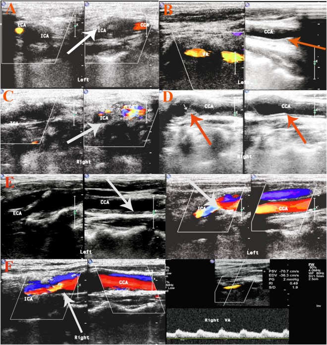
Radiotherapy (RT) serves as the most efficient treatment for nasopharyngeal carcinoma (NPC) and can cause carotid stenosis. This work compared the incidence of significant carotid stenosis between intensity-modulated radiotherapy (IMRT) and two-dimensional conventional radiotherapy (2D-RT) for NPC and explored the risk factors. We retrospectively reviewed 233 cases with NPC who underwent carotid ultrasound post IMRT or 2D-RT from 2006 to 2015. The incidence of significant stenosis after RT was 19.3%. Significant stenosis was identified in 20 (14.6%) of 137 patients treated with IMRT and 25 (26.0%) of 96 patients with 2D-RT, respectively (p = 0.035). Multivariate logistic analysis indicated age (odds ratio = 1.054, 95% CI = 1.011-1.099, p = 0.014), radiation technique (IMRT) (odds ratio = 0.471, 95%CI = 0.241-0.919, p = 0.027) and time interval (odds ratio = 1.068, 95%CI = 1.033-1.105, p = 0.001) as independent predictors for significant carotid stenosis. Our study suggests that IMRT was associated with decreased incidence of significant carotid stenosis versus 2D-RT for NPC. Prevention and carotid ultrasound should be considered for older NPC survivors with longer interval from RT, especially those treated with 2D-RT.
Conflict of interest statement
The authors declare no competing interests.
Figures
References
-
- Fang, W. et al. Late-onset cystic brain necrosis after radiotherapy for nasopharyngeal carcinoma. Jpn J Clin Oncol, 1–6, 10.1093/jjco/hyx028 (2017). - PubMed
Publication types
MeSH terms
Grants and funding
- 201803010013/Guangzhou Science and Technology Program key projects/International
- 201604020100/Guangzhou Science and Technology Program key projects/International
- 81372919/National Natural Science Foundation of China (National Science Foundation of China)/International
- 17ykjc17/Sun Yat-sen University (SYSY)/International
LinkOut - more resources
Full Text Sources
Other Literature Sources
