. 2018 Sep 17;8(1):13906.
doi: 10.1038/s41598-018-32125-7.

sox9b is required in cardiomyocytes for cardiac morphogenesis and function

Affiliations

sox9b is required in cardiomyocytes for cardiac morphogenesis and function

Joseph C Gawdzik et al. Sci Rep. .

Abstract

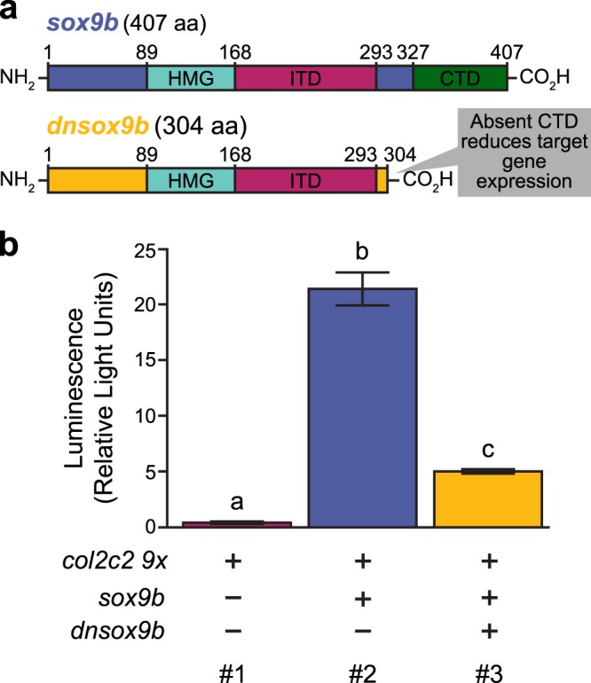
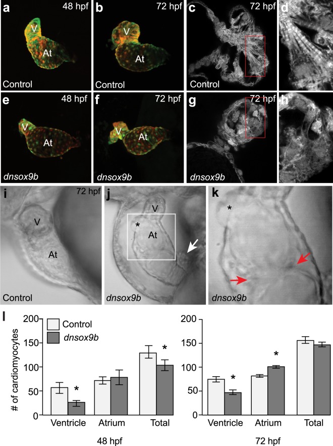
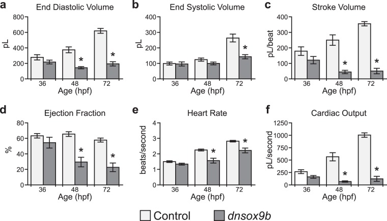
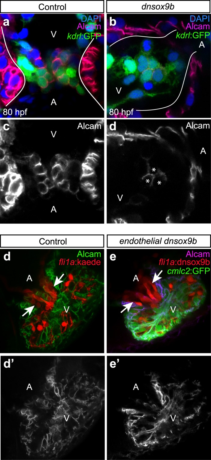
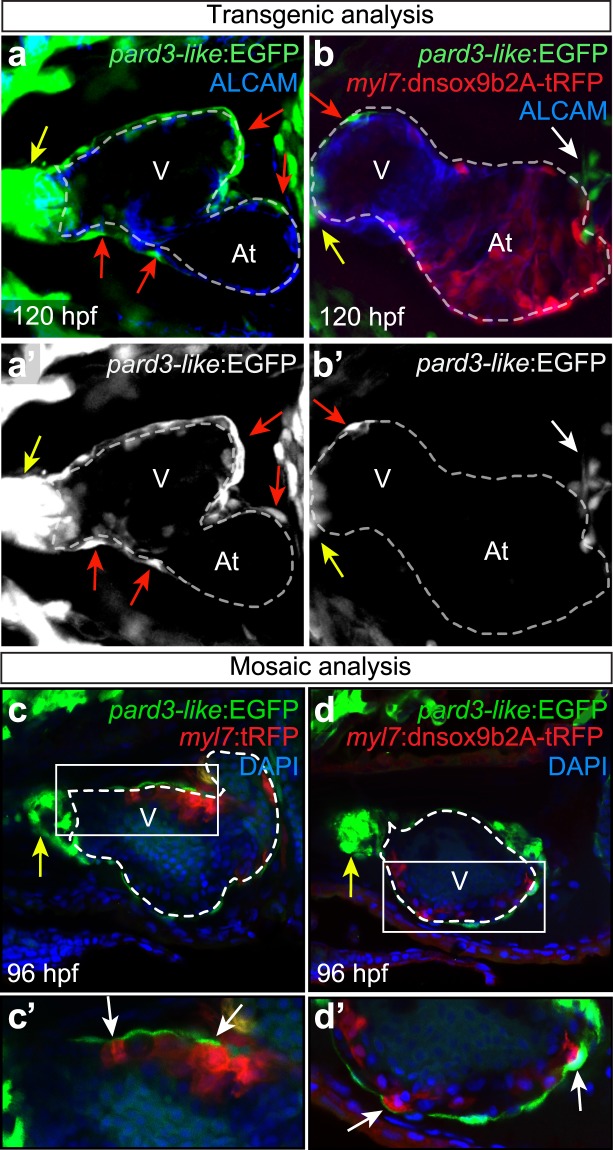
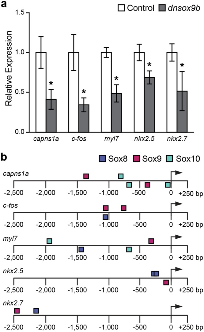
The high mobility group transcription factor SOX9 is expressed in stem cells, progenitor cells, and differentiated cell-types in developing and mature organs. Exposure to a variety of toxicants including dioxin, di(2-ethylhexyl) phthalate, 6:2 chlorinated polyfluorinated ether sulfonate, and chlorpyrifos results in the downregulation of tetrapod Sox9 and/or zebrafish sox9b. Disruption of Sox9/sox9b function through environmental exposures or genetic mutations produce a wide range of phenotypes and adversely affect organ development and health. We generated a dominant-negative sox9b (dnsox9b) to inhibit sox9b target gene expression and used the Gal4/UAS system to drive dnsox9b specifically in cardiomyocytes. Cardiomyocyte-specific inhibition of sox9b function resulted in a decrease in ventricular cardiomyocytes, an increase in atrial cardiomyocytes, hypoplastic endothelial cushions, and impaired epicardial development, ultimately culminating in heart failure. Cardiomyocyte-specific dnsox9b expression significantly reduced end diastolic volume, which corresponded with a decrease in stroke volume, ejection fraction, and cardiac output. Further analysis of isolated cardiac tissue by RT-qPCR revealed cardiomyocyte-specific inhibition of sox9b function significantly decreased the expression of the critical cardiac development genes nkx2.5, nkx2.7, and myl7, as well as c-fos, an immediate early gene necessary for cardiomyocyte progenitor differentiation. Together our studies indicate sox9b transcriptional regulation is necessary for cardiomyocyte development and function.


Conflict of interest statement

The authors declare no competing interests.

Figures

Figure 1
Figure 1
Design and in vitro validation of a dominant-negative sox9b (dnsox9b). (a) Schematics of the coding sequences of sox9b and dnsox9b. The DNA-binding HMG domain (HMG), internal transactivation domain (ITD), and C-terminal transactivation domain (CTD) are indicated. Numbers give amino acid positions. (b) In vitro HEK293T cell luciferase expression assay. Column #1: transfection of the col2c2-9x plasmid alone is not sufficient to induce luciferase activity. Column #2: Co-transfection of the col2c2-9x and sox9b-2A-tRFP plasmids robustly induces luciferase activity. Column #3: When dnsox9b-2A-tRFP is transfected along with sox9b-2A-tRFP and the col2c2-9x plasmid, luciferase reporter activity is significantly reduced. Average luciferase activity is shown relative to a transfection control. Error bars represent standard error of the mean. A one-way ANOVA was used to compare groups. Letters indicate significant differences between groups (p < 0.05), n = 4 for each condition.
Figure 2
Figure 2
Cardiomyocyte-specific inhibition of sox9b function disrupts cardiac morphogenesis. Live control (a,b and i; Tg(myl7:Gal4VP16;UAS:tRFP)) zebrafish and zebrafish with cardiomyocyte-specific inhibition of sox9b function (e,f,j and k; Tg(myl7:Gal4VP16;UAS:dnsox9b-2A-tRFP)). (a,b,e,f) Embryos and larvae were anesthetized in Tricaine, treated with 20 mM 2,3-butanedione 2-monoxime to temporarily stop heartbeat, then mounted in low melting point agarose for confocal imaging. Ventral images were collected at 48 hpf (a,e) and 72 hpf (b,f). Control (Tg(myl7:Gal4VP16;UAS:tRFP)) embryos had looped hearts and exhibit time dependent chamber growth. Embryos and larvae with cardiomyocyte-specific inhibition of sox9b function (Tg(myl7:Gal4VP16;UAS:dnsox9b-2A-tRFP)) had small, compacted ventricles and unlooped heart chambers. (c,d,g,h) Super-resolution images of fixed control (c and d; Tg(myl7:Gal4VP16;UAS:tRFP)) and experimental larvae (g,h; Tg(myl7:Gal4VP16;UAS:dnsox9b-2A-tRFP)). Cardiomyocyte-specific inhibition of sox9b function disrupted the development of myofibrillar bundles and z-lines were notably absent. (i–k) Still images from Supplemental Movies. (k) Boxed area in j. Pericardial edema, holes in the heart (asterisks in j,k), and ectopic endothelial cushions (red arrows in k) were observed in larvae with cardiomyocyte-specific inhibition of sox9b function. Proepicardial cells were observed adjacent to the myocardium (white arrow in j). (l) Quantification of ventricular and atrial cardiomyocytes at 48 and 72 hpf. Cardiomyocyte-specific inhibition of sox9b function resulted in a decrease in ventricular cardiomyocytes and an increase in atrial cardiomyocytes. Light bars indicate control group and dark bars indicate the experimental group. Asterisks indicate significant differences between groups as determined by Student’s t-test (p < 0.05), n = 8–10 per group. Ventricle (V) and atrium (At) are abbreviated as indicated.
Figure 3
Figure 3
sox9b function is necessary in cardiomyocytes for proper cardiac function. (a–f) Videos of control (Tg(myl7:Gal4VP16;UAS:tRFP)) zebrafish and zebrafish with cardiomyocyte-specific loss of sox9b function (Tg(myl7:Gal4VP16;UAS:dnsox9b-2A-tRFP) were collected and cardiac function was analyzed at 36, 48, and 72 hpf. End-diastolic (EDV) and end-systolic (ESV) volumes were approximated by applying “The Method of Discs” or “Simpson’s Method” to still images of ventricles at peak diastole and systole. EDV and ESV were used to calculate stroke volume (SV; SV = EDV − ESV) and ejection fraction (EF; EF = (EDV − ESV)/EDV × 100). Heart rate was also measured and, along with stroke volume, used to calculate cardiac output (CO; CO = SV x HR). Error bars represent standard error of the mean. Asterisks indicate significant differences between groups as determined by Student’s t-test. (p < 0.05), n = 7 per group. Light bars indicate control group and dark bars indicate the experimental group.
Figure 4
Figure 4
Cardiomyocyte-specific but not endothelial-specific inhibition of sox9b function results in hypoplastic atrioventricular cushions. Control larvae (a,c,d,d’; Tg(myl7:Gal4VP16;kdrl:GFP)) and larvae with cardiomyocyte-specific loss of sox9b function (b,d,e,e’; Tg(myl7:Gal4VP16;kdrl:GFP;UAS:dnsox9b-2A-tRFP)) were fixed at 80 hpf and processed for fluorescent immunohistochemistry using antibodies against activated leukocyte cell adhesion molecule (Alcam; purple). (a,b) Larvae were mounted ventrally in low melting point agarose and imaged at 40x magnification with a confocal microscope. (c,d) Boxed areas in a & b showing endocardial expressing Alcam, a marker of differentiated endocardial cushion cells. (a,c) AV cushions form normally in control larvae, as indicated by a coalescence of Alcam + endocardial cells at the junction between the atrium and ventricle (boxed area in a). (b,d) Larvae with in cardiomyocyte-specific inhibition of sox9b had hypoplastic cushions with Alcam + endocardial cells. White arrow indicates endocardium pushing through a hole in myocardium. Ventricle (V) and atrium (At) are abbreviated as indicated. Images are representative phenotypes, n = 5 per group. (e-f’) Control larvae (Tg(fli1a:Gal4ff;UAS:Kaede)) and larvae with endothelial-specific loss of sox9b function Tg(fli1a:Gal4ff;UAS:dnsox9b-2A-tRFP; UAS:Kaede)) were fixed at 120 hpf, processed for fluorescent immunohistochemistry using antibodies against Alcam, and scored for the presence of endothelial cushion. Endothelial cushions clearly formed in both control larvae (e) and larvae with endothelial-specific loss of sox9b function (f). Images are representative phenotypes, n = 8 per group.
Figure 5
Figure 5
Impaired epicardium formation following cardiomyocyte-specific inhibition of sox9b function. Control (Tg(myl7:Gal4VP16;pard3-like:EGFP)) larvae and larvae with cardiomyocyte-specific loss of sox9b function (Tg(myl7:Gal4VP16;pard3-like:EGFP;UAS:dnsox9b-2A-tRFP)) were fixed at 120 hpf and processed for fluorescent immunohistochemistry using antibodies against tRFP (red) and Alcam (blue). Larvae were mounted ventrally in low melting point agarose and imaged at 40x magnification with a confocal microscope. The myocardium is indicated by a dashed line. (a,a’) The epicardium forms normally in control larvae, as indicated by EGFP-labeled epicardial cells on the surface of the ventricular and atrial myocardium (red arrows). (b,b’) Epicardium formation is impaired when sox9b function is lost in cardiomyocytes. Very few EGFP-labeled epicardial cells can be seen on the ventricular myocardium (red arrow) and none are present on the atrial myocardium. A cluster of proepicardial progenitors is visible in the pericardial space (white arrow). (c,d) pard3-like:EGFP zebrafish embryos were injected with either a control plasmid (Tg(myl7:tRFP)) or plasmid with the dnsox9b fused to a cardiomyocyte specific promoter (Tg(myl7:dnsox9b-2A-tRFP)). Injections resulted in mosaic expression of the constructs in cardiomyocytes. Samples were fixed at 96 hpf and processed for immunohistochemistry using an antibody for tRFP (red) and DAPI to label nuclei (blue). With mosaic dnsox9b expression, epicardial cells (red arrows) are found overlying dnSox9b+ myocardial cells. Ventricle (V) and atrium (At) are abbreviated as indicated, and the outflow tract (bulbus arteriousus) is indicated by the yellow arrow. Images are representative phenotypes, n = 8 per group.
Figure 6
Figure 6
Cardiomyocyte-specific inhibition of sox9b function alters the expression of critical cardiac development genes. (a) Expression of selected genes from hearts isolated from control (Tg(myl7:Gal4VP16;UAS:tRFP)) embryos and embryos with cardiomyocyte-specific loss of sox9b function (Tg(myl7:Gal4VP16;UAS:dnsox9b-2A-tRFP)) at 50 hpf. Expression of each transcript was analyzed using RT-qPCR normalized to actb1. The expression of these genes in control samples was set to 1. Asterisks represent a significant difference in expression between control (light gray) and experimental hearts (dark gray), *p < 0.05 and False Discovery Rate (FDR) < 0.25, n = 4 for each group. Error bars represent standard error of the mean. (b) A schematic depicting the −2.5 kb upstream DNA sequence of genes in panel A shows the locations of SOX8, SOX9, and SOX10 binding sites in this region. The sequence is shown as a solid black line with distance relative to the TSS (arrow) indicated. The position of each oval above or below the sequence refers to the strand each binding site resides on (top = positive strand, bottom = minus strand).

References

    1. Blue GM, et al. Advances in the Genetics of Congenital Heart Disease: A Clinician’s Guide. Journal of the American College of Cardiology. 2017;69:859–870. doi: 10.1016/j.jacc.2016.11.060. - DOI - PubMed
    1. Jenkins KJ, et al. Noninherited risk factors and congenital cardiovascular defects: current knowledge: a scientific statement from the American Heart Association Council on Cardiovascular Disease in the Young: endorsed by the American Academy of Pediatrics. Circulation. 2007;115:2995–3014. doi: 10.1161/CIRCULATIONAHA.106.183216. - DOI - PubMed
    1. Rosamond W, et al. Heart disease and stroke statistics–2007 update: a report from the American Heart Association Statistics Committee and Stroke Statistics Subcommittee. Circulation. 2007;115:e69–171. doi: 10.1161/CIRCULATIONAHA.106.179918. - DOI - PubMed
    1. Goldmuntz E. The epidemiology and genetics of congenital heart disease. Clinics in perinatology. 2001;28:1–10. doi: 10.1016/S0095-5108(05)70067-1. - DOI - PubMed
    1. Jo A, et al. The versatile functions of Sox9 in development, stem cells, and human diseases. Genes & diseases. 2014;1:149–161. doi: 10.1016/j.gendis.2014.09.004. - DOI - PMC - PubMed

Publication types

Substances